|
Nervus Spinosus
The meningeal branch of the mandibular nerve (also known as the nervus spinosus) is a sensory branch of the mandibular nerve (CN V3) that enters the middle cranial fossa through either the foramen spinosum or foramen ovale to innervate the meninges of this fossa as well as the mastoid air cells. Anatomy Branches It divides into two branches - anterior and posterior - which accompany the main divisions of the middle meningeal artery and supply the dura mater: * The ''anterior branch'' communicates with the meningeal branch of the maxillary nerve. * The ''posterior branch'' also supplies the mucous lining of the mastoid cells The mastoid cells (also called air cells of Lenoir or mastoid cells of Lenoir) are air-filled cavities within the mastoid process of the temporal bone of the cranium. The mastoid cells are a form of skeletal pneumaticity. Infection in these cells .... References External links Overview at tufts.edu* Mandibular nerve Meninges {{Neuro ... [...More Info...] [...Related Items...] OR: [Wikipedia] [Google] [Baidu] |
Sphenoid Bone
The sphenoid bone is an unpaired bone of the neurocranium. It is situated in the middle of the skull towards the front, in front of the basilar part of occipital bone, basilar part of the occipital bone. The sphenoid bone is one of the seven bones that articulate to form the orbit (anatomy), orbit. Its shape somewhat resembles that of a butterfly, bat or wasp with its wings extended. The name presumably originates from this shape, since () means in Ancient Greek. Structure It is divided into the following parts: * a median portion, known as the body of sphenoid bone, containing the sella turcica, which houses the pituitary gland as well as the paired paranasal sinuses, the sphenoidal sinuses * two Greater wing of sphenoid bone, greater wings on the lateral side of the body and two Lesser wing of sphenoid bone, lesser wings from the anterior side. * Pterygoid processes of the sphenoides, directed downwards from the junction of the body and the greater wings. Two sphenoidal co ... [...More Info...] [...Related Items...] OR: [Wikipedia] [Google] [Baidu] |
Mandibular Nerve
In neuroanatomy, the mandibular nerve (V) is the largest of the three divisions of the trigeminal nerve, the fifth Cranial nerves, cranial nerve (CN V). Unlike the other divisions of the trigeminal nerve (ophthalmic nerve, maxillary nerve) which contain only Afferent nerve fiber, afferent fibers, the mandibular nerve contains both afferent and Efferent nerve fiber, efferent fibers. These nerve fibers innervate structures of the lower jaw and face, such as the tongue, lower lip, and chin. The mandibular nerve also innervates the muscles of mastication. Structure Course The large sensory root of mandibular nerve emerges from the lateral part of the trigeminal ganglion and exits the cranial cavity through the Foramen ovale (skull), foramen ovale. The motor root (Latin: ''radix motoria'' s. ''portio minor''), the small motor root of the trigeminal nerve, passes under the trigeminal ganglion and through the Foramen ovale (skull), foramen ovale to unite with the sensory root just out ... [...More Info...] [...Related Items...] OR: [Wikipedia] [Google] [Baidu] |
Middle Cranial Fossa
The middle cranial fossa is formed by the sphenoid bones, and the temporal bones. It lodges the temporal lobes, and the pituitary gland. It is deeper than the anterior cranial fossa, is narrow medially and widens laterally to the sides of the skull. It is separated from the posterior cranial fossa by the clivus and the petrous crest. It is bounded in front by the posterior margins of the lesser wings of the sphenoid bone, the anterior clinoid processes, and the ridge forming the anterior margin of the chiasmatic groove; behind, by the superior angles of the petrous portions of the temporal bones and the dorsum sellae; laterally by the temporal squamae, sphenoidal angles of the parietals, and greater wings of the sphenoid. It is traversed by the squamosal, sphenoparietal, sphenosquamosal, and sphenopetrosal sutures. Anatomy Features Middle part The middle part of the fossa presents, in front, the chiasmatic groove and tuberculum sellae; the chiasmatic groove ends o ... [...More Info...] [...Related Items...] OR: [Wikipedia] [Google] [Baidu] |
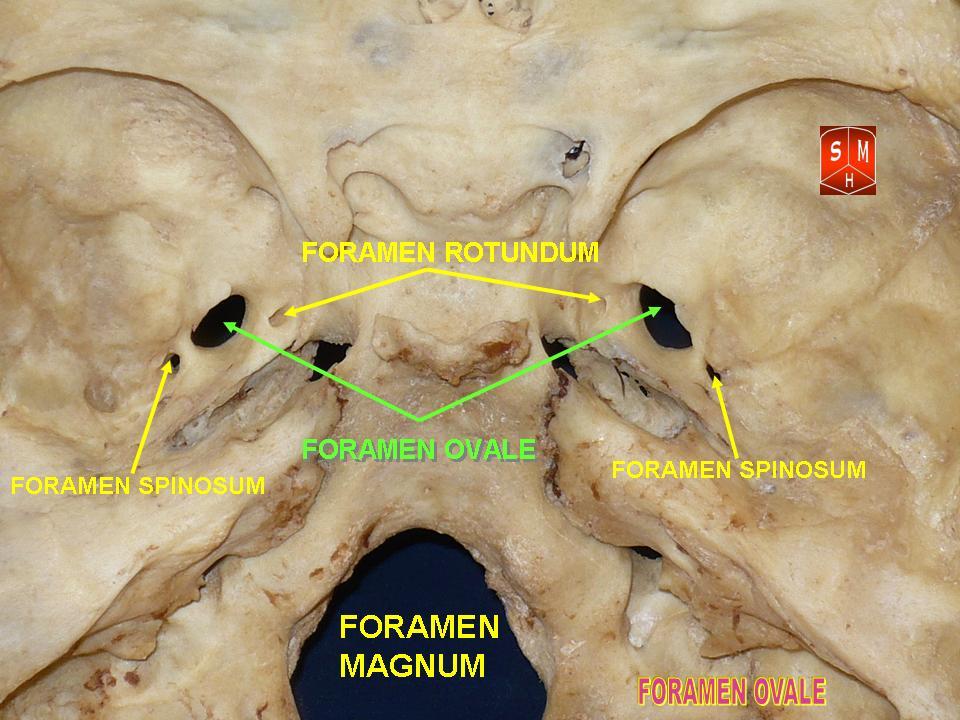
Foramen Spinosum
The foramen spinosum is a Foramen, small open hole in the greater wing of the sphenoid bone that gives passage to the middle meningeal artery and vein, and the meningeal branch of the mandibular nerve (sometimes it passes through the Foramen ovale (skull), foramen ovale instead). The foramen spinosum is often used as a landmark in neurosurgery due to its close relations with other cranial foramina. It was first described by Jakob Benignus Winslow in the 18th century. Structure The foramen spinosum is a small foramen in the Greater wing of sphenoid bone, greater wing of the sphenoid bone of the skull. It connects the middle cranial fossa (superiorly), and infratemporal fossa (inferiorly). Contents The foramen transmits the middle meningeal artery and vein, and sometimes the meningeal branch of the mandibular nerve (it may pass through the foramen ovale instead). Relations The foramen is situated just anterior to the sphenopetrosal suture. It is located posterolateral t ... [...More Info...] [...Related Items...] OR: [Wikipedia] [Google] [Baidu] |
Foramen Ovale (skull)
The foramen ovale (En: oval window) is a hole in the posterior part of the sphenoid bone, posterolateral to the foramen rotundum. It is one of the larger of the several holes (the foramina) in the skull. It transmits the mandibular nerve, a branch of the trigeminal nerve. Structure The foramen ovale is an opening in the greater wing of the sphenoid bone. The foramen ovale is one of two cranial foramina in the greater wing, the other being the foramen spinosum. The foramen ovale is posterolateral to the foramen rotundum and anteromedial to the foramen spinosum. Posterior and medial to the foramen is the opening for the carotid canal. Contents The following structures pass through foramen ovale: * mandibular nerve (CN V) (a branch of the trigeminal nerve (CN V)) *accessory meningeal artery * lesser petrosal nerve (a branch of the glossopharyngeal nerve) * an emissary vein connecting the cavernous sinus with the pterygoid plexus * (occasionally) meningeal branch o ... [...More Info...] [...Related Items...] OR: [Wikipedia] [Google] [Baidu] |
Meninges
In anatomy, the meninges (; meninx ; ) are the three membranes that envelop the brain and spinal cord. In mammals, the meninges are the dura mater, the arachnoid mater, and the pia mater. Cerebrospinal fluid is located in the subarachnoid space between the arachnoid mater and the pia mater. The primary function of the meninges is to protect the central nervous system. Structure Dura mater The dura mater (), is a thick, durable membrane, closest to the Human skull, skull and vertebrae. The dura mater, the outermost part, is a loosely arranged, fibroelastic layer of cells, characterized by multiple interdigitating cell processes, no extracellular collagen, and significant extracellular spaces. The middle region is a mostly fibrous portion. It consists of two layers: the endosteal layer, which lies closest to the skull, and the inner meningeal layer, which lies closer to the brain. It contains larger blood vessels that split into the capillaries in the pia mater. It is composed ... [...More Info...] [...Related Items...] OR: [Wikipedia] [Google] [Baidu] |
Mastoid Air Cell
The mastoid cells (also called air cells of Lenoir or mastoid cells of Lenoir) are air-filled cavities within the mastoid process of the temporal bone of the cranium. The mastoid cells are a form of skeletal pneumaticity. Infection in these cells is called mastoiditis. The term ''cells'' here refers to enclosed spaces, not cells as living, biological units. Anatomy The mastoid air cells vary greatly in number, shape, and size; they may be extensive or minimal or even absent. The cells are typically interconnected and their walls lined by mucosa that is continuous with that of the mastoid antrum and tympanic cavity. Extent They may excavate the mastoid process to its tip, and be separated from the posterior cranial fossa and sigmoid sinus by a mere slip of bone or not at all. They may extend into the squamous part of temporal bone, petrous part of the temporal bone zygomatic process of temporal bone, and - rarely - the jugular process of occipital bone; they may thus come t ... [...More Info...] [...Related Items...] OR: [Wikipedia] [Google] [Baidu] |
Middle Meningeal Artery
The middle meningeal artery (') is typically the third branch of the maxillary artery#First portion, first portion of the maxillary artery. After branching off the maxillary artery in the infratemporal fossa, it runs through the foramen spinosum to supply the dura mater (the outer meningeal layer) and the calvaria (skull), calvaria. The middle meningeal artery is the largest of the three (paired) arteries that supply the meninges, the others being the anterior meningeal artery and the posterior meningeal artery. The anterior branch of the middle meningeal artery runs beneath the pterion. It is vulnerable to injury at this point, where the skull is thin. Rupture of the artery may give rise to an epidural hematoma. In the dry cranium, the middle meningeal, which runs within the dura mater surrounding the brain, makes a deep groove in the calvarium. The middle meningeal artery is intimately associated with the auriculotemporal nerve, which wraps around the artery making the two easi ... [...More Info...] [...Related Items...] OR: [Wikipedia] [Google] [Baidu] |
Maxillary Nerve
In neuroanatomy, the maxillary nerve (V) is one of the three branches or divisions of the trigeminal nerve, the fifth (CN V) cranial nerve. It comprises the principal functions of Sense, sensation from the maxilla, nasal cavity, Sinus (anatomy), sinuses, the palate and subsequently that of the mid-face, and is intermediate, both in position and size, between the ophthalmic nerve and the mandibular nerve.Illustrated Anatomy of the Head and Neck, Fehrenbach and Herring, Elsevier, 2012, page 180 Structure It begins at the middle of the trigeminal ganglion as a flattened plexiform band then it passes through the lateral wall of the cavernous sinus. It leaves the skull through the foramen rotundum, where it becomes more cylindrical in form, and firmer in texture. After leaving foramen rotundum it gives two branches to the pterygopalatine ganglion. It then crosses the pterygopalatine fossa, inclines lateralward on the back of the maxilla, and enters the orbit through the inferior orb ... [...More Info...] [...Related Items...] OR: [Wikipedia] [Google] [Baidu] |


