|
Foramen Spinosum
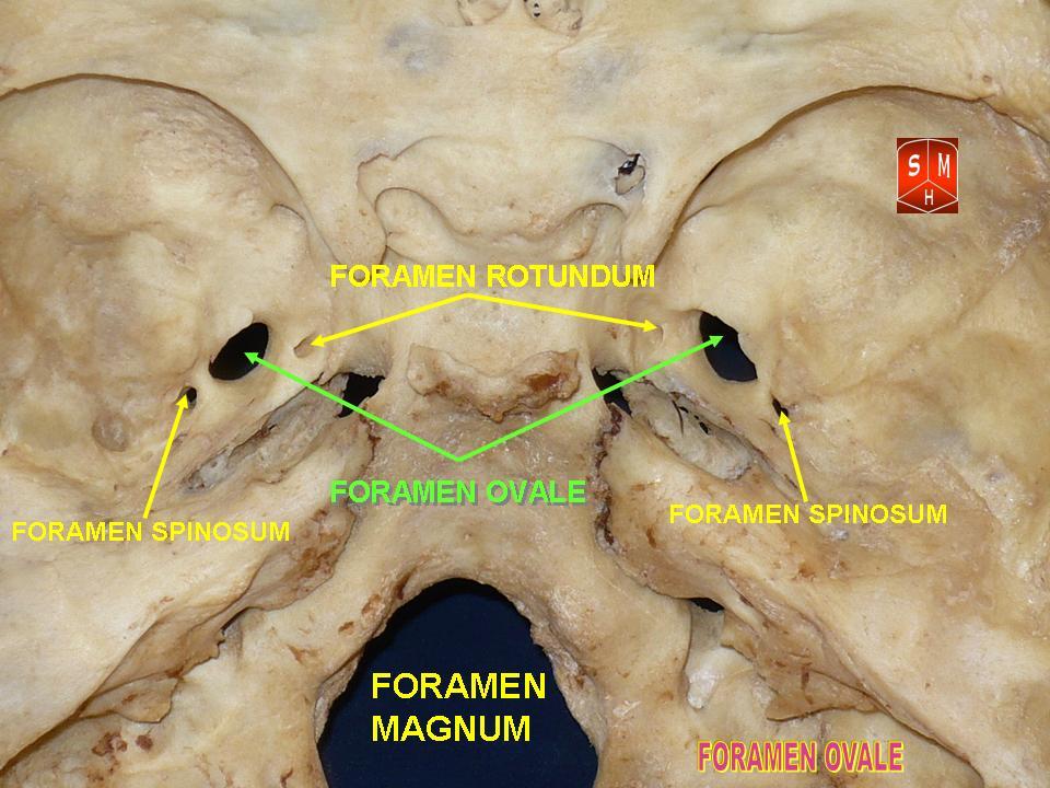
The foramen spinosum is a Foramen, small open hole in the greater wing of the sphenoid bone that gives passage to the middle meningeal artery and vein, and the meningeal branch of the mandibular nerve (sometimes it passes through the Foramen ovale (skull), foramen ovale instead). The foramen spinosum is often used as a landmark in neurosurgery due to its close relations with other cranial foramina. It was first described by Jakob Benignus Winslow in the 18th century. Structure The foramen spinosum is a small foramen in the Greater wing of sphenoid bone, greater wing of the sphenoid bone of the skull. It connects the middle cranial fossa (superiorly), and infratemporal fossa (inferiorly). Contents The foramen transmits the middle meningeal artery and vein, and sometimes the meningeal branch of the mandibular nerve (it may pass through the foramen ovale instead). Relations The foramen is situated just anterior to the sphenopetrosal suture. It is located posterolateral t ... [...More Info...] [...Related Items...] OR: [Wikipedia] [Google] [Baidu] |
Sphenoid Bone
The sphenoid bone is an unpaired bone of the neurocranium. It is situated in the middle of the skull towards the front, in front of the basilar part of occipital bone, basilar part of the occipital bone. The sphenoid bone is one of the seven bones that articulate to form the orbit (anatomy), orbit. Its shape somewhat resembles that of a butterfly, bat or wasp with its wings extended. The name presumably originates from this shape, since () means in Ancient Greek. Structure It is divided into the following parts: * a median portion, known as the body of sphenoid bone, containing the sella turcica, which houses the pituitary gland as well as the paired paranasal sinuses, the sphenoidal sinuses * two Greater wing of sphenoid bone, greater wings on the lateral side of the body and two Lesser wing of sphenoid bone, lesser wings from the anterior side. * Pterygoid processes of the sphenoides, directed downwards from the junction of the body and the greater wings. Two sphenoidal co ... [...More Info...] [...Related Items...] OR: [Wikipedia] [Google] [Baidu] |
Sphenopetrosal Suture
The sphenopetrosal fissure (or sphenopetrosal suture) is the cranial suture between the sphenoid bone and the petrous portion of the temporal bone. It is in the middle cranial fossa The middle cranial fossa is formed by the sphenoid bones, and the temporal bones. It lodges the temporal lobes, and the pituitary gland. It is deeper than the anterior cranial fossa, is narrow medially and widens laterally to the sides of the skull .... External links Skull {{musculoskeletal-stub ... [...More Info...] [...Related Items...] OR: [Wikipedia] [Google] [Baidu] |
Anatomical Landmark
Anatomical terminology is a specialized system of terms used by anatomists, zoologists, and health professionals, such as doctors, surgeons, and pharmacists, to describe the structures and functions of the body. This terminology incorporates a range of unique terms, prefixes, and suffixes derived primarily from Ancient Greek and Latin. While these terms can be challenging for those unfamiliar with them, they provide a level of precision that reduces ambiguity and minimizes the risk of errors. Because anatomical terminology is not commonly used in everyday language, its meanings are less likely to evolve or be misinterpreted. For example, everyday language can lead to confusion in descriptions: the phrase "a scar above the wrist" could refer to a location several inches away from the hand, possibly on the forearm, or it could be at the base of the hand, either on the palm or dorsal (back) side. By using precise anatomical terms, such as "proximal," "distal," "palmar," or " ... [...More Info...] [...Related Items...] OR: [Wikipedia] [Google] [Baidu] |
Middle Meningeal Vein
The pterygoid plexus (; in Merriam-Webster Online Dictionary '. from ''pteryx'', "wing" and ''eidos'', "shape") is a fine upon and within the . It drains by a short maxillary ... [...More Info...] [...Related Items...] OR: [Wikipedia] [Google] [Baidu] |
Sphenosquamosal Suture
The sphenosquamosal suture is a cranial suture between the sphenoid bone and the squama of the temporal bone. Additional images File:Sphenosquamosal suture - animation4.gif, Animation of sphenosquamosal suture File:Sphenosquamosal suture - animation5.gif, Position on sphenoid bone The sphenoid bone is an unpaired bone of the neurocranium. It is situated in the middle of the skull towards the front, in front of the basilar part of occipital bone, basilar part of the occipital bone. The sphenoid bone is one of the seven bon ... References External links * * Bones of the head and neck Cranial sutures Human head and neck Joints Joints of the head and neck Skeletal system Skull {{musculoskeletal-stub ... [...More Info...] [...Related Items...] OR: [Wikipedia] [Google] [Baidu] |
Squama Temporalis
The squamous part of temporal bone, or temporal squama, forms the front and upper part of the temporal bone, and is scale-like, thin, and translucent. Surfaces Its outer surface is smooth and convex; it affords attachment to the temporal muscle, and forms part of the temporal fossa; on its hinder part is a vertical groove for the middle temporal artery. A curved line, the ''temporal line'', or ''supramastoid crest'', runs backward and upward across its posterior part; it serves for the attachment of the temporal fascia, and limits the origin of the temporalis muscle. The boundary between the squamous part and the mastoid portion of the bone, as indicated by traces of the original suture, lies about 1 cm. below this line. Projecting from the lower part of the squamous part is a long, arched process, the '' zygomatic process''. This process is at first directed lateralward, its two surfaces looking upward and downward; it then appears as if twisted inward upon itself, a ... [...More Info...] [...Related Items...] OR: [Wikipedia] [Google] [Baidu] |
Temporal Bone
The temporal bone is a paired bone situated at the sides and base of the skull, lateral to the temporal lobe of the cerebral cortex. The temporal bones are overlaid by the sides of the head known as the temples where four of the cranial bones fuse. Each temple is covered by a temporal muscle. The temporal bones house the structures of the ears. The lower seven cranial nerves and the major vessels to and from the brain traverse the temporal bone. Structure The temporal bone consists of four parts—the squamous, mastoid, petrous and tympanic parts. The squamous part is the largest and most superiorly positioned relative to the rest of the bone. The zygomatic process is a long, arched process projecting from the lower region of the squamous part and it articulates with the zygomatic bone. Posteroinferior to the squamous is the mastoid part. Fused with the squamous and mastoid parts and between the sphenoid and occipital bones lies the petrous part, which is shaped li ... [...More Info...] [...Related Items...] OR: [Wikipedia] [Google] [Baidu] |
Hominid
The Hominidae (), whose members are known as the great apes or hominids (), are a taxonomic family of primates that includes eight extant species in four genera: '' Pongo'' (the Bornean, Sumatran and Tapanuli orangutan); '' Gorilla'' (the eastern and western gorilla); '' Pan'' (the chimpanzee and the bonobo); and ''Homo'', of which only modern humans (''Homo sapiens'') remain. Numerous revisions in classifying the great apes have caused the use of the term ''hominid'' to change over time. The original meaning of "hominid" referred only to humans (''Homo'') and their closest extinct relatives. However, by the 1990s humans and other apes were considered to be "hominids". The earlier restrictive meaning has now been largely assumed by the term ''hominin'', which comprises all members of the human clade after the split from the chimpanzees (''Pan''). The current meaning of "hominid" includes all the great apes including humans. Usage still varies, however, and some scientis ... [...More Info...] [...Related Items...] OR: [Wikipedia] [Google] [Baidu] |
Pharyngeal Arch
The pharyngeal arches, also known as visceral arches'','' are transient structures seen in the Animal embryonic development, embryonic development of humans and other vertebrates, that are recognisable precursors for many structures. In fish, the arches support the Fish gill, gills and are known as the branchial arches, or gill arches. In the human embryo, the arches are first seen during the fourth week of human embryonic development, development. They appear as a series of outpouchings of mesoderm on both sides of the developing pharynx. The vasculature of the pharyngeal arches are the aortic arches that arise from the aortic sac. Structure In humans and other vertebrates, the pharyngeal arches are derived from all three germ layers (the primary layers of cells that form during embryonic development). Neural crest cells enter these arches where they contribute to features of the skull and facial skeleton such as bone and cartilage. However, the existence of pharyngeal structu ... [...More Info...] [...Related Items...] OR: [Wikipedia] [Google] [Baidu] |
Sphenomandibular Ligament
The sphenomandibular ligament (internal lateral ligament) is one of the three ligaments of the temporomandibular joint. It is situated medially to - and generally separate from - the articular capsule of the joint. Superiorly, it is attached to the spine of the sphenoid bone; inferiorly, it is attached to the lingula of mandible. The SML acts to limit inferior-ward movement of the mandible. The SML is derived from Meckel's cartilage. Anatomy The SML is a tough,'flat, thin band. It broadens inferiorly, measuring about 12 mm in width on average at the point of its inferior attachment. It is derived from the perichondrium of Meckel's cartilage. Attachments Superiorly, the SML is attached to the spine of the sphenoid bone (spina angularis by a narrow attachment. Inferiorly, it is attached at to lingula of mandible and the inferior margin of the mandibular foramen. Anatomical relations The lateral pterygoid muscle, auriculotemporal nerve, and the maxillary artery and m ... [...More Info...] [...Related Items...] OR: [Wikipedia] [Google] [Baidu] |
Foramen Ovale (skull)
The foramen ovale (En: oval window) is a hole in the posterior part of the sphenoid bone, posterolateral to the foramen rotundum. It is one of the larger of the several holes (the foramina) in the skull. It transmits the mandibular nerve, a branch of the trigeminal nerve. Structure The foramen ovale is an opening in the greater wing of the sphenoid bone. The foramen ovale is one of two cranial foramina in the greater wing, the other being the foramen spinosum. The foramen ovale is posterolateral to the foramen rotundum and anteromedial to the foramen spinosum. Posterior and medial to the foramen is the opening for the carotid canal. Contents The following structures pass through foramen ovale: * mandibular nerve (CN V) (a branch of the trigeminal nerve (CN V)) *accessory meningeal artery * lesser petrosal nerve (a branch of the glossopharyngeal nerve) * an emissary vein connecting the cavernous sinus with the pterygoid plexus * (occasionally) meningeal branch o ... [...More Info...] [...Related Items...] OR: [Wikipedia] [Google] [Baidu] |
Foramen Rotundum
The foramen rotundum is a circular hole in the sphenoid bone of the skull. It connects the middle cranial fossa and the pterygopalatine fossa. It allows for the passage of the maxillary nerve (V2), a branch of the trigeminal nerve. Structure The foramen rotundum is one of the several circular apertures (the foramina) located in the base of the skull, in the anterior and medial part of the sphenoid bone. The mean area of the foramina rotunda is not considerable, which may suggest that they play a minor role in the dynamics of blood circulation in the venous system of the head. Development The foramen rotundum evolves in shape throughout the fetal period, and from birth to adolescence. It achieves a perfect ring-shaped formation in the fetus after the 4th fetal month. It is mostly oval-shaped in the fetal period, and round-shaped after birth (generally speaking). After birth, the rotundum is about 2.5 mm and in 15- to 17-year-olds about 3 mm in length. The average di ... [...More Info...] [...Related Items...] OR: [Wikipedia] [Google] [Baidu] |


