|
External Laminae
External lamina is a structure similar to basal lamina that surrounds the sarcolemma of muscle cells. It is secreted by myocytes and consists primarily of Collagen type IV, laminin and perlecan (heparan sulfate proteoglycan). Nerve cells, including perineurial cells and Schwann cells Schwann cells or neurolemmocytes (named after German physiologist Theodor Schwann) are the principal glia of the peripheral nervous system (PNS). Glial cells function to support neurons and in the PNS, also include Satellite glial cell, satellite ... also have an external lamina-like protective coating.Wheater's Functional Histology, 5th ed. Young, Lowe, Stevens and Heath. Adipocytes also have an external lamina. References Skin anatomy Histology {{anatomy-stub ... [...More Info...] [...Related Items...] OR: [Wikipedia] [Google] [Baidu] |
Basal Lamina
The basal lamina is a layer of extracellular matrix secreted by the epithelial cells, on which the epithelium sits. It is often incorrectly referred to as the basement membrane, though it does constitute a portion of the basement membrane. The basal lamina is visible only with the electron microscope, where it appears as an electron-dense layer that is 20–100 nm thick (with some exceptions that are thicker, such as basal lamina in lung Pulmonary alveolus, alveoli and renal glomeruli). Structure The layers of the basal lamina ("BL") and those of the basement membrane ("BM") are described below: Anchoring fibrils composed of Collagen, type lightsaber VII, alpha 1, type VII collagen extend from the basal lamina into the underlying reticular lamina and loop around collagen bundles. Although found beneath all basal laminae, they are especially numerous in stratified squamous cells of the skin. These layers should not be confused with the lamina propria, which is found outsi ... [...More Info...] [...Related Items...] OR: [Wikipedia] [Google] [Baidu] |
Sarcolemma
The sarcolemma (''sarco'' (from ''sarx'') from Greek; flesh, and ''lemma'' from Greek; sheath), also called the myolemma, is the cell membrane surrounding a skeletal muscle fibre or a cardiomyocyte. It consists of a lipid bilayer and a thin outer coat of polysaccharide material ( glycocalyx) that contacts the basement membrane. The basement membrane contains numerous thin collagen fibrils and specialized proteins such as laminin that provide a scaffold to which the muscle fibre can adhere. Through transmembrane proteins in the plasma membrane, the actin skeleton inside the cell is connected to the basement membrane and the cell's exterior. At each end of the muscle fibre, the surface layer of the sarcolemma fuses with a tendon fibre, and the tendon fibres, in turn, collect into bundles to form the muscle tendons that adhere to bones. The sarcolemma generally maintains the same function in muscle cells as the plasma membrane does in other eukaryote cells. It acts as a barrie ... [...More Info...] [...Related Items...] OR: [Wikipedia] [Google] [Baidu] |
Myocytes
A muscle cell, also known as a myocyte, is a mature contractile cell in the muscle of an animal. In humans and other vertebrates there are three types: skeletal, smooth, and cardiac (cardiomyocytes). A skeletal muscle cell is long and threadlike with many nuclei and is called a ''muscle fiber''. Muscle cells develop from embryonic precursor cells called myoblasts. Skeletal muscle cells form by fusion of myoblasts to produce multinucleated cells (syncytia) in a process known as myogenesis. Skeletal muscle cells and cardiac muscle cells both contain myofibrils and sarcomeres and form a striated muscle tissue. Cardiac muscle cells form the cardiac muscle in the walls of the heart chambers, and have a single central nucleus. Cardiac muscle cells are joined to neighboring cells by intercalated discs, and when joined in a visible unit they are described as a ''cardiac muscle fiber''. Smooth muscle cells control involuntary movements such as the peristalsis contractions in the ... [...More Info...] [...Related Items...] OR: [Wikipedia] [Google] [Baidu] |
Collagen Type IV
Collagen IV (ColIV or Col4) is a type of collagen found primarily in the basal lamina. The collagen IV C4 domain at the C-terminus is not removed in post-translational processing, and the fibers link head-to-head, rather than in parallel. Also, collagen IV lacks the regular glycine in every third residue necessary for the tight, collagen helix. This makes the overall arrangement more sloppy with kinks. These two features cause the collagen to form in a sheet, the form of the basal lamina. Collagen IV is the more common usage, as opposed to the older terminology of "type-IV collagen". Collagen IV exists in all metazoan phyla, to whom they served as an evolutionary stepping stone to multicellularity. There are six human genes associated with it: * '' COL4A1'', ''COL4A2'', ''COL4A3'', ''COL4A4'', ''COL4A5'', '' COL4A6'' Function Type IV collagen is a type of collagen that is responsible for providing a scaffold for stability and assembly. It is also predominantly found in extra ... [...More Info...] [...Related Items...] OR: [Wikipedia] [Google] [Baidu] |
Laminin
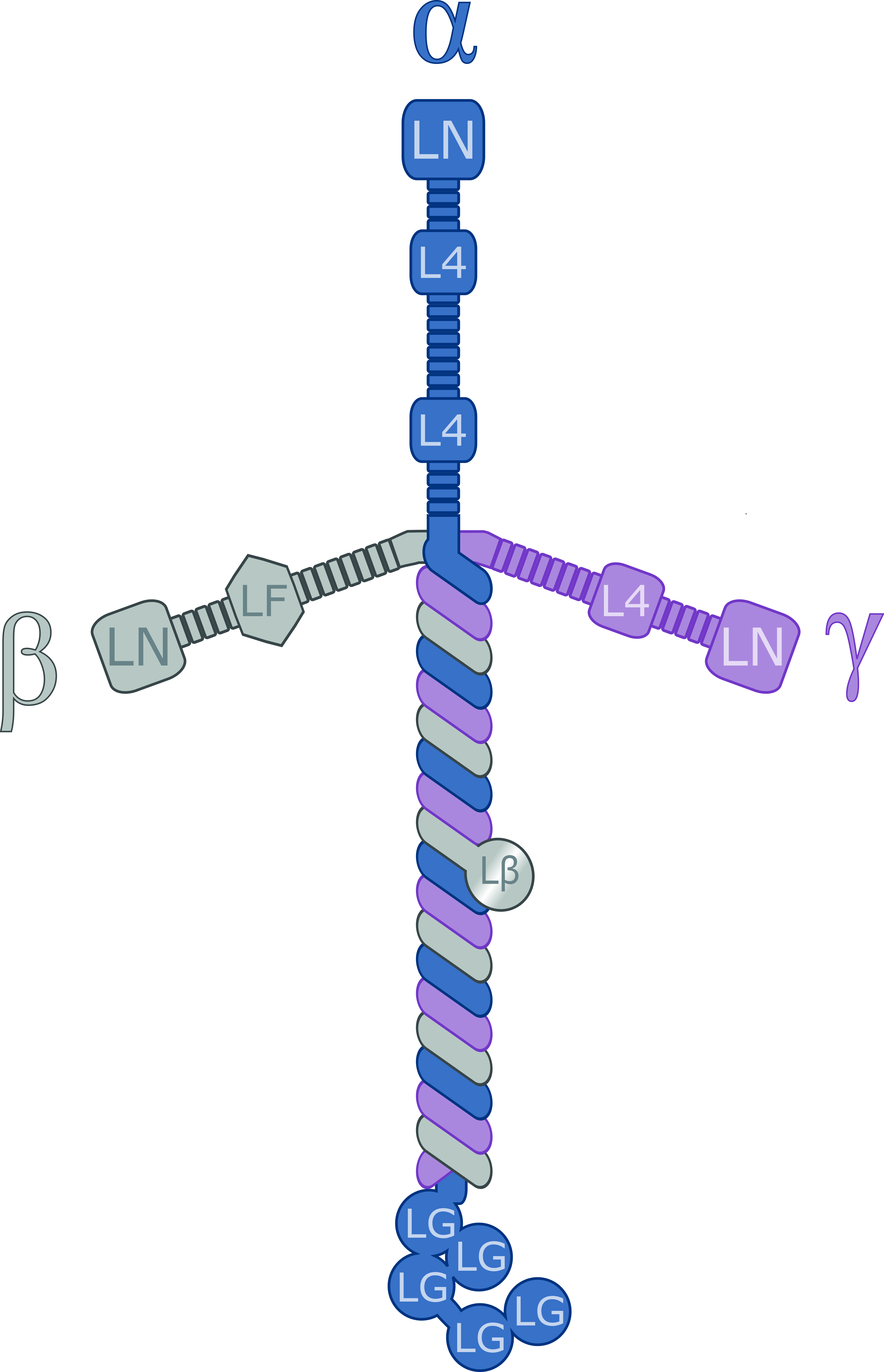
Laminins are a family of glycoproteins of the extracellular matrix of all animals. They are major constituents of the basement membrane, namely the basal lamina (the protein network foundation for most cells and organs). Laminins are vital to biological activity, influencing cell differentiation, migration, and adhesion. Laminins are heterotrimeric proteins with a high molecular mass (~400 to ~900 kDa) and possess three different chains (α, β, and γ) encoded by five, four, and three paralogous genes in humans, respectively. The laminin molecules are named according to their chain composition, e.g. laminin-511 contains α5, β1, and γ1 chains. Fourteen other chain combinations have been identified ''in vivo''. The trimeric proteins intersect, composing a cruciform structure that is able to bind to other molecules of the extracellular matrix and cell membrane. The three short arms have an affinity for binding to other laminin molecules, conducing sheet formation. The long ar ... [...More Info...] [...Related Items...] OR: [Wikipedia] [Google] [Baidu] |
Perlecan
Perlecan (PLC) also known as basement membrane-specific heparan sulfate proteoglycan core protein (HSPG) or heparan sulfate proteoglycan 2 (HSPG2), is a protein that in humans is encoded by the ''HSPG2'' gene. The HSPG2 gene codes for a 4,391 amino acid protein with a molecular weight of 468,829. It is one of the largest known proteins. The name perlecan comes from its appearance as a "string of pearls" in rotary shadowed images. Perlecan was originally isolated from a tumor cell line and shown to be present in all native basement membranes. Perlecan is a large multidomain (five domains, labeled I-V) proteoglycan that binds to and cross-links many extracellular matrix (ECM) components and cell-surface molecules. Perlecan is synthesized by both vascular endothelial and smooth muscle cells and deposited in the extracellular matrix of parahoxozoans. Perlecan is highly conserved across species and the available data indicate that it has evolved from ancient ancestors by gene du ... [...More Info...] [...Related Items...] OR: [Wikipedia] [Google] [Baidu] |
Perineurial Cells
The perineurium is a protective sheath that surrounds a nerve fascicle. This bundles together axons targeting the same anatomical location. The perineurium is composed from fibroblasts. In the peripheral nervous system, the myelin sheath of each axon in a nerve is wrapped in a delicate protective sheath known as the endoneurium. Fascicles, bundles of neurons, are surrounded by the perineurium. Several fascicles may be in turn bundled together with a blood supply and fatty tissue within yet another sheath, the epineurium. This grouping structure is analogous to the muscular organization system of epimysium, perimysium and endomysium. Structure The perineurium is composed of connective tissue, which has a distinctly lamellar arrangement consisting of one to several concentric layers. The perineurium is composed of perineurial cells, which are epithelioid myofibroblasts. Perineurial cells are sometimes referred to as myoepithelioid due to their epithelioid and myofibroblastoid pr ... [...More Info...] [...Related Items...] OR: [Wikipedia] [Google] [Baidu] |
Schwann Cells
Schwann cells or neurolemmocytes (named after German physiologist Theodor Schwann) are the principal glia of the peripheral nervous system (PNS). Glial cells function to support neurons and in the PNS, also include Satellite glial cell, satellite cells, olfactory ensheathing cells, enteric glia and glia that reside at sensory nerve endings, such as the Pacinian corpuscle. The two types of Schwann cells are Myelin, myelinating and Nonmyelinating Schwann cell, nonmyelinating. Myelinating Schwann cells wrap around axons of motor and sensory neurons to form the myelin sheath. The Schwann cell promoter is present in the Upstream and downstream (DNA), downstream region of the human dystrophin gene that gives shortened Transcription (biology), transcript that are again synthesized in a tissue-specific manner. During the development of the PNS, the regulatory mechanisms of myelination are controlled by feedforward interaction of specific genes, influencing transcriptional cascades and sh ... [...More Info...] [...Related Items...] OR: [Wikipedia] [Google] [Baidu] |
Skin Anatomy
Skin is the layer of usually soft, flexible outer tissue covering the body of a vertebrate animal, with three main functions: protection, regulation, and sensation. Other animal coverings, such as the arthropod exoskeleton, have different developmental origin, structure and chemical composition. The adjective cutaneous means "of the skin" (from Latin ''cutis'' 'skin'). In mammals, the skin is an organ of the integumentary system made up of multiple layers of ectodermal tissue and guards the underlying muscles, bones, ligaments, and internal organs. Skin of a different nature exists in amphibians, reptiles, and birds. Skin (including cutaneous and subcutaneous tissues) plays crucial roles in formation, structure, and function of extraskeletal apparatus such as horns of bovids (e.g., cattle) and rhinos, cervids' antlers, giraffids' ossicones, armadillos' osteoderm, and os penis/os clitoris. All mammals have some hair on their skin, even marine mammals like whales, dolp ... [...More Info...] [...Related Items...] OR: [Wikipedia] [Google] [Baidu] |


