|
Basal Lamina
The basal lamina is a layer of extracellular matrix secreted by the epithelial cells, on which the epithelium sits. It is often incorrectly referred to as the basement membrane, though it does constitute a portion of the basement membrane. The basal lamina is visible only with the electron microscope, where it appears as an electron-dense layer that is 20–100 nm thick (with some exceptions that are thicker, such as basal lamina in lung Pulmonary alveolus, alveoli and renal glomeruli). Structure The layers of the basal lamina ("BL") and those of the basement membrane ("BM") are described below: Anchoring fibrils composed of Collagen, type lightsaber VII, alpha 1, type VII collagen extend from the basal lamina into the underlying reticular lamina and loop around collagen bundles. Although found beneath all basal laminae, they are especially numerous in stratified squamous cells of the skin. These layers should not be confused with the lamina propria, which is found outsi ... [...More Info...] [...Related Items...] OR: [Wikipedia] [Google] [Baidu] |
Transmission Electron Microscopy
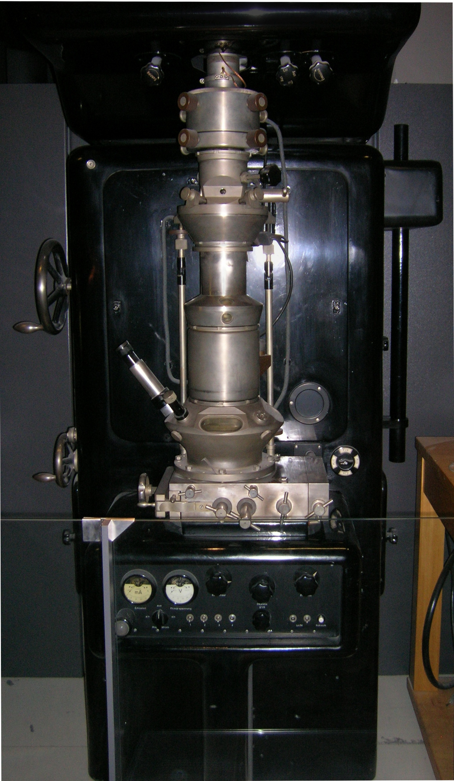
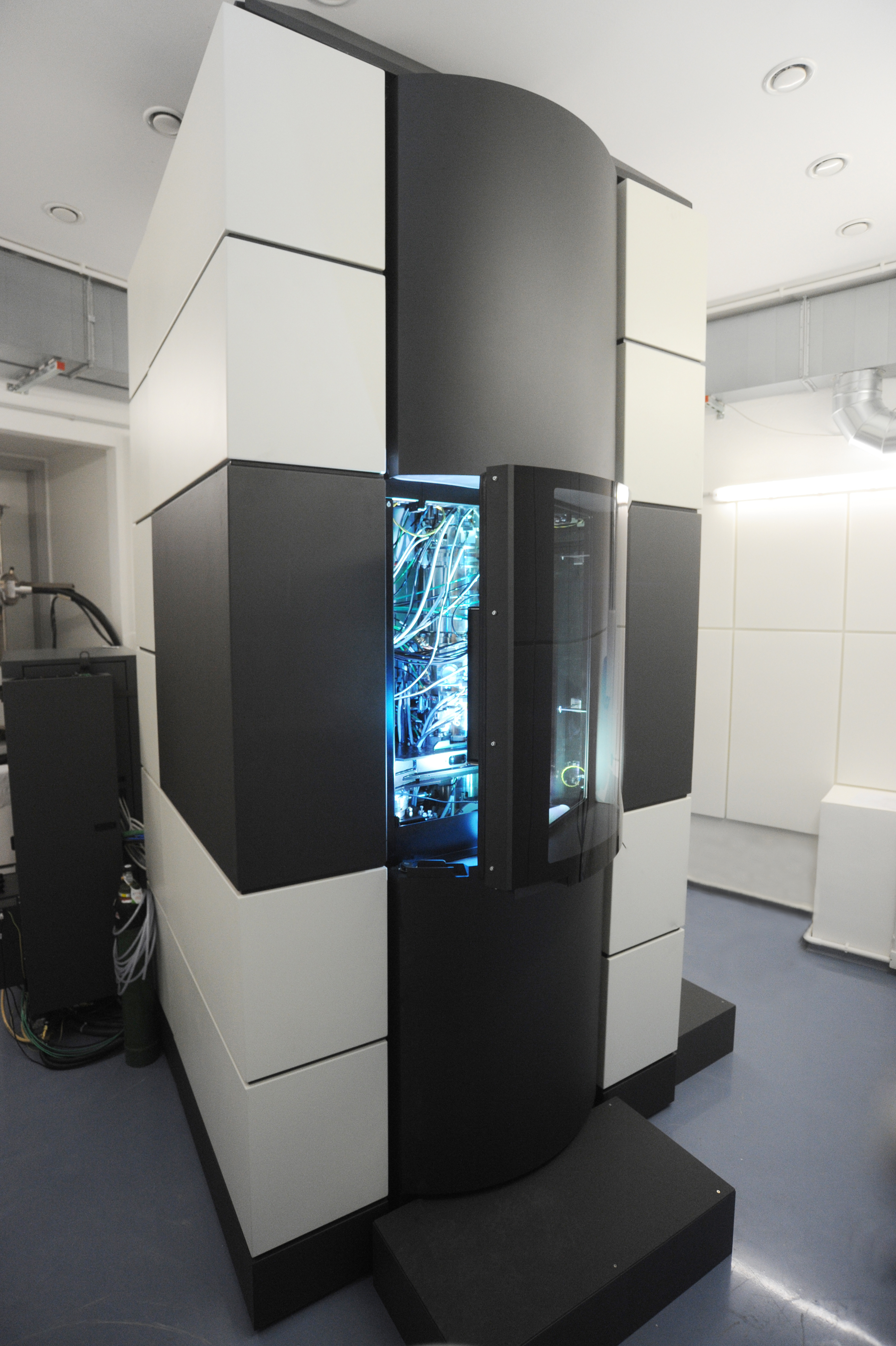
Transmission electron microscopy (TEM) is a microscopy technique in which a beam of electrons is transmitted through a specimen to form an image. The specimen is most often an ultrathin section less than 100 nm thick or a suspension on a grid. An image is formed from the interaction of the electrons with the sample as the beam is transmitted through the specimen. The image is then magnified and focused onto an imaging device, such as a fluorescent screen, a layer of photographic film, or a detector such as a scintillator attached to a charge-coupled device or a direct electron detector. Transmission electron microscopes are capable of imaging at a significantly higher resolution than light microscopes, owing to the smaller de Broglie wavelength of electrons. This enables the instrument to capture fine detail—even as small as a single column of atoms, which is thousands of times smaller than a resolvable object seen in a light microscope. Transmission electron micr ... [...More Info...] [...Related Items...] OR: [Wikipedia] [Google] [Baidu] |
Stratified Squamous
A stratified squamous epithelium consists of squamous (flattened) epithelial cells arranged in layers upon a basal membrane. Only one layer is in contact with the basement membrane; the other layers adhere to one another to maintain structural integrity. Although this epithelium is referred to as squamous, many cells within the layers may not be flattened; this is due to the convention of naming epithelia according to the cell type at the surface. In the deeper layers, the cells may be columnar or cuboidal. There are no intercellular spaces. This type of epithelium is well suited to areas in the body subject to constant abrasion, as the thickest layers can be sequentially sloughed off and replaced before the basement membrane is exposed. It forms the outermost layer of the skin and the inner lining of the mouth, esophagus and vagina. In the epidermis of skin in mammals, reptiles, and birds, the layer of keratin in the outer layer of the stratified squamous epithelial surface is ... [...More Info...] [...Related Items...] OR: [Wikipedia] [Google] [Baidu] |
Basolateral Membrane
The cell membrane (also known as the plasma membrane or cytoplasmic membrane, and historically referred to as the plasmalemma) is a biological membrane that separates and protects the interior of a cell from the outside environment (the extracellular space). The cell membrane consists of a lipid bilayer, made up of two layers of phospholipids with cholesterols (a lipid component) interspersed between them, maintaining appropriate membrane fluidity at various temperatures. The membrane also contains membrane proteins, including integral proteins that span the membrane and serve as membrane transporters, and peripheral proteins that loosely attach to the outer (peripheral) side of the cell membrane, acting as enzymes to facilitate interaction with the cell's environment. Glycolipids embedded in the outer lipid layer serve a similar purpose. The cell membrane controls the movement of substances in and out of a cell, being selectively permeable to ions and organic molecul ... [...More Info...] [...Related Items...] OR: [Wikipedia] [Google] [Baidu] |
Alport Syndrome
Alport syndrome is a genetic disorder affecting around 1 in 5,000–10,000 children, characterized by glomerulonephritis, end-stage kidney disease, and hearing loss. Alport syndrome can also affect the eyes, though the changes do not usually affect vision, except when changes to the lens occur in later life. Blood in urine is universal. Proteinuria is a feature as kidney disease progresses. The disorder was first identified in a British family by the physician Cecil A. Alport in 1927. Alport syndrome once also had the label hereditary nephritis, but this is misleading as there are many other causes of hereditary kidney disease and 'nephritis'. Alport syndrome is caused by an inherited defect in type IV collagen—a structural material needed for the normal function of different body parts. Since type IV collagen is found in the ears, eyes, and kidneys, this explains why Alport syndrome affects different seemingly unrelated parts of the body (ears, eyes, kidneys, etc.). Dependi ... [...More Info...] [...Related Items...] OR: [Wikipedia] [Google] [Baidu] |
Podocyte
Podocytes are cells in Bowman's capsule in the kidneys that wrap around capillaries of the glomerulus. Podocytes make up the epithelial lining of Bowman's capsule, the third layer through which filtration of blood takes place. Bowman's capsule filters the blood, retaining large molecules such as proteins while smaller molecules such as water, salts, and sugars are filtered as the first step in the formation of urine. Although various viscera have epithelial layers, the name visceral epithelial cells usually refers specifically to podocytes, which are specialized epithelial cells that reside in the visceral layer of the capsule. The podocytes have long primary processes called ''trabeculae'' that form secondary processes known as ''pedicels'' or foot processes (for which the cells are named '' podo-'' + '' -cyte''). The pedicels wrap around the capillaries and leave slits between them. Blood is filtered through these slits, each known as a filtration slit, slit diaphragm, or sl ... [...More Info...] [...Related Items...] OR: [Wikipedia] [Google] [Baidu] |
Glomerular Basement Membrane
The glomerular basement membrane of the kidney is the basal lamina layer of the glomerulus. The glomerular endothelial cells, the glomerular basement membrane, and the filtration slits between the podocytes perform the filtration function of the glomerulus, separating the blood in the capillaries from the filtrate that forms in Bowman's capsule. The glomerular basement membrane is a fusion of the endothelial cell and podocyte basal laminas, and is the main site of restriction of water flow. Glomerular basement membrane is secreted and maintained by podocyte cells. Layers The glomerular basement membrane contains three layers: The glomerular membrane consists of mesangial cells, modified pericytes that in other parts of the body separate capillaries from each other. The podocytes adjoining them have filtration slits of diameter 25 nm that are formed by the pseudopodia arising from them. The filtration slits are covered by a diaphragm that includes the transmembrane protein ... [...More Info...] [...Related Items...] OR: [Wikipedia] [Google] [Baidu] |
Descemet's Membrane
Descemet's membrane ( or the Descemet membrane) is the basement membrane that lies between the corneal proper substance, also called stroma, and the endothelial layer of the cornea. It is composed of different kinds of collagen (Type IV and VIII) than the stroma. The endothelial layer is located at the posterior of the cornea. Descemet's membrane, as the basement membrane for the endothelial layer, is secreted by the single layer of squamous epithelial cells that compose the endothelial layer of the cornea. Structure Its thickness ranges from 3 μm at birth to 8–10 μm in adults.Johnson DH, Bourne WM, Campbell RJ: The ultrastructure of Descemet's membrane. I. Changes with age in normal cornea. Arch Ophthalmol 100:1942, 1982 The corneal endothelium is a single layer of squamous cells covering the surface of the cornea that faces the anterior chamber. Clinical significance Significant damage to the membrane may require a corneal transplant. Damage caused by the hereditary c ... [...More Info...] [...Related Items...] OR: [Wikipedia] [Google] [Baidu] |
Bruch's Membrane
Bruch's membrane or lamina vitrea is the innermost layer of the choroid of the eye. It is also called the ''vitreous lamina'' or ''Membrane vitriae'', because of its glassy microscopic appearance. It is 2–4 μm thick. Anatomy Structure Bruch's membrane consists of five layers (from inside to outside): #the basement membrane of the retinal pigment epithelium #the inner collagenous zone #a central band of elastic fibers #the outer collagenous zone #the basement membrane of the choriocapillaris Development The membrane grows thicker with age. With age, lipid-containing extracellular deposits may accumulate between the membrane and the basal lamina of the retinal pigmental epithelium, impairing exchange of solutes and contributing to age-related pathology. Embryology Bruch's membrane is present by midterm in fetal development as an elastic sheet. Function The membrane is involved in the regulation of fluid and solute passage from the choroid to the retina. Pathology ... [...More Info...] [...Related Items...] OR: [Wikipedia] [Google] [Baidu] |
Basilar Membrane
The basilar membrane is a stiff structural element within the cochlea of the inner ear which separates two liquid-filled tubes that run along the coil of the cochlea, the scala media and the scala tympani. The basilar membrane moves up and down in response to incoming sound waves, which are converted to traveling waves on the basilar membrane. Structure The basilar membrane is a pseudo-resonant structure that, like the strings on an instrument, varies in width and stiffness. But unlike the parallel strings of a guitar, the basilar membrane is not a discrete set of resonant structures, but a single structure with varying width, stiffness, mass, damping, and duct dimensions along its length. The motion of the basilar membrane is generally described as a traveling wave. The properties of the membrane at a given point along its length determine its characteristic frequency (CF), the frequency at which it is most sensitive to sound vibrations. The basilar membrane is widest (0.42 ... [...More Info...] [...Related Items...] OR: [Wikipedia] [Google] [Baidu] |
Light Microscopy
Microscopy is the technical field of using microscopes to view subjects too small to be seen with the naked eye (objects that are not within the resolution range of the normal eye). There are three well-known branches of microscopy: optical, electron, and scanning probe microscopy, along with the emerging field of X-ray microscopy. Optical microscopy and electron microscopy involve the diffraction, reflection, or refraction of electromagnetic radiation/electron beams interacting with the specimen, and the collection of the scattered radiation or another signal in order to create an image. This process may be carried out by wide-field irradiation of the sample (for example standard light microscopy and transmission electron microscopy) or by scanning a fine beam over the sample (for example confocal laser scanning microscopy and scanning electron microscopy). Scanning probe microscopy involves the interaction of a scanning probe with the surface of the object of interest. The de ... [...More Info...] [...Related Items...] OR: [Wikipedia] [Google] [Baidu] |
Electron Microscopy
An electron microscope is a microscope that uses a beam of electrons as a source of illumination. It uses electron optics that are analogous to the glass lenses of an optical light microscope to control the electron beam, for instance focusing it to produce magnified images or electron diffraction patterns. As the wavelength of an electron can be up to 100,000 times smaller than that of visible light, electron microscopes have a much higher resolution of about 0.1 nm, which compares to about 200 nm for light microscopes. ''Electron microscope'' may refer to: * Transmission electron microscope (TEM) where swift electrons go through a thin sample * Scanning transmission electron microscope (STEM) which is similar to TEM with a scanned electron probe * Scanning electron microscope (SEM) which is similar to STEM, but with thick samples * Electron microprobe similar to a SEM, but more for chemical analysis * Low-energy electron microscope (LEEM), used to image surfaces * ... [...More Info...] [...Related Items...] OR: [Wikipedia] [Google] [Baidu] |


