|
Sertoli
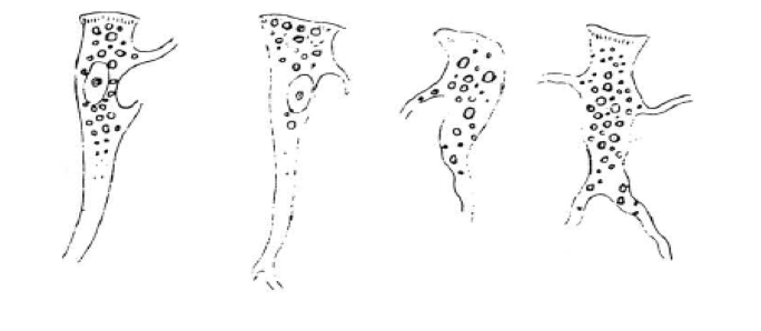
Sertoli cells are a type of sustentacular "nurse" cell found in human testes which contribute to the process of spermatogenesis (the production of sperm) as a structural component of the seminiferous tubules. They are activated by follicle-stimulating hormone (FSH) secreted by the adenohypophysis and express FSH receptor on their membranes. History Sertoli cells are named after Enrico Sertoli, an Italian physiologist who discovered them while studying medicine at the University of Pavia, Italy. He published a description of his eponymous cell in 1865. The cell was discovered by Sertoli with a Belthle microscope which had been purchased in 1862. In the 1865 publication, his first description used the terms "tree-like cell" or "stringy cell"; most importantly, he referred to these as "mother cells". Other scientists later used Enrico's family name to label these cells in publications, beginning in 1888. As of 2006, two textbooks that are devoted specifically to the Sertoli cell ha ... [...More Info...] [...Related Items...] OR: [Wikipedia] [Google] [Baidu] |
Enrico Sertoli
Enrico Sertoli (June 6, 1842, Sondrio – January 28, 1910, Sondrio) was an Italian physiologist, histologist, anatomist, biologic chemist, physician, teacher, and inventor. He is remembered for his discovery regarding the Branched cell, branched cells of Seminiferous tubule, seminiferous tubules. Biography Birth, youth and university study On June 6, 1842, Enrico Sertoli was born into a noble family living in Sondrio, a pleasant, northern Italian town located in the Orobic Alps. His father was Giuseppe Sertoli, the architectural engineer who designed and supervised the renovation of Sts. Gervaso and Protasio Church. Enrico spent his childhood and adolescence in Sondrio, the second of five brothers. He received a classical undergraduate education in Sondrio. When he was 18 years old, he matriculated in the Department of Medicine and Surgery of the University of Pavia, where he became a resident student under the supervision of the physiologist and histologist Eusebio Oehl, a ... [...More Info...] [...Related Items...] OR: [Wikipedia] [Google] [Baidu] |
Spermatogenesis
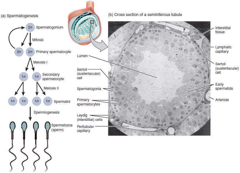
Spermatogenesis is the process by which haploid spermatozoa develop from germ cells in the seminiferous tubules of the testicle. This process starts with the Mitosis, mitotic division of the stem cells located close to the basement membrane of the tubules. These cells are called Spermatogonial Stem Cells, spermatogonial stem cells. The mitotic division of these produces two types of cells. Type A cells replenish the stem cells, and type B cells differentiate into primary spermatocytes. The primary spermatocyte divides meiotically (Meiosis I) into two secondary spermatocytes; each secondary spermatocyte divides into two equal haploid spermatids by Meiosis II. The spermatids are transformed into spermatozoa (sperm) by the process of spermiogenesis. These develop into mature spermatozoa, also known as sperm, sperm cells. Thus, the primary spermatocyte gives rise to two cells, the secondary spermatocytes, and the two secondary spermatocytes by their subdivision produce four spermatoz ... [...More Info...] [...Related Items...] OR: [Wikipedia] [Google] [Baidu] |
Testicle
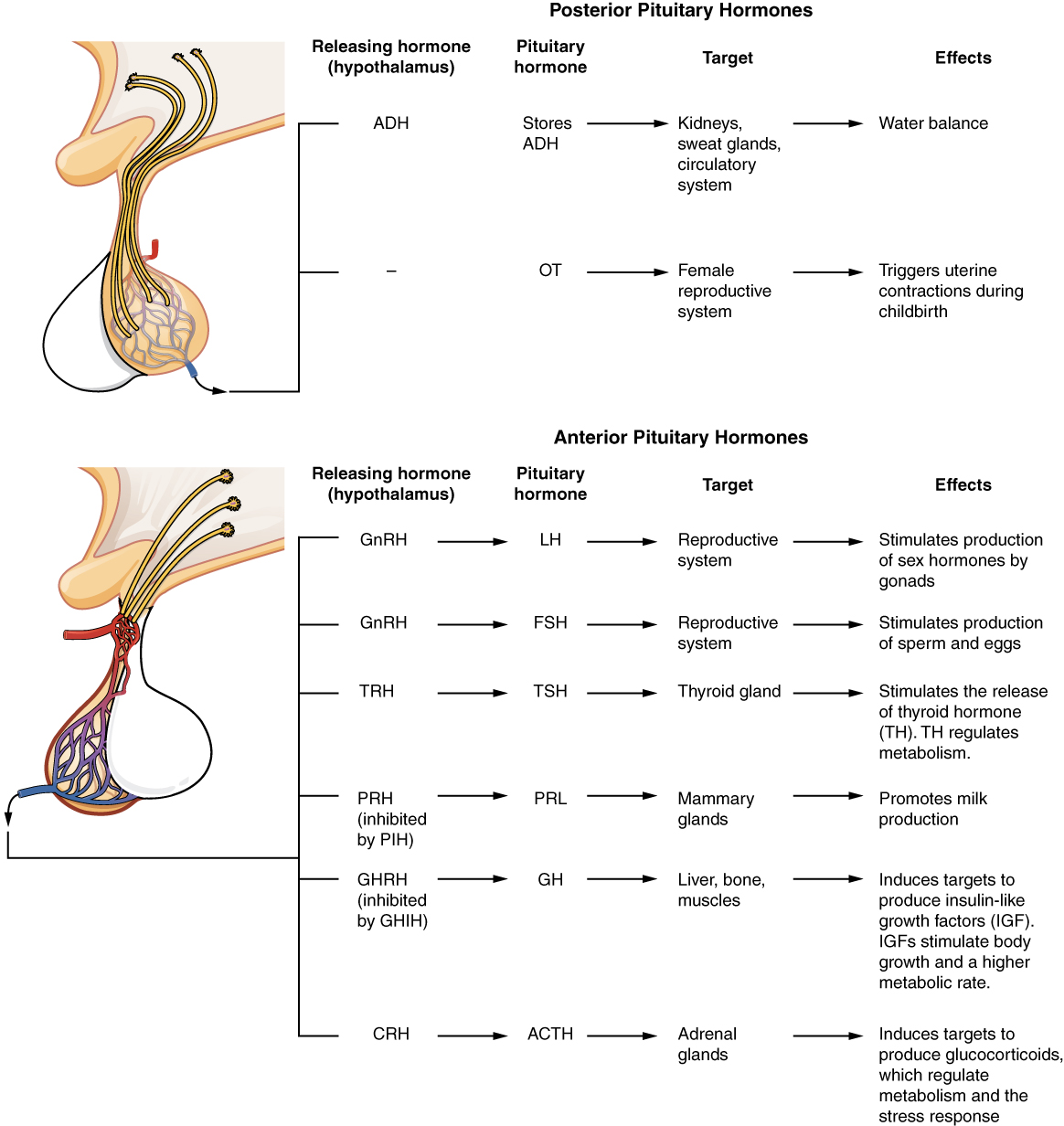
A testicle or testis ( testes) is the gonad in all male bilaterians, including humans, and is Homology (biology), homologous to the ovary in females. Its primary functions are the production of sperm and the secretion of Androgen, androgens, primarily testosterone. The release of testosterone is regulated by luteinizing hormone (LH) from the anterior pituitary gland. Sperm production is controlled by follicle-stimulating hormone (FSH) from the anterior pituitary gland and by testosterone produced within the gonads. Structure Appearance Males have two testicles of similar size contained within the scrotum, which is an extension of the abdominal wall. Scrotal asymmetry, in which one testicle extends farther down into the scrotum than the other, is common. This is because of the differences in the vasculature's anatomy. For 85% of men, the right testis hangs lower than the left one. Measurement and volume The volume of the testicle can be estimated by palpating it and compari ... [...More Info...] [...Related Items...] OR: [Wikipedia] [Google] [Baidu] |
Testes
A testicle or testis ( testes) is the gonad in all male bilaterians, including humans, and is homologous to the ovary in females. Its primary functions are the production of sperm and the secretion of androgens, primarily testosterone. The release of testosterone is regulated by luteinizing hormone (LH) from the anterior pituitary gland. Sperm production is controlled by follicle-stimulating hormone (FSH) from the anterior pituitary gland and by testosterone produced within the gonads. Structure Appearance Males have two testicles of similar size contained within the scrotum, which is an extension of the abdominal wall. Scrotal asymmetry, in which one testicle extends farther down into the scrotum than the other, is common. This is because of the differences in the vasculature's anatomy. For 85% of men, the right testis hangs lower than the left one. Measurement and volume The volume of the testicle can be estimated by palpating it and comparing it to ellipsoids (an ... [...More Info...] [...Related Items...] OR: [Wikipedia] [Google] [Baidu] |
Leydig Cell
Leydig cells, also known as interstitial cells of the testes and interstitial cells of Leydig, are found adjacent to the seminiferous tubules in the testicle and produce testosterone in the presence of luteinizing hormone (LH). They are polyhedral in shape and have a large, prominent nucleus, an eosinophilic cytoplasm, and numerous lipid-filled vesicles. Males have two types of leydig cells that appear in two distinct stages of development: the fetal type and the adult type. Structure The mammalian Leydig cell is a polyhedral epithelioid cell with a single eccentrically located ovoid nucleus. The nucleus contains one to three prominent nucleoli and large amounts of dark-staining peripheral heterochromatin. The acidophilic cytoplasm usually contains numerous membrane-bound lipid droplets and large amounts of smooth endoplasmic reticulum (SER). Besides the abundance of SER with scattered patches of rough endoplasmic reticulum, several mitochondria are also prominent within the cy ... [...More Info...] [...Related Items...] OR: [Wikipedia] [Google] [Baidu] |
Spermatogonia
A spermatogonium (plural: ''spermatogonia'') is an undifferentiated male germ cell. Spermatogonia undergo spermatogenesis to form mature spermatozoa in the seminiferous tubules of the testicles. There are three subtypes of spermatogonia in humans: Type A (dark) cells, with dark nuclei. These cells are reserve spermatogonial stem cells which do not usually undergo active mitosis. Type A (pale) cells, with pale nuclei. These are the spermatogonial stem cells that undergo active mitosis. These cells divide to produce Type B cells. Type B cells, which undergo growth and become primary spermatocytes. Types of spermatogonia Spermatogonia are often classified into different types depending on their stage in the differentiation process. In humans and most mammals, spermatogonia are divided into two types, A and B, but this can differ for other organisms. There are three subtypes of spermatogonia in humans: *Type A (dark) cells, with dark nuclei. These cells are reserve spermatogonia ... [...More Info...] [...Related Items...] OR: [Wikipedia] [Google] [Baidu] |
Follicle-stimulating Hormone
Follicle-stimulating hormone (FSH) is a gonadotropin, a glycoprotein polypeptide hormone. FSH is synthesized and secreted by the gonadotropic cells of the anterior pituitary gland and regulates the development, growth, puberty, pubertal maturation, and reproductive processes of the body. FSH and luteinizing hormone (LH) work together in the reproductive system. Structure FSH is a 35.5 kDa glycoprotein protein dimer, heterodimer, consisting of two polypeptide units, alpha and beta. Its structure is similar to those of luteinizing hormone (LH), thyroid-stimulating hormone (TSH), and human chorionic gonadotropin (hCG). The chorionic gonadotropin alpha, alpha subunits of the glycoproteins LH, FSH, TSH, and hCG are identical and consist of 96 amino acids, while the beta subunits vary. Both subunits are required for biological activity. FSH has a beta subunit of 111 amino acids (FSH β), which confers its specific biologic action, and is responsible for interaction with the follicl ... [...More Info...] [...Related Items...] OR: [Wikipedia] [Google] [Baidu] |
Spermatocyte
Spermatocytes are a type of male gametocyte in animals. They derive from immature germ cells called spermatogonia. They are found in the testis, in a structure known as the seminiferous tubules. There are two types of spermatocytes, primary and secondary spermatocytes. Primary and secondary spermatocytes are formed through the process of spermatocytogenesis. Primary spermatocytes are diploid (2N) cells. After meiosis I, two secondary spermatocytes are formed. Secondary spermatocytes are haploid (N) cells that contain half the number of chromosomes. In all animals, Male, males produce spermatocytes, even hermaphrodites such as Caenorhabditis elegans, ''C. elegans'', which exist as a male or hermaphrodite. In hermaphrodite ''C. elegans'', sperm production occurs first and is then stored in the spermatheca. Once the egg cell, eggs are formed, they are able to self-fertilize and produce up to 350 offspring, progeny. Development At puberty, spermatogonia located along the walls of th ... [...More Info...] [...Related Items...] OR: [Wikipedia] [Google] [Baidu] |
Tubulus Seminiferus Contortus
Seminiferous tubules are located within the testicles, and are the specific location of meiosis, and the subsequent creation of male gametes, namely spermatozoa. Structure The epithelium of the tubule consists of a type of sustentacular cells known as Sertoli cells, which are tall, columnar type cells that line the tubule. In between the Sertoli cells are spermatogenic cells, which differentiate through meiosis to sperm cells. Sertoli cells function to nourish the developing sperm cells. They secrete androgen-binding protein, a binding protein which increases the concentration of testosterone. There are two types: convoluted and straight, convoluted toward the lateral side, and straight as the tubule comes medially to form ducts that will exit the testis. The seminiferous tubules are formed from the testis cords that develop from the primitive gonadal cords, formed from the gonadal ridge. Function Spermatogenesis, the process for producing spermatozoa, takes place in the ... [...More Info...] [...Related Items...] OR: [Wikipedia] [Google] [Baidu] |
Spermatocytes
Spermatocytes are a type of male gametocyte in animals. They derive from immature germ cells called spermatogonia. They are found in the testis, in a structure known as the seminiferous tubules. There are two types of spermatocytes, primary and secondary spermatocytes. Primary and secondary spermatocytes are formed through the process of spermatocytogenesis. Primary spermatocytes are diploid (2N) cells. After meiosis I, two secondary spermatocytes are formed. Secondary spermatocytes are haploid (N) cells that contain half the number of chromosomes. In all animals, males produce spermatocytes, even hermaphrodites such as ''C. elegans'', which exist as a male or hermaphrodite. In hermaphrodite ''C. elegans'', sperm production occurs first and is then stored in the spermatheca. Once the eggs are formed, they are able to self-fertilize and produce up to 350 progeny. Development At puberty, spermatogonia located along the walls of the seminiferous tubules within the testis will be ... [...More Info...] [...Related Items...] OR: [Wikipedia] [Google] [Baidu] |


