|
Spermatogonia
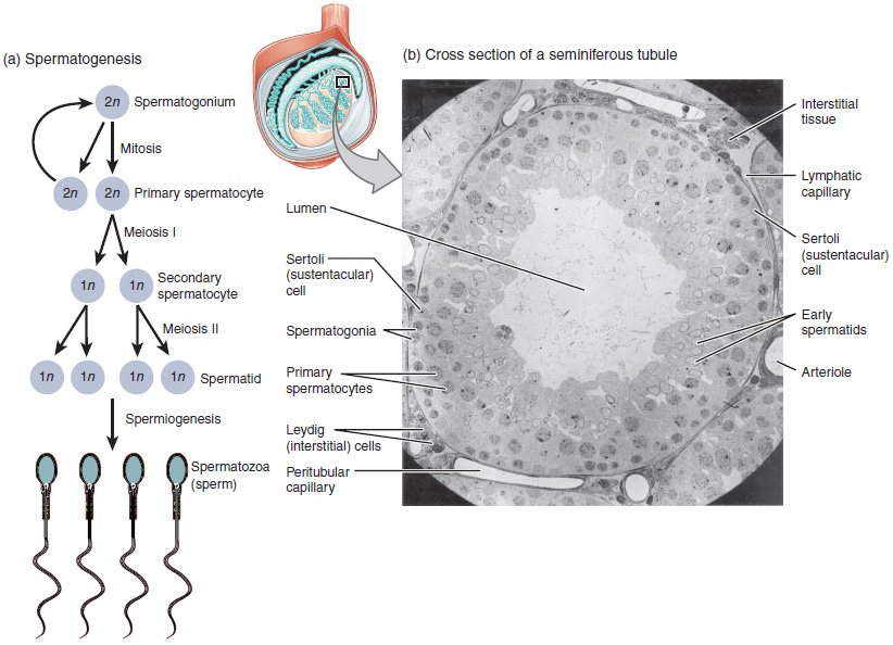
A spermatogonium (plural: ''spermatogonia'') is an undifferentiated male germ cell. Spermatogonia undergo spermatogenesis to form mature spermatozoa in the seminiferous tubules of the testicles. There are three subtypes of spermatogonia in humans: Type A (dark) cells, with dark nuclei. These cells are reserve spermatogonial stem cells which do not usually undergo active mitosis. Type A (pale) cells, with pale nuclei. These are the spermatogonial stem cells that undergo active mitosis. These cells divide to produce Type B cells. Type B cells, which undergo growth and become primary spermatocytes. Types of spermatogonia Spermatogonia are often classified into different types depending on their stage in the differentiation process. In humans and most mammals, spermatogonia are divided into two types, A and B, but this can differ for other organisms. There are three subtypes of spermatogonia in humans: *Type A (dark) cells, with dark nuclei. These cells are reserve spermatogonia ... [...More Info...] [...Related Items...] OR: [Wikipedia] [Google] [Baidu] |
Spermatogonial Stem Cell
A spermatogonial stem cell (SSC), also known as a type A spermatogonium, is a spermatogonium that does not differentiate into a spermatocyte, a precursor of sperm cells. Instead, they continue dividing into other spermatogonia or remain dormant to maintain a reserve of spermatogonia. Type B spermatogonia, on the other hand, differentiate into spermatocytes, which in turn undergo meiosis to eventually form mature sperm cells. Spermatogonial stem cells in the testis During fetal development, gonocytes develop from primordial germ cells, and following this SSCs develop from gonocytes in the testis. SSCs are the early precursor for Spermatozoon, spermatozoa and are responsible for the continuation of spermatogenesis in adult mammals. The stem cells are capable of dividing into more SSCs which is vital for maintaining the stem cell pool. Alternatively, they go on to differentiate into spermatocytes, spermatids, and finally spermatozoa. One SSC is the precursor for multiple spermat ... [...More Info...] [...Related Items...] OR: [Wikipedia] [Google] [Baidu] |
Spermatogonia
A spermatogonium (plural: ''spermatogonia'') is an undifferentiated male germ cell. Spermatogonia undergo spermatogenesis to form mature spermatozoa in the seminiferous tubules of the testicles. There are three subtypes of spermatogonia in humans: Type A (dark) cells, with dark nuclei. These cells are reserve spermatogonial stem cells which do not usually undergo active mitosis. Type A (pale) cells, with pale nuclei. These are the spermatogonial stem cells that undergo active mitosis. These cells divide to produce Type B cells. Type B cells, which undergo growth and become primary spermatocytes. Types of spermatogonia Spermatogonia are often classified into different types depending on their stage in the differentiation process. In humans and most mammals, spermatogonia are divided into two types, A and B, but this can differ for other organisms. There are three subtypes of spermatogonia in humans: *Type A (dark) cells, with dark nuclei. These cells are reserve spermatogonia ... [...More Info...] [...Related Items...] OR: [Wikipedia] [Google] [Baidu] |
Primary Spermatocytes
Spermatocytes are a type of male gametocyte in animals. They derive from immature germ cells called spermatogonia. They are found in the testis, in a structure known as the seminiferous tubules. There are two types of spermatocytes, primary and secondary spermatocytes. Primary and secondary spermatocytes are formed through the process of spermatocytogenesis. Primary spermatocytes are diploid (2N) cells. After meiosis I, two secondary spermatocytes are formed. Secondary spermatocytes are haploid (N) cells that contain half the number of chromosomes. In all animals, males produce spermatocytes, even hermaphrodites such as ''C. elegans'', which exist as a male or hermaphrodite. In hermaphrodite ''C. elegans'', sperm production occurs first and is then stored in the spermatheca. Once the eggs are formed, they are able to self-fertilize and produce up to 350 progeny. Development At puberty, spermatogonia located along the walls of the seminiferous tubules within the testis will be ... [...More Info...] [...Related Items...] OR: [Wikipedia] [Google] [Baidu] |
Sertoli Cell
Sertoli cells are a type of sustentacular "nurse" cell found in human testes which contribute to the process of spermatogenesis (the production of sperm) as a structural component of the seminiferous tubules. They are activated by follicle-stimulating hormone (FSH) secreted by the adenohypophysis and express FSH receptor on their membranes. History Sertoli cells are named after Enrico Sertoli, an Italian physiologist who discovered them while studying medicine at the University of Pavia, Italy. He published a description of his eponymous cell in 1865. The cell was discovered by Sertoli with a Belthle microscope which had been purchased in 1862. In the 1865 publication, his first description used the terms "tree-like cell" or "stringy cell"; most importantly, he referred to these as "mother cells". Other scientists later used Enrico's family name to label these cells in publications, beginning in 1888. As of 2006, two textbooks that are devoted specifically to the Sertoli cell ... [...More Info...] [...Related Items...] OR: [Wikipedia] [Google] [Baidu] |
Spermatogenesis
Spermatogenesis is the process by which haploid spermatozoa develop from germ cells in the seminiferous tubules of the testicle. This process starts with the Mitosis, mitotic division of the stem cells located close to the basement membrane of the tubules. These cells are called Spermatogonial Stem Cells, spermatogonial stem cells. The mitotic division of these produces two types of cells. Type A cells replenish the stem cells, and type B cells differentiate into primary spermatocytes. The primary spermatocyte divides meiotically (Meiosis I) into two secondary spermatocytes; each secondary spermatocyte divides into two equal haploid spermatids by Meiosis II. The spermatids are transformed into spermatozoa (sperm) by the process of spermiogenesis. These develop into mature spermatozoa, also known as sperm, sperm cells. Thus, the primary spermatocyte gives rise to two cells, the secondary spermatocytes, and the two secondary spermatocytes by their subdivision produce four spermatoz ... [...More Info...] [...Related Items...] OR: [Wikipedia] [Google] [Baidu] |
Testicle
A testicle or testis ( testes) is the gonad in all male bilaterians, including humans, and is Homology (biology), homologous to the ovary in females. Its primary functions are the production of sperm and the secretion of Androgen, androgens, primarily testosterone. The release of testosterone is regulated by luteinizing hormone (LH) from the anterior pituitary gland. Sperm production is controlled by follicle-stimulating hormone (FSH) from the anterior pituitary gland and by testosterone produced within the gonads. Structure Appearance Males have two testicles of similar size contained within the scrotum, which is an extension of the abdominal wall. Scrotal asymmetry, in which one testicle extends farther down into the scrotum than the other, is common. This is because of the differences in the vasculature's anatomy. For 85% of men, the right testis hangs lower than the left one. Measurement and volume The volume of the testicle can be estimated by palpating it and compari ... [...More Info...] [...Related Items...] OR: [Wikipedia] [Google] [Baidu] |
Meiosis
Meiosis () is a special type of cell division of germ cells in sexually-reproducing organisms that produces the gametes, the sperm or egg cells. It involves two rounds of division that ultimately result in four cells, each with only one copy of each chromosome (haploid). Additionally, prior to the division, genetic material from the paternal and maternal copies of each chromosome is crossed over, creating new combinations of code on each chromosome. Later on, during fertilisation, the haploid cells produced by meiosis from a male and a female will fuse to create a zygote, a cell with two copies of each chromosome. Errors in meiosis resulting in aneuploidy (an abnormal number of chromosomes) are the leading known cause of miscarriage and the most frequent genetic cause of developmental disabilities. In meiosis, DNA replication is followed by two rounds of cell division to produce four daughter cells, each with half the number of chromosomes as the original parent cell. ... [...More Info...] [...Related Items...] OR: [Wikipedia] [Google] [Baidu] |
Germinal Epithelium (male)
The germinal epithelium is the epithelial layer of the seminiferous tubules of the testicles. It is also known as the wall of the seminiferous tubules. The cells in the epithelium are connected via tight junctions. There are two types of cells in the germinal epithelium. The large Sertoli cells (not dividing) function as supportive cells to the developing sperm. The second cell type is the cells belonging to the spermatogenic cell lineage. These eventually develop into sperm cells (spermatozoon A spermatozoon (; also spelled spermatozoön; : spermatozoa; ) is a motile sperm cell (biology), cell produced by male animals relying on internal fertilization. A spermatozoon is a moving form of the ploidy, haploid cell (biology), cell that is ...). Typically, the spermatogenic cells will make four to eight layers in the germinal epithelium.Junqueira's Basic Histology: Text and Atlas, Thirteenth Edition References External links * - "Male Reproductive System: testis, germinal ep ... [...More Info...] [...Related Items...] OR: [Wikipedia] [Google] [Baidu] |
Leydig Cell
Leydig cells, also known as interstitial cells of the testes and interstitial cells of Leydig, are found adjacent to the seminiferous tubules in the testicle and produce testosterone in the presence of luteinizing hormone (LH). They are polyhedral in shape and have a large, prominent nucleus, an eosinophilic cytoplasm, and numerous lipid-filled vesicles. Males have two types of leydig cells that appear in two distinct stages of development: the fetal type and the adult type. Structure The mammalian Leydig cell is a polyhedral epithelioid cell with a single eccentrically located ovoid nucleus. The nucleus contains one to three prominent nucleoli and large amounts of dark-staining peripheral heterochromatin. The acidophilic cytoplasm usually contains numerous membrane-bound lipid droplets and large amounts of smooth endoplasmic reticulum (SER). Besides the abundance of SER with scattered patches of rough endoplasmic reticulum, several mitochondria are also prominent within the cy ... [...More Info...] [...Related Items...] OR: [Wikipedia] [Google] [Baidu] |
Myofibroblast
A myofibroblast is a cell phenotype that was first described as being in a state between a fibroblast and a smooth muscle cell. Structure Myofibroblasts are contractile web-like fusiform cells that are identifiable by their expression of α-smooth muscle actin within their cytoplasmic stress fibers. In the gastrointestinal and genitourinary tracts, myofibroblasts are found subepithelially in mucosal surfaces. Here they not only act as a regulator of the shape of the crypts and villi, but also act as stem-niche cells in the intestinal crypts and as parts of atypical antigen-presenting cells. They have both support as well as paracrine function in most places. Location Myofibroblasts were first identified in granulation tissue during skin wound healing. Typically, these cells are found in granulation tissue, scar tissue (fibrosis) and the stroma of tumours. They also line the gastrointestinal tract, wherein they regulate the shapes of crypts and villi. Markers Myofibroblas ... [...More Info...] [...Related Items...] OR: [Wikipedia] [Google] [Baidu] |


