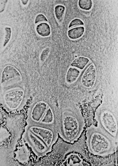
Endochondral Bone on:
[Wikipedia]
[Google]
[Amazon]
Endochondral ossification is one of the two essential pathways by which
 In developing bones, ossification commences within the primary ossification center located in the center of the
 During endochondral ossification, five distinct zones can be seen at the light-microscope level:


File:Proximal tibia Masson Goldner Trikrom rabbit 600x growth zone.jpg, Masson Goldner trichrome stain of growth plate in a rabbit tibia.
File:Gray79.png, Section of fetal bone of cat. ir. Irruption of the subperiosteal tissue. p. Fibrous layer of the periosteum. o. Layer of osteoblasts. im. Subperiosteal bony deposit.
File:Endochondral CCN.jpg, Process of endochondral ossification.
File:Gray80.png, Drawing of part of a longitudinal section of the developing femur of a rabbit. a. Flattened cartilage cells. b. Enlarged cartilage cells. c, d. Newly formed bone. e. Osteoblasts. f. Giant cells or osteoclasts. g, h. Shrunken cartilage cells.
bone tissue
A bone is a Stiffness, rigid Organ (biology), organ that constitutes part of the skeleton in most vertebrate animals. Bones protect the various other organs of the body, produce red blood cell, red and white blood cells, store minerals, provi ...
is produced during fetal development
Prenatal development () involves the development of the embryo and of the fetus during a viviparous animal's gestation. Prenatal development starts with fertilization, in the germinal stage of embryonic development, and continues in fetal deve ...
and bone repair of the mammal
A mammal () is a vertebrate animal of the Class (biology), class Mammalia (). Mammals are characterised by the presence of milk-producing mammary glands for feeding their young, a broad neocortex region of the brain, fur or hair, and three ...
ian skeletal system
A skeleton is the structural frame that supports the body of most animals. There are several types of skeletons, including the exoskeleton, which is a rigid outer shell that holds up an organism's shape; the endoskeleton, a rigid internal fra ...
, the other pathway being intramembranous ossification
Intramembranous ossification is one of the two essential processes during fetal development of the gnathostome (excluding chondrichthyans such as sharks) skeletal system by which rudimentary bone tissue is created.
Intramembranous ossification i ...
. Both endochondral and intramembranous processes initiate from a precursor mesenchymal tissue, but their transformations into bone are different. In intramembranous ossification, mesenchymal tissue is directly converted into bone. On the other hand, endochondral ossification starts with mesenchymal tissue turning into an intermediate cartilage stage, which is eventually substituted by bone.
Endochondral ossification is responsible for development of most bones including long
Long may refer to:
Measurement
* Long, characteristic of something of great duration
* Long, characteristic of something of great length
* Longitude (abbreviation: long.), a geographic coordinate
* Longa (music), note value in early music mens ...
and short bones, the bones of the axial (ribs
The rib cage or thoracic cage is an endoskeletal enclosure in the thorax of most vertebrates that comprises the ribs, vertebral column and sternum, which protect the vital organs of the thoracic cavity, such as the heart, lungs and great vessels ...
and vertebrae
Each vertebra (: vertebrae) is an irregular bone with a complex structure composed of bone and some hyaline cartilage, that make up the vertebral column or spine, of vertebrates. The proportions of the vertebrae differ according to their spinal ...
) and the appendicular skeleton (e.g. upper and lower
Lower may refer to:
* ''Lower'' (album), 2025 album by Benjamin Booker
*Lower (surname)
*Lower Township, New Jersey
*Lower Receiver (firearms)
*Lower Wick
Lower Wick is a small hamlet located in the county of Gloucestershire, England. It is sit ...
limbs), the bones of the skull base
The base of skull, also known as the cranial base or the cranial floor, is the most Anatomical terms of location#Superior and inferior, inferior area of the human skull, skull. It is composed of the endocranium and the lower parts of the Calvaria ...
(including the ethmoid
The ethmoid bone (; from ) is an unpaired bone in the skull that separates the nasal cavity from the brain. It is located at the roof of the nose, between the two orbit (anatomy), orbits. The cubical (cube-shaped) bone is lightweight due to a sp ...
and sphenoid bones) and the medial end of the clavicle
The clavicle, collarbone, or keybone is a slender, S-shaped long bone approximately long that serves as a strut between the scapula, shoulder blade and the sternum (breastbone). There are two clavicles, one on each side of the body. The clavic ...
. In addition, endochondral ossification is not exclusively confined to embryonic development; it also plays a crucial role in the healing of fractures.
Formation of the cartilage model
The initiation of endochondral ossification starts by proliferation and condensation of mesenchymal cells in the area where the bone will eventually be formed. Subsequently, these mesenchymal progenitor cells differentiate intochondroblasts
Chondroblasts, or perichondrial cells, is the name given to mesenchymal progenitor cells in situ which, from endochondral ossification, will form chondrocytes in the growing cartilage matrix. Another name for them is subchondral cortico-spongio ...
, which actively synthesize cartilage matrix components. Thus, the initial hyaline cartilage template is formed, which has the same basic shape and outline as the future bone.
Primary center of ossification
 In developing bones, ossification commences within the primary ossification center located in the center of the
 diaphysis
The diaphysis (: diaphyses) is the main or midsection (shaft) of a long bone. It is made up of cortical bone and usually contains bone marrow and adipose tissue (fat).
It is a middle tubular part composed of compact bone which surrounds a centr ...
(bone shaft), where the following changes occur:
- The perichondrium surrounding the cartilage model transforms into the periosteum The periosteum is a membrane that covers the outer surface of all bones, except at the articular surfaces (i.e. the parts within a joint space) of long bones. (At the joints of long bones the bone's outer surface is lined with "articular cartila .... During this transformation, special cells within the perichondrium switch gears. Instead of becoming cartilage cells (chondrocytes Chondrocytes (, ) are the only cells found in healthy cartilage. They produce and maintain the cartilaginous matrix, which consists mainly of collagen and proteoglycans. Although the word '' chondroblast'' is commonly used to describe an immatu ...), they mature into bone-building osteoblasts. This newly formed bone can be called "periosteal bone" as it originates from the transformed periosteum. However, considering its developmental pathway, it could be classified as "intramembranous bone".
- After the formation of the periosteum, chondrocytes in the primary center of ossification begin to grow ( hypertrophy). They begin secreting:
- When chondrocytes die, matrix metalloproteinases result in catabolism of various components within the extracellular matrix and the physical boundaries between neighboring lacunae (the spaces housing chondrocytes) weaken. This can lead to the merging of these lacunae, creating larger empty spaces.
- Blood vessels arising from the periosteum invade these empty spaces and mesenchymal stem cells migrate guided by penetrating blood vessels. Following the invading blood vessels, mesenchymal stem cells reach these empty spaces and undergo differentiation into osteoprogenitor cells. These progenitors further mature into osteoblasts, that deposit unmineralized bone matrix, termed osteoid. Mineralization subsequently follows leading to formation of bone trabeculae (Endochondral bone formation).

Secondary center of ossification
During the postnatal life, a secondary ossification center appears in each end (epiphysis
An epiphysis (; : epiphyses) is one of the rounded ends or tips of a long bone that ossify from one or more secondary centers of ossification. Between the epiphysis and diaphysis (the long midsection of the long bone) lies the metaphysis, inc ...
) of long bones. In these secondary centers, cartilage is converted to bone similarly to that occurring in a primary ossification center. As the secondary ossification centers enlarge, residual cartilage persists in two distinct locations:
At the end of an individual’s growth period, the production of new cartilage in the epiphyseal plate stops. After this point, existing cartilage within the plate turns into mature bone tissue.
Histology
 During endochondral ossification, five distinct zones can be seen at the light-microscope level:

Fracture healing
For complete recovery of a fractured bone’s biomechanical functionality, thebone healing
Bone healing, or fracture healing, is a proliferative physiological process in which the body facilitates the repair of a bone fracture.
Generally, bone fracture treatment consists of a doctor reducing (pushing) displaced bones back into place ...
process needs to culminate in the formation of lamellar bone at the fracture site to withstand the same forces and stresses it did before the fracture. Indirect fracture healing, the most common type of bone repair, relies heavily on endochondral ossification. In this type of healing, endochondral ossification occurs within the fracture gap and external to the periosteum. In contrast, intramembranous ossification takes place directly beneath the periosteum, adjacent to the broken bone’s ends.

Additional images
References
{{Bone and cartilage Animal developmental biology Skeletal system