|
Ethmoid Bone
The ethmoid bone (; from ) is an unpaired bone in the skull that separates the nasal cavity from the brain. It is located at the roof of the nose, between the two orbits. The cubical (cube-shaped) bone is lightweight due to a spongy construction. The ethmoid bone is one of the bones that make up the orbit of the eye. Structure The ethmoid bone is an anterior cranial bone located between the eyes. It contributes to the medial wall of the orbit, the nasal cavity, and the nasal septum. The ethmoid has three parts: cribriform plate, ethmoidal labyrinth, and perpendicular plate. The cribriform plate forms the roof of the nasal cavity and also contributes to formation of the anterior cranial fossa, the ethmoidal labyrinth consists of a large mass on either side of the perpendicular plate, and the perpendicular plate forms the superior two-thirds of the nasal septum. Between the orbital plate and the nasal conchae are the ethmoidal sinuses or ethmoidal air cells, which are a var ... [...More Info...] [...Related Items...] OR: [Wikipedia] [Google] [Baidu] |
Orbit (anatomy)
In anatomy Anatomy () is the branch of morphology concerned with the study of the internal structure of organisms and their parts. Anatomy is a branch of natural science that deals with the structural organization of living things. It is an old scien ..., the orbit is the Body cavity, cavity or socket/hole of the skull in which the eye and Accessory visual structures, its appendages are situated. "Orbit" can refer to the bony socket, or it can also be used to imply the contents. In the adult human, the volume of the orbit is about , of which the eye occupies . The orbital contents comprise the eye, the Orbital fascia, orbital and retrobulbar fascia, extraocular muscles, cranial nerves optic nerve, II, oculomotor nerve, III, trochlear nerve, IV, trigeminal nerve, V, and abducens nerve, VI, blood vessels, fat, the lacrimal gland with its Lacrimal sac, sac and nasolacrimal duct, duct, the eyelids, Medial palpebral ligament, medial and Lateral palpebral raphe, lateral palpebr ... [...More Info...] [...Related Items...] OR: [Wikipedia] [Google] [Baidu] |
Sphenoidal Conchae
The sphenoidal conchae (sphenoidal turbinated processes) are two thin, curved plates, situated at the anterior and lower part of the body of the sphenoid. An aperture of variable size exists in the anterior wall of each, and through this the sphenoidal sinus opens into the nasal cavity. ''General Anatomy and Osteology of Head and Neck'' (I. K. International Pvt Ltd, 2009; by Mahdi Hasan)- Retrieved 2018-08-29 Each is irregular in form, and tapers to a point behind, being broader and thinner in front. Its upper surface is concave, and looks toward the cavity of the sinus; its under surface is convex, and forms part of the roof of the corresponding nasal cavity. Each bone articulates in front with the ethmoid, laterally with the Hard palate, palatine; its pointed poste ... [...More Info...] [...Related Items...] OR: [Wikipedia] [Google] [Baidu] |
Magnetite
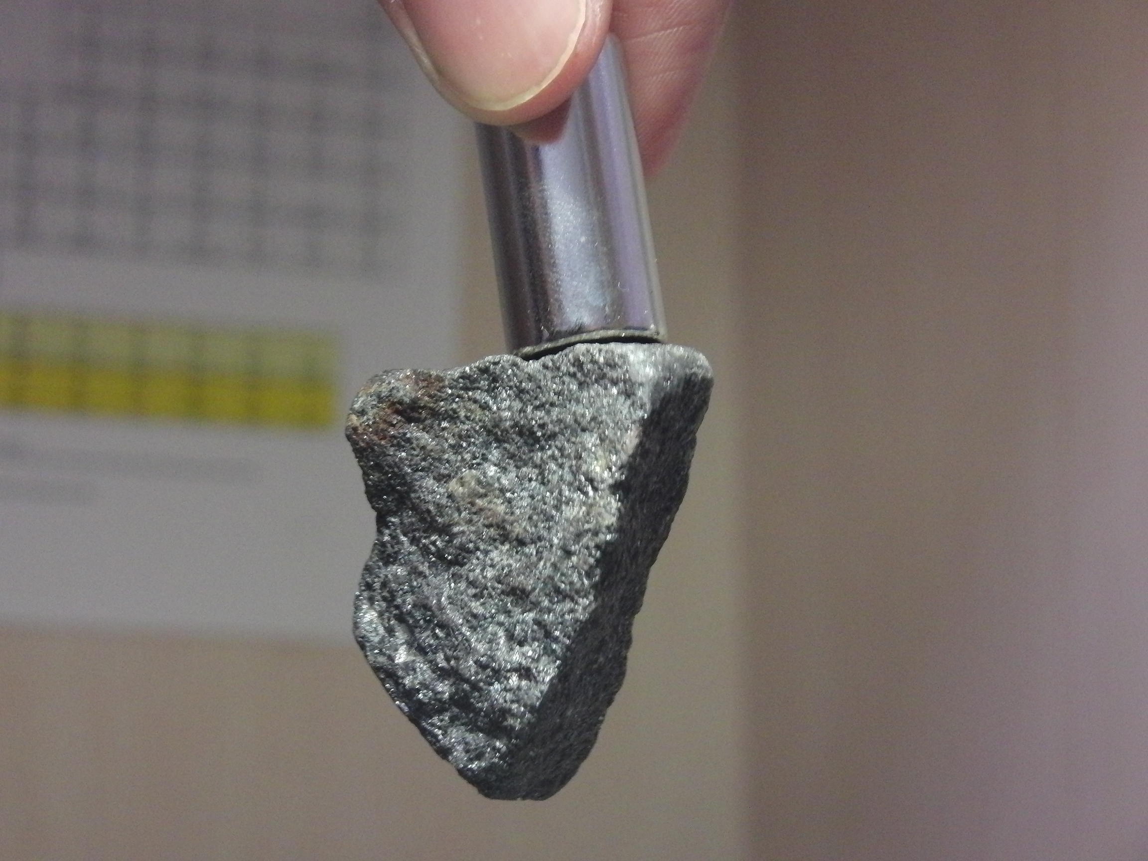
Magnetite is a mineral and one of the main iron ores, with the chemical formula . It is one of the iron oxide, oxides of iron, and is ferrimagnetism, ferrimagnetic; it is attracted to a magnet and can be magnetization, magnetized to become a permanent magnet itself. With the exception of extremely rare native iron deposits, it is the most magnetic of all the naturally occurring minerals on Earth. Naturally magnetized pieces of magnetite, called lodestone, will attract small pieces of iron, which is how ancient peoples first discovered the property of magnetism. Magnetite is black or brownish-black with a metallic luster, has a Mohs scale of mineral hardness, Mohs hardness of 5–6 and leaves a black streak (mineralogy), streak. Small grains of magnetite are very common in igneous rocks, igneous and metamorphic rocks. The chemical IUPAC name is iron(II,III) oxide and the common chemical name is ''ferrous-ferric oxide''. Properties In addition to igneous rocks, magnetite als ... [...More Info...] [...Related Items...] OR: [Wikipedia] [Google] [Baidu] |
Bird
Birds are a group of warm-blooded vertebrates constituting the class (biology), class Aves (), characterised by feathers, toothless beaked jaws, the Oviparity, laying of Eggshell, hard-shelled eggs, a high Metabolism, metabolic rate, a four-chambered heart, and a strong yet lightweight Bird skeleton, skeleton. Birds live worldwide and range in size from the bee hummingbird to the common ostrich. There are over 11,000 living species and they are split into 44 Order (biology), orders. More than half are passerine or "perching" birds. Birds have Bird wing, wings whose development varies according to species; the only known groups without wings are the extinct moa and elephant birds. Wings, which are modified forelimbs, gave birds the ability to fly, although further evolution has led to the Flightless bird, loss of flight in some birds, including ratites, penguins, and diverse endemism, endemic island species. The digestive and respiratory systems of birds are also uniquely a ... [...More Info...] [...Related Items...] OR: [Wikipedia] [Google] [Baidu] |
Crista Galli
The crista galli (Latin: "crest of the rooster") is a wedge-shaped, vertical, midline upward continuation of the perpendicular plate of the ethmoid bone of the skull, projecting above the cribriform plate into the cranial cavity. It serves as an attachment for the membranes surrounding the brain. Structure Attachments The falx cerebri (a fold of the dura mater surrounding the brain) attaches to the crista galli. Relations The olfactory bulbs of the olfactory nerve The olfactory nerve, also known as the first cranial nerve, cranial nerve I, or simply CN I, is a cranial nerve that contains sensory nerve fibers relating to the sense of smell. The afferent nerve fibers of the olfactory receptor neurons t ... lie on either side of the crista galli on top of the cribriform plate. Variation The base of crista galli varies in height. A bifid crista galli may suggest intracranial extension of a nasal dermoid cyst. References External links * * * * Bones ... [...More Info...] [...Related Items...] OR: [Wikipedia] [Google] [Baidu] |
Turbinate
In anatomy, a nasal concha (; : conchae; ; Latin for 'shell'), also called a nasal turbinate or turbinal, is a long, narrow, curled shelf of bone that protrudes into the breathing passage of the nose in humans and various other animals. The conchae are shaped like an elongated seashell, which gave them their name (Latin ''concha'' from Greek ''κόγχη''). A concha is any of the scrolled spongy bones of the nasal passages in vertebrates.''Anatomy of the Human Body'' Gray, Henry (1918) The Nasal Cavity. In humans, the conchae divide the nasal airway into four groove-like air passages, and are responsible for forcing inhaled air to flow in a steady, regular pattern around the largest possible ... [...More Info...] [...Related Items...] OR: [Wikipedia] [Google] [Baidu] |
Fetus
A fetus or foetus (; : fetuses, foetuses, rarely feti or foeti) is the unborn offspring of a viviparous animal that develops from an embryo. Following the embryonic development, embryonic stage, the fetal stage of development takes place. Prenatal development is a continuum, with no clear defining feature distinguishing an embryo from a fetus. However, in general a fetus is characterized by the presence of all the major body organs, though they will not yet be fully developed and functional, and some may not yet be situated in their final Anatomy, anatomical location. In human prenatal development, fetal development begins from the ninth week after Human fertilization, fertilization (which is the eleventh week of Gestational age (obstetrics), gestational age) and continues until the childbirth, birth of a newborn. Etymology The word ''wikt:fetus#English, fetus'' (plural ''wikt:fetuses#English, fetuses'' or rarely, the solecism ''wikt:feti#English, feti''''Oxford English Dict ... [...More Info...] [...Related Items...] OR: [Wikipedia] [Google] [Baidu] |
Lamina Papyracea
The orbital lamina of ethmoid bone (or lamina papyracea or orbital lamina) is a smooth, oblong, paper-thin bone plate which forms the lateral wall of the labyrinth of the ethmoid bone. It covers the middle and posterior ethmoidal cells, and forms a large part of the medial wall of the orbit. It articulates above with the orbital plate of the frontal bone, below with the maxilla In vertebrates, the maxilla (: maxillae ) is the upper fixed (not fixed in Neopterygii) bone of the jaw formed from the fusion of two maxillary bones. In humans, the upper jaw includes the hard palate in the front of the mouth. The two maxil ... and the orbital process of palatine bone, in front with the lacrimal, and behind with the sphenoid. Its name lamina papyracea is an appropriate description, as this part of the ethmoid bone is paper-thin and fractures easily. A fracture here could cause entrapment of the medial rectus muscle. Additional images File:Slide4fen.JPG, Orbital lamina of et ... [...More Info...] [...Related Items...] OR: [Wikipedia] [Google] [Baidu] |
Ossified
Ossification (also called osteogenesis or bone mineralization) in bone remodeling is the process of laying down new bone material by cells named osteoblasts. It is synonymous with bone tissue formation. There are two processes resulting in the formation of normal, healthy bone tissue: Intramembranous ossification is the direct laying down of bone into the primitive connective tissue ( mesenchyme), while endochondral ossification involves cartilage as a precursor. In fracture healing, endochondral osteogenesis is the most commonly occurring process, for example in fractures of long bones treated by plaster of Paris, whereas fractures treated by open reduction and internal fixation with metal plates, screws, pins, rods and nails may heal by intramembranous osteogenesis. Heterotopic ossification is a process resulting in the formation of bone tissue that is often atypical, at an extraskeletal location. Calcification is often confused with ossification. Calcification ... [...More Info...] [...Related Items...] OR: [Wikipedia] [Google] [Baidu] |
Vomer
The vomer (; ) is one of the unpaired facial bones of the skull. It is located in the midsagittal line, and articulates with the sphenoid, the ethmoid, the left and right palatine bones, and the left and right maxillary bones. The vomer forms the inferior part of the nasal septum in humans, with the superior part formed by the perpendicular plate of the ethmoid bone. The name is derived from the Latin word for a ploughshare and the shape of the bone. In humans The vomer is situated in the median plane, but its anterior portion is frequently bent to one side. It is thin, somewhat quadrilateral in shape, and forms the hinder and lower part of the nasal septum; it has two surfaces and four borders. The surfaces are marked by small furrows for blood vessels, and on each is the nasopalatine groove, which runs obliquely downward and forward, and lodges the nasopalatine nerve and vessels. Borders The ''superior border'', the thickest, presents a deep furrow, bounded on eith ... [...More Info...] [...Related Items...] OR: [Wikipedia] [Google] [Baidu] |
Inferior Nasal Concha
The inferior nasal concha (inferior turbinated bone or inferior turbinal/turbinate) is one of the three paired nasal conchae in the human nose, nose. It extends horizontally along the lateral wall of the nasal cavity and consists of a wikt:lamina, lamina of spongy bone, curled upon itself like a scroll, (''turbinate'' meaning inverted cone). The inferior nasal conchae are considered a pair of facial bones. As the air passes through the turbinates, the air is churned against these mucosa-lined bones in order to receive warmth, moisture and cleansing. Superior to inferior nasal concha are the middle nasal concha and superior nasal concha which both arise from the ethmoid bone, of the cranial portion of the skull. Hence, these two are considered as a part of the cranial bones. It has two surfaces, two borders, and two extremities. Structure Surfaces The medial surface is wikt:convex, convex, perforated by numerous apertures, and traversed by longitudinal grooves for the lodgem ... [...More Info...] [...Related Items...] OR: [Wikipedia] [Google] [Baidu] |
Palatine Bone
In anatomy, the palatine bones (; derived from the Latin ''palatum'') are two irregular bones of the facial skeleton in many animal species, located above the uvula in the throat. Together with the maxilla, they comprise the hard palate. Structure The palatine bones are situated at the back of the nasal cavity between the maxilla and the pterygoid process of the sphenoid bone. They contribute to the walls of three cavities: the floor and lateral walls of the nasal cavity, the roof of the mouth, and the floor of the orbits. They help to form the pterygopalatine and pterygoid fossae, and the inferior orbital fissures. Each palatine bone somewhat resembles the letter L, and consists of a horizontal plate, a perpendicular plate, and three projecting processes—the pyramidal process, which is directed backward and lateral from the junction of the two parts, and the orbital and sphenoidal processes, which surmount the vertical part, and are separated by a deep notch, the s ... [...More Info...] [...Related Items...] OR: [Wikipedia] [Google] [Baidu] |


