|
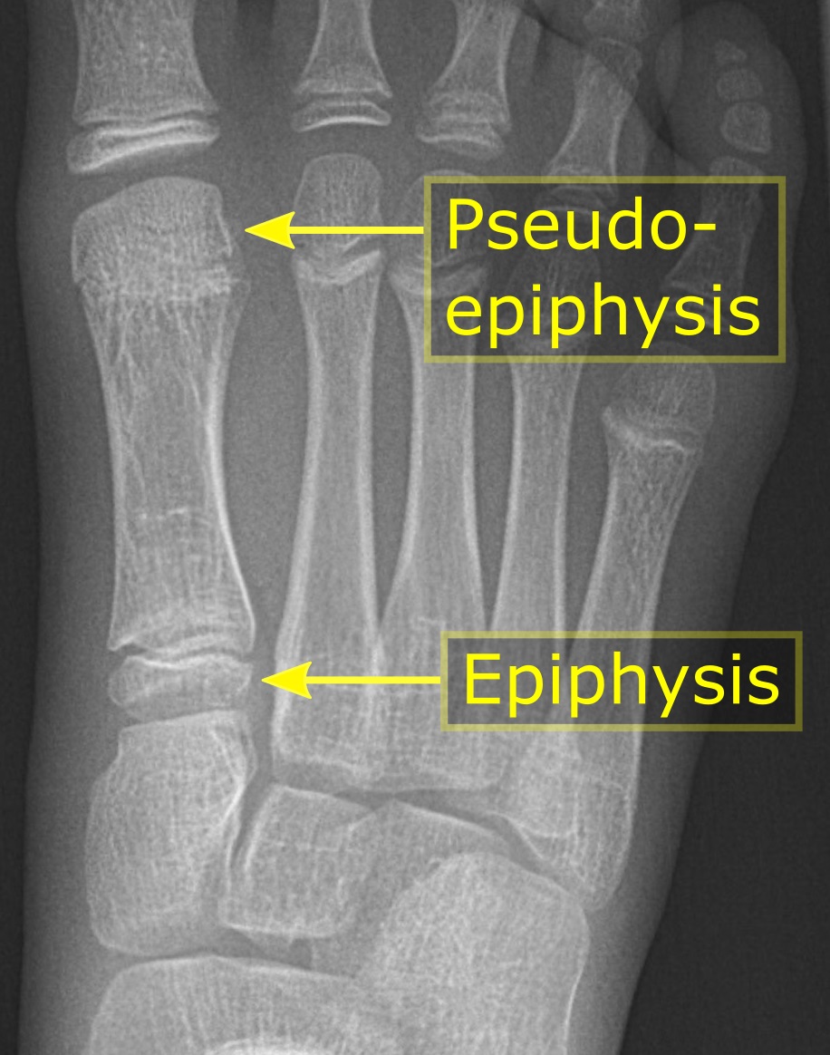
Epiphysis
An epiphysis (; : epiphyses) is one of the rounded ends or tips of a long bone that ossify from one or more secondary centers of ossification. Between the epiphysis and diaphysis (the long midsection of the long bone) lies the metaphysis, including the epiphyseal plate (growth plate). During formation of the secondary ossification center, vascular canals (epiphysial canals) stemming from the perichondrium invade the epiphysis, supplying nutrients to the developing secondary centers of ossification. At the joint, the epiphysis is covered with articular cartilage; below that covering is a zone similar to the epiphyseal plate, known as Wikt:subchondral, subchondral bone. The epiphysis is mostly found in mammals but it is also present in some lizards. However, the secondary center of ossification may have evolved multiple times, having been found in the Jurassic sphenodont ''Sapheosaurus'' as well as in the therapsid ''Niassodon, Niassodon mfumukasi.'' The epiphysis is filled wi ... [...More Info...] [...Related Items...] OR: [Wikipedia] [Google] [Baidu] |
Humerus
The humerus (; : humeri) is a long bone in the arm that runs from the shoulder to the elbow. It connects the scapula and the two bones of the lower arm, the radius (bone), radius and ulna, and consists of three sections. The humeral upper extremity of humerus, upper extremity consists of a rounded head, a narrow neck, and two short processes (tubercles, sometimes called tuberosities). The body of humerus, body is cylindrical in its upper portion, and more prism (geometry), prismatic below. The lower extremity of humerus, lower extremity consists of 2 epicondyles, 2 processes (trochlea of the humerus, trochlea and capitulum of the humerus, capitulum), and 3 fossae (radial fossa, coronoid fossa, and olecranon fossa). As well as its true anatomical neck, the constriction below the greater and lesser tubercles of the humerus is referred to as its Surgical neck of the humerus, surgical neck due to its tendency to fracture, thus often becoming the focus of surgeons. Etymology The word ... [...More Info...] [...Related Items...] OR: [Wikipedia] [Google] [Baidu] |
Ossification
Ossification (also called osteogenesis or bone mineralization) in bone remodeling is the process of laying down new bone material by cells named osteoblasts. It is synonymous with bone tissue formation. There are two processes resulting in the formation of normal, healthy bone tissue: Intramembranous ossification is the direct laying down of bone into the primitive connective tissue ( mesenchyme), while endochondral ossification involves cartilage as a precursor. In fracture healing, endochondral osteogenesis is the most commonly occurring process, for example in fractures of long bones treated by plaster of Paris, whereas fractures treated by open reduction and internal fixation with metal plates, screws, pins, rods and nails may heal by intramembranous osteogenesis. Heterotopic ossification is a process resulting in the formation of bone tissue that is often atypical, at an extraskeletal location. Calcification is often confused with ossification. Calcificatio ... [...More Info...] [...Related Items...] OR: [Wikipedia] [Google] [Baidu] |
Metaphysis
The metaphysis (: metaphyses) is the neck portion of a long bone between the epiphysis and the diaphysis. It contains the growth plate, the part of the bone that grows during childhood, and as it grows it ossifies near the diaphysis and the epiphyses. The metaphysis contains a diverse population of cells including mesenchymal stem cells, which give rise to bone and fat cells, as well as hematopoietic stem cells which give rise to a variety of blood cells as well as bone-destroying cells called osteoclasts. Thus the metaphysis contains a highly metabolic set of tissues including trabecular (spongy) bone, blood vessels, as well as marrow adipose tissue (MAT). The metaphysis may be divided anatomically into three components based on tissue content: a cartilaginous component (epiphyseal plate), a bony component (metaphysis) and a fibrous component surrounding the periphery of the plate. The growth plate synchronizes chondrogenesis with osteogenesis or interstitial carti ... [...More Info...] [...Related Items...] OR: [Wikipedia] [Google] [Baidu] |
Epiphyseal Plate
The epiphyseal plate, epiphysial plate, physis, or growth plate is a hyaline cartilage plate in the metaphysis at each end of a long bone. It is the part of a long bone where new bone growth takes place; that is, the whole bone is alive, with maintenance bone remodeling, remodeling throughout its existing bone tissue, but the growth plate is the place where the long bone grows longer (adds length). The plate is only found in children and adolescents; in adults, who have stopped growing, the plate is replaced by an ''epiphyseal line''. This replacement is known as epiphyseal closure or growth plate fusion. Complete fusion can occur as early as 12 for girls (with the most common being 14–15 years for girls) and as early as 14 for boys (with the most common being 15–17 years for boys). Structure Development Endochondral ossification is responsible for the initial bone development from cartilage Uterus, in utero and infants and the longitudinal growth of long bones in the epiph ... [...More Info...] [...Related Items...] OR: [Wikipedia] [Google] [Baidu] |
Long Bone
The long bones are those that are longer than they are wide. They are one of five types of bones: long, short, flat, irregular and sesamoid. Long bones, especially the femur and tibia, are subjected to most of the load during daily activities and they are crucial for skeletal mobility. They grow primarily by elongation of the diaphysis, with an epiphysis at each end of the growing bone. The ends of epiphyses are covered with hyaline cartilage ("articular cartilage"). The longitudinal growth of long bones is a result of endochondral ossification at the epiphyseal plate. Bone growth in length is stimulated by the production of growth hormone (GH), a secretion of the anterior lobe of the pituitary gland. The long bone category includes the femora, tibiae, and fibulae of the legs; the humeri, radii, and ulnae of the arms; metacarpals and metatarsals of the hands and feet, the phalanges of the fingers and toes, and the clavicles or collar bones. The long bones of the ... [...More Info...] [...Related Items...] OR: [Wikipedia] [Google] [Baidu] |
Giant-cell Tumor Of Bone
Giant-cell tumor of the bone (GCTOB) is a relatively uncommon bone tumor characterized by the presence of multinucleated giant cells (osteoclast-like cells). Malignancy in giant-cell tumor is uncommon and occurs in about 2% of all cases. However, if malignant degeneration does occur, it is likely to metastasize to the lungs. Giant-cell tumors are normally benign, with unpredictable behavior. It is a heterogeneous tumor composed of different cell populations. The giant-cell tumour stromal cells (GCTSC) constitute the neoplastic cells, which are from a mesenchymal stem cell origin and are classified based on expression of osteoblast cell markers such as alkaline phosphatase and osteocalcin. In contrast, the mononuclear osteoclast precursor cells giving rise to multinucleated giant cells (MNGC) are secondarily recruited and comprise the non-neoplastic cell population. They are derived from an hematopoietic monocyte/ macrophage lineage determined primarily by expression of ''CD68'', ... [...More Info...] [...Related Items...] OR: [Wikipedia] [Google] [Baidu] |
Chondroblastoma
Chondroblastoma is a rare, benign, locally aggressive bone tumor that typically affects the epiphyses or apophyses of long bones. It is thought to arise from an outgrowth of immature cartilage cells ( chondroblasts) from secondary ossification centers, originating from the epiphyseal plate or some remnant of it. Chondroblastoma is very uncommon, accounting less than 1% of all bone tumors. (The chances of having this condition are roughly one in a million.) It affects mostly children and young adults with most patients being less than 20 years of age. Chondroblastoma shows a predilection towards the male sex, with a ratio of male to female patients of 2:1. The most commonly affected site is the femur, followed by the humerus and tibia. Less commonly affected sites include the talus and calcaneus of the foot and flat bones. Signs and symptoms The most common symptom is mild to severe pain that is gradually progressive in the affected region and may be initially attributed to a ... [...More Info...] [...Related Items...] OR: [Wikipedia] [Google] [Baidu] |
Osteochondritis Dissecans
Osteochondritis dissecans (OCD or OD) is a joint disorder primarily of the subchondral bone in which cracks form in the articular cartilage and the underlying subchondral bone. OCD usually causes pain during and after sports. In later stages of the disorder there will be Swelling (medical), swelling of the affected joint that catches and locks during movement. Physical examination in the early stages does only show pain as symptom, in later stages there could be an Joint effusion, effusion, tenderness, and a crepitus, crackling sound with joint movement. OCD is caused by blood deprivation of the secondary physes around the bone core of the femoral condyle. This happens to the epiphyseal vessels under the influence of repetitive overloading of the joint during running and jumping sports. During growth such chondronecrotic areas grow into the subchondral bone. There it will show as bone defect area under articular cartilage. The bone will then possibly heal to the surrounding co ... [...More Info...] [...Related Items...] OR: [Wikipedia] [Google] [Baidu] |
Avascular Necrosis
Avascular necrosis (AVN), also called osteonecrosis or bone infarction, is death of bone tissue due to interruption of the blood supply. Early on, there may be no symptoms. Gradually joint pain may develop, which may limit the person's ability to move. Complications may include collapse of the bone or nearby joint surface. Risk factors include bone fractures, joint dislocations, alcoholism, and the use of high-dose steroids. The condition may also occur without any clear reason. The most commonly affected bone is the femur (thigh bone). Other relatively common sites include the upper arm bone, knee, shoulder, and ankle. Diagnosis is typically by medical imaging such as X-ray, CT scan, or MRI. Rarely biopsy may be used. Treatments may include medication, not walking on the affected leg, stretching, and surgery. Most of the time surgery is eventually required and may include core decompression, osteotomy, bone grafts, or joint replacement. About 15,000 cases occur per year ... [...More Info...] [...Related Items...] OR: [Wikipedia] [Google] [Baidu] |
Metatarsal Pseudo-epiphysis
The metatarsal bones or metatarsus (: metatarsi) are a group of five long bones in the midfoot, located between the tarsal bones (which form the heel and the ankle) and the phalanges (toes). Lacking individual names, the metatarsal bones are numbered from the medial side (the side of the great toe): the first, second, third, fourth, and fifth metatarsal (often depicted with Roman numerals). The metatarsals are analogous to the metacarpal bones of the hand. The lengths of the metatarsal bones in humans are, in descending order, second, third, fourth, fifth, and first. A bovine hind leg has two metatarsals. Structure The five metatarsals are dorsal convex long bones consisting of a shaft or body, a base (proximally), and a head (distally).Platzer 2004, p. 220 The body is prismoid in form, tapers gradually from the tarsal to the phalangeal extremity, and is curved longitudinally, so as to be concave below, slightly convex above. The base or posterior extremity is wedge-sh ... [...More Info...] [...Related Items...] OR: [Wikipedia] [Google] [Baidu] |
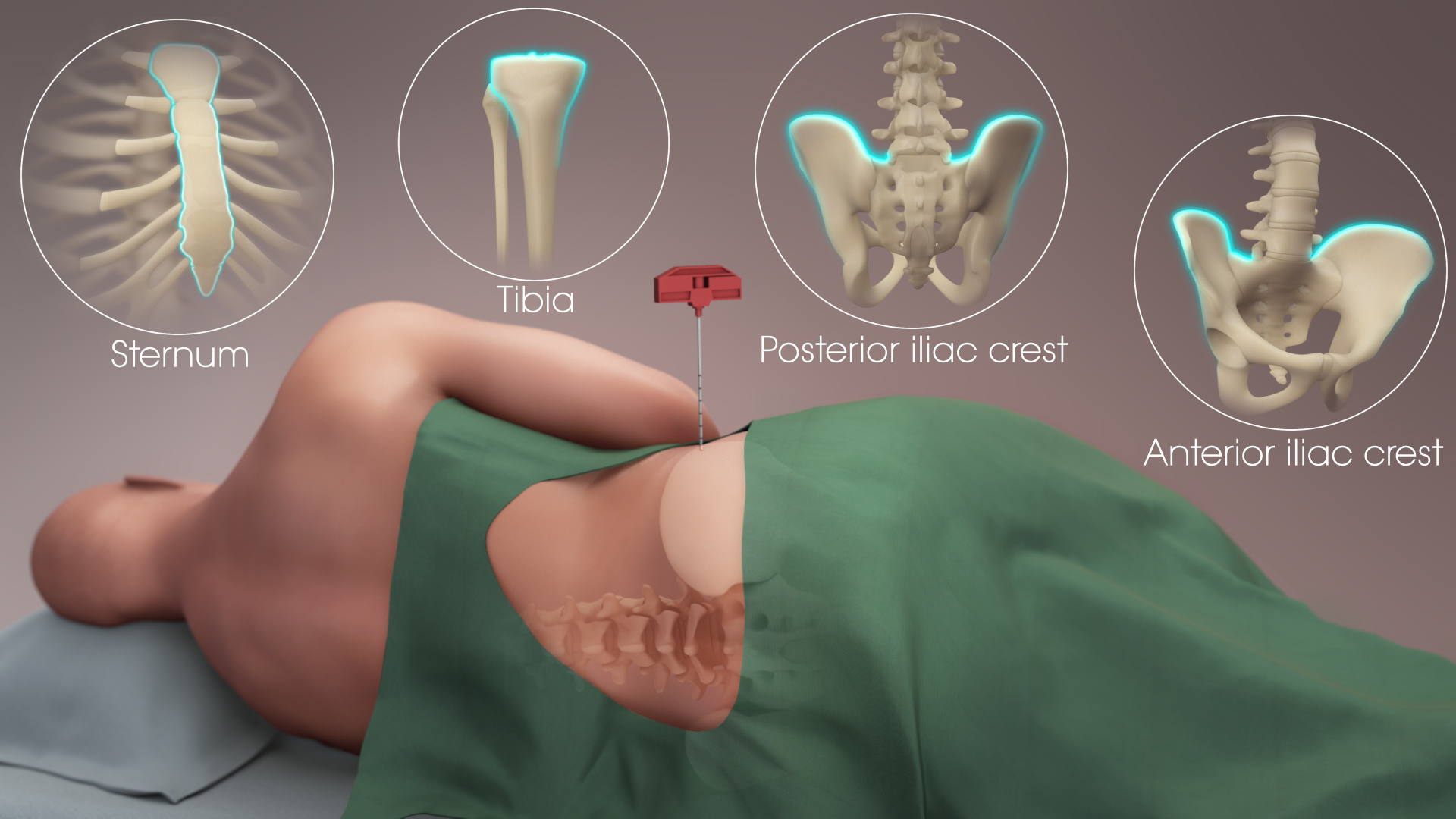
Bone Marrow
Bone marrow is a semi-solid biological tissue, tissue found within the Spongy bone, spongy (also known as cancellous) portions of bones. In birds and mammals, bone marrow is the primary site of new blood cell production (or haematopoiesis). It is composed of Blood cell, hematopoietic cells, marrow adipose tissue, and supportive stromal cells. In adult humans, bone marrow is primarily located in the Rib cage, ribs, vertebrae, sternum, and Pelvis, bones of the pelvis. Bone marrow comprises approximately 5% of total body mass in healthy adult humans, such that a person weighing 73 kg (161 lbs) will have around 3.7 kg (8 lbs) of bone marrow. Human marrow produces approximately 500 billion blood cells per day, which join the Circulatory system, systemic circulation via permeable vasculature sinusoids within the medullary cavity. All types of Hematopoietic cell, hematopoietic cells, including both Myeloid tissue, myeloid and Lymphocyte, lymphoid lineages, are create ... [...More Info...] [...Related Items...] OR: [Wikipedia] [Google] [Baidu] |


