|
Subependymoma
A subependymoma is a type of brain tumor; specifically, it is a rare form of ependymal tumor. They are usually in middle aged people. Earlier, they were called subependymal astrocytomas. The prognosis for a subependymoma is better than for most ependymal tumors, and it is considered a grade I tumor in the World Health Organization (WHO) classification. They are classically found within the fourth ventricle, typically have a well demarcated interface to normal tissue and do not usually extend into the brain parenchyma, like ependymomas often do. Symptoms and signs Patients are often asymptomatic, and are incidentally diagnosed. Larger tumours are often with increased intracranial pressure. Pathology These tumours are small, no more than two centimeters across, coming from the ependyma. The best way to distinguish it from a subependymal giant cell astrocytoma is the size. Diagnosis The diagnosis is based on tissue, e.g. a biopsy. Histologically subependymomas consistent ... [...More Info...] [...Related Items...] OR: [Wikipedia] [Google] [Baidu] |
Brain Tumor
A brain tumor (sometimes referred to as brain cancer) occurs when a group of cells within the Human brain, brain turn cancerous and grow out of control, creating a mass. There are two main types of tumors: malignant (cancerous) tumors and benign tumor, benign (non-cancerous) tumors. These can be further classified as primary tumors, which start within the brain, and metastasis, secondary tumors, which most commonly have spread from tumors located outside the brain, known as brain metastasis tumors. All types of brain tumors may produce symptoms that vary depending on the size of the tumor and the part of the brain that is involved. Where symptoms exist, they may include headaches, seizures, problems with visual perception, vision, vomiting and cognition, mental changes. Other symptoms may include difficulty walking, speaking, with sensations, or unconsciousness. The cause of most brain tumors is unknown, though up to 4% of brain cancers may be caused by CT scan radiation. Uncommo ... [...More Info...] [...Related Items...] OR: [Wikipedia] [Google] [Baidu] |
Astrocytomas
Astrocytoma is a type of brain tumor. Astrocytomas (also astrocytomata) originate from a specific kind of star-shaped glial cell in the cerebrum called an astrocyte. This type of tumor does not usually spread outside the brain and spinal cord, and it does not usually affect other organs. After glioblastomas, astrocytomas are the second most common glioma and can occur in most parts of the brain and occasionally in the spinal cord. Within the astrocytomas, two broad classes are recognized in literature, those with: * Narrow zones of infiltration (mostly noninvasive tumors; e.g., pilocytic astrocytoma, subependymal giant cell astrocytoma, pleomorphic xanthoastrocytoma), that often are clearly outlined on diagnostic images * Diffuse zones of infiltration (e.g., high-grade astrocytoma), that share various features, including the ability to arise at any location in the central nervous system, but with a preference for the cerebral hemispheres; they occur usually in adults, and have an ... [...More Info...] [...Related Items...] OR: [Wikipedia] [Google] [Baidu] |
Micrograph
A micrograph is an image, captured photographically or digitally, taken through a microscope or similar device to show a magnify, magnified image of an object. This is opposed to a macrograph or photomacrograph, an image which is also taken on a microscope but is only slightly magnified, usually less than 10 times. Micrography is the practice or art of using microscopes to make photographs. A photographic micrograph is a photomicrograph, and one taken with an electron microscope is an electron micrograph. A micrograph contains extensive details of microstructure. A wealth of information can be obtained from a simple micrograph like behavior of the material under different conditions, the phases found in the system, failure analysis, grain size estimation, elemental analysis and so on. Micrographs are widely used in all fields of microscopy. Types Photomicrograph A light micrograph or photomicrograph is a micrograph prepared using an optical microscope, a process referred to ... [...More Info...] [...Related Items...] OR: [Wikipedia] [Google] [Baidu] |
Nuclear Atypia
Nuclear atypia refers to abnormal appearance of cell nuclei. It is a term used in cytopathology and histopathology. Atypical nuclei are often pleomorphic. Nuclear atypia can be seen in reactive changes, pre-neoplastic changes and malignancy. Severe nuclear atypia is, in most cases, considered an indicator of malignancy. See also * Arias-Stella reaction *NC ratio NC may refer to: People * Naga Chaitanya, an Indian Telugu film actor; sometimes nicknamed by the initials of his first and middle name, NC * Nathan Connolly, lead guitarist for Snow Patrol * Nostalgia Critic, the alter ego of Internet comedian ... * Nuclear pleomorphism {{pathology-stub Pathology ... [...More Info...] [...Related Items...] OR: [Wikipedia] [Google] [Baidu] |
Histology
Histology, also known as microscopic anatomy or microanatomy, is the branch of biology that studies the microscopic anatomy of biological tissue (biology), tissues. Histology is the microscopic counterpart to gross anatomy, which looks at larger structures visible without a microscope. Although one may divide microscopic anatomy into ''organology'', the study of organs, ''histology'', the study of tissues, and ''cytology'', the study of cell (biology), cells, modern usage places all of these topics under the field of histology. In medicine, histopathology is the branch of histology that includes the microscopic identification and study of diseased tissue. In the field of paleontology, the term paleohistology refers to the histology of fossil organisms. Biological tissues Animal tissue classification There are four basic types of animal tissues: muscle tissue, nervous tissue, connective tissue, and epithelial tissue. All animal tissues are considered to be subtypes of these ... [...More Info...] [...Related Items...] OR: [Wikipedia] [Google] [Baidu] |
Brain Biopsy
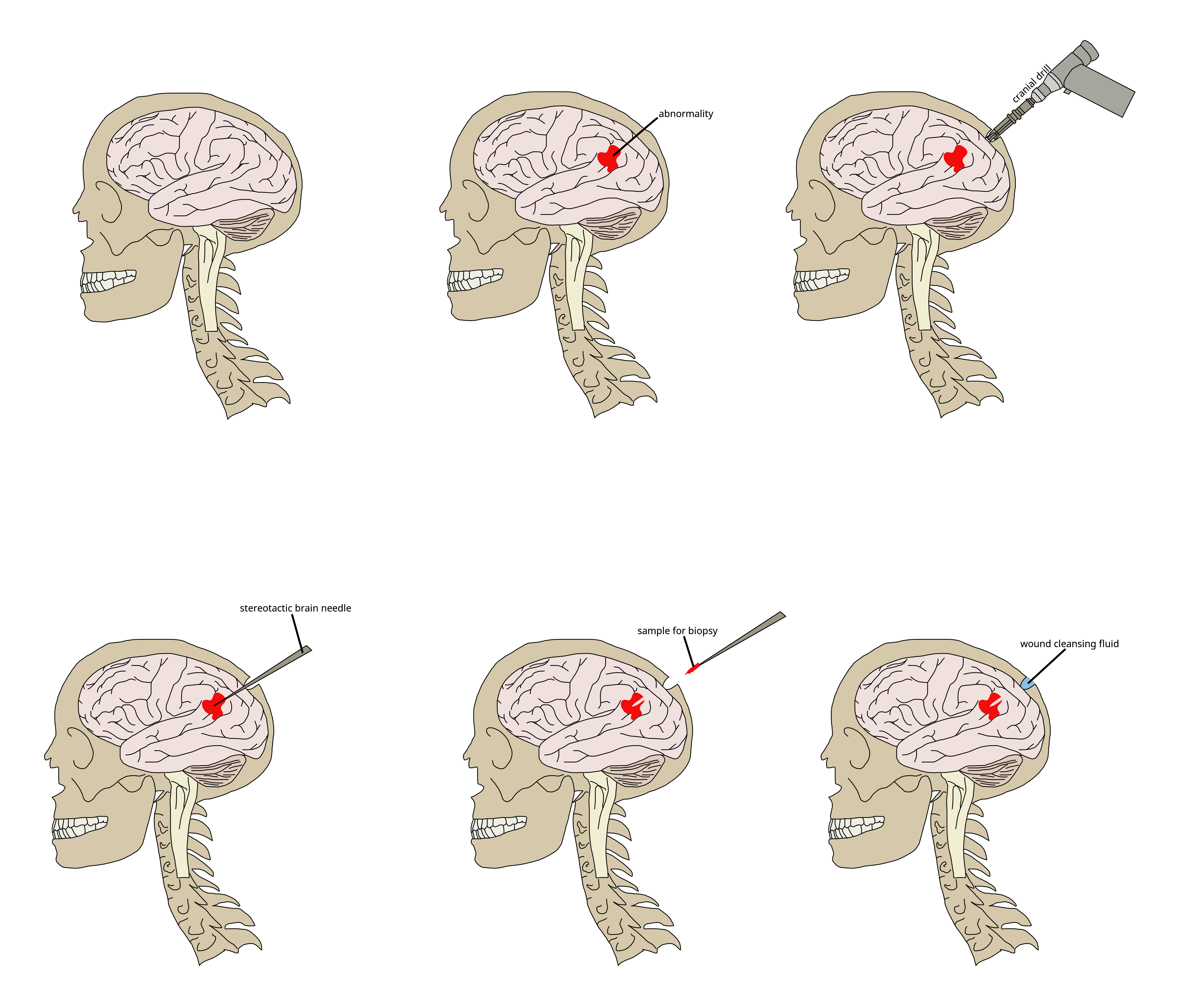
Brain biopsy is the removal of a small piece of brain tissue for the diagnosis of abnormalities of the brain. It is used to diagnose tumors, infection, inflammation, and other brain disorders. By examining the tissue sample under a microscope, the biopsy sample provides information about the appropriate diagnosis and treatment. Indications Given the potential risks surrounding the procedure, cerebral biopsy is indicated only if other diagnostic approaches (e.g. magnetic resonance imaging) have been insufficient in showing the cause of symptoms, and if it is felt that the benefits of histological diagnosis will influence the treatment plan. If the person has a brain tumor, biopsy is 95% sensitive. The procedure can also be valuable in people who are immunocompromised and who have evidence of brain lesions that could be caused by opportunistic infection An opportunistic infection is an infection that occurs most commonly in individuals with an immunodeficiency disorder and acts ... [...More Info...] [...Related Items...] OR: [Wikipedia] [Google] [Baidu] |
Subependymal Giant Cell Astrocytoma
Subependymal giant cell astrocytoma (SEGA, SGCA, or SGCT) is a low-grade astrocytic brain tumor (astrocytoma) that arises within the ventricles of the brain. It is most commonly associated with tuberous sclerosis complex (TSC). Although it is a low-grade tumor, its location can potentially obstruct the ventricles and lead to hydrocephalus. Signs and symptoms Individuals with this type of tumor may have no symptoms if cerebrospinal fluid (CSF) flow remains open. Obstruction of CSF flow will result in the symptoms associated with increased CSF pressure: nausea, vomiting, headache (often positional), lethargy, blurry or double vision, new or worsened seizures, and personality change. Diagnosis Diagnosis is made by imaging with a contrast-enhanced MRI or CT scan of the brain. Screening It is recommended that children with TSC be screened for SEGA with neuroimaging every 1–3 years. Treatment Pharmacotherapy Two related drugs have been shown to shrink or stabilize subependyma ... [...More Info...] [...Related Items...] OR: [Wikipedia] [Google] [Baidu] |
Intracranial Pressure
Intracranial pressure (ICP) is the pressure exerted by fluids such as cerebrospinal fluid (CSF) inside the skull and on the brain tissue. ICP is measured in millimeters of mercury ( mmHg) and at rest, is normally 7–15 mmHg for a supine adult. This equals to 9–20 cmH2O, which is a common scale used in lumbar punctures. The body has various mechanisms by which it keeps the ICP stable, with CSF pressures varying by about 1 mmHg in normal adults through shifts in production and absorption of CSF. Changes in ICP are attributed to volume changes in one or more of the constituents contained in the cranium. CSF pressure has been shown to be influenced by abrupt changes in intrathoracic pressure during coughing (which is induced by contraction of the diaphragm and abdominal wall muscles, the latter of which also increases intra-abdominal pressure), the valsalva maneuver, and communication with the vasculature ( venous and arterial systems). Intracranial hypertension (IH), ... [...More Info...] [...Related Items...] OR: [Wikipedia] [Google] [Baidu] |
Parenchyma
upright=1.6, Lung parenchyma showing damage due to large subpleural bullae. Parenchyma () is the bulk of functional substance in an animal organ such as the brain or lungs, or a structure such as a tumour. In zoology, it is the tissue that fills the interior of flatworms. In botany, it is some layers in the cross-section of the leaf. Etymology The term ''parenchyma'' is Neo-Latin from the Ancient Greek word meaning 'visceral flesh', and from meaning 'to pour in' from 'beside' + 'in' + 'to pour'. Originally, Erasistratus and other anatomists used it for certain human tissues. Later, it was also applied to plant tissues by Nehemiah Grew. Structure The parenchyma is the ''functional'' parts of an organ, or of a structure such as a tumour in the body. This is in contrast to the stroma, which refers to the ''structural'' tissue of organs or of structures, namely, the connective tissues. Brain The brain parenchyma refers to the functional tissue in th ... [...More Info...] [...Related Items...] OR: [Wikipedia] [Google] [Baidu] |
Ependymoma
An ependymoma is a tumor that arises from the ependyma, a tissue of the central nervous system. Usually, in pediatric cases the location is intracranial, while in adults it is spinal. The common location of intracranial ependymomas is the floor of the fourth ventricle. Rarely, ependymomas can occur in the pelvic cavity. Syringomyelia can be caused by an ependymoma. Ependymomas are also seen with neurofibromatosis type II. Signs and symptoms Source: Symptoms are dependent on the location and severity of the tumor. Intracranial ependymomas: * severe headache * nausea * vomiting * visual loss (due to papilledema) * loss of balance * vertigo * hydrocephalus * drowsiness (after several hours of the above symptoms) Spinal ependymomas: * bilateral Babinski sign * gait change (rotation of feet when walking) * impaction/constipation * back flexibility Morphology Ependymomas are composed of cells with regular, round to oval nuclei. There is a variably dense fibrillary background. ... [...More Info...] [...Related Items...] OR: [Wikipedia] [Google] [Baidu] |
H&E Stain
Hematoxylin and eosin stain ( or haematoxylin and eosin stain or hematoxylin–eosin stain; often abbreviated as H&E stain or HE stain) is one of the principal tissue stains used in histology. It is the most widely used stain in medical diagnosis and is often the ''gold standard.'' For example, when a pathologist looks at a biopsy of a suspected cancer, the histological section is likely to be stained with H&E. H&E is the combination of two histological stains: hematoxylin and eosin. The hematoxylin stains cell nuclei a purplish blue, and eosin stains the extracellular matrix and cytoplasm pink, with other structures taking on different shades, hues, and combinations of these colors. Hence a pathologist can easily differentiate between the nuclear and cytoplasmic parts of a cell, and additionally, the overall patterns of coloration from the stain show the general layout and distribution of cells and provides a general overview of a tissue sample's structure. Thus, patte ... [...More Info...] [...Related Items...] OR: [Wikipedia] [Google] [Baidu] |


