Brain Biopsy on:
[Wikipedia]
[Google]
[Amazon]
Brain biopsy is the removal of a small piece of brain tissue for the diagnosis of abnormalities of the
 Procedures are categorized into stereotactic, needle, and open. Stereotactic is the least invasive and open is the most invasive.
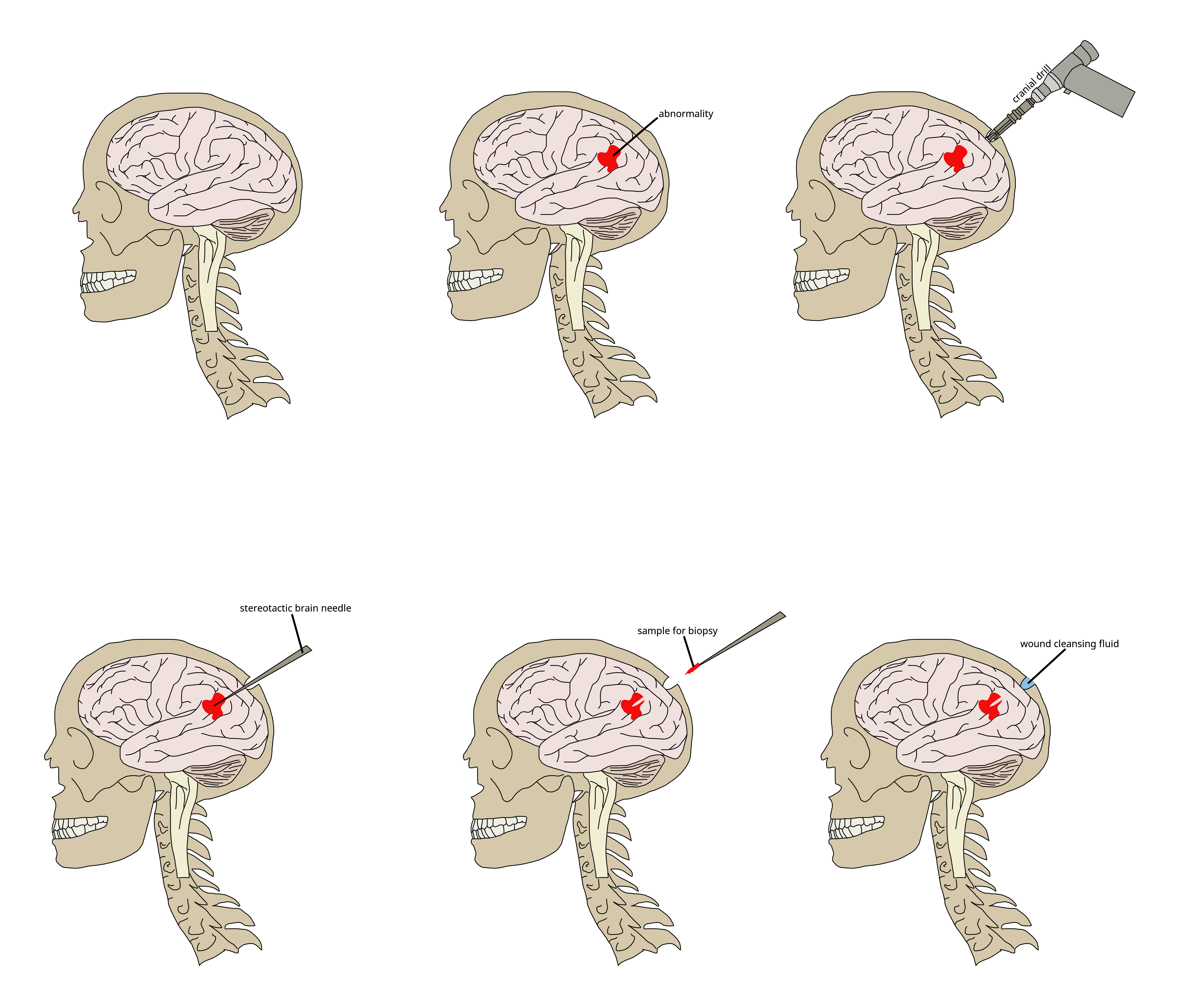
When an abnormality of the brain is suspected, stereotactic (probing in three dimensions) brain needle biopsy is performed and guided precisely by a computer system to avoid serious complications. A small hole is drilled into the skull, and a needle is inserted into the brain tissue guided by computer-assisted imaging techniques (CT or MRI scans). Historically, the patient's head was held in a rigid frame to direct the probe into the brain; however, since the early 1990s, it has been possible to perform these biopsies without the frame. Since the frame was attached to the skull with screws, this advancement is less invasive and better tolerated by the patient. The doctor (pathologist) prepares the sample for analysis and studies it further under a microscope.
brain
The brain is an organ (biology), organ that serves as the center of the nervous system in all vertebrate and most invertebrate animals. It consists of nervous tissue and is typically located in the head (cephalization), usually near organs for ...
. It is used to diagnose tumor
A neoplasm () is a type of abnormal and excessive growth of tissue. The process that occurs to form or produce a neoplasm is called neoplasia. The growth of a neoplasm is uncoordinated with that of the normal surrounding tissue, and persists ...
s, infection
An infection is the invasion of tissue (biology), tissues by pathogens, their multiplication, and the reaction of host (biology), host tissues to the infectious agent and the toxins they produce. An infectious disease, also known as a transmis ...
, inflammation
Inflammation (from ) is part of the biological response of body tissues to harmful stimuli, such as pathogens, damaged cells, or irritants. The five cardinal signs are heat, pain, redness, swelling, and loss of function (Latin ''calor'', '' ...
, and other brain disorders. By examining the tissue sample under a microscope
A microscope () is a laboratory equipment, laboratory instrument used to examine objects that are too small to be seen by the naked eye. Microscopy is the science of investigating small objects and structures using a microscope. Microscopic ...
, the biopsy
A biopsy is a medical test commonly performed by a surgeon, interventional radiologist, an interventional radiologist, or an interventional cardiology, interventional cardiologist. The process involves the extraction of sampling (medicine), sample ...
sample provides information about the appropriate diagnosis
Diagnosis (: diagnoses) is the identification of the nature and cause of a certain phenomenon. Diagnosis is used in a lot of different academic discipline, disciplines, with variations in the use of logic, analytics, and experience, to determine " ...
and treatment.
Indications
Given the potential risks surrounding the procedure, cerebral biopsy is indicated only if other diagnostic approaches (e.g.magnetic resonance imaging
Magnetic resonance imaging (MRI) is a medical imaging technique used in radiology to generate pictures of the anatomy and the physiological processes inside the body. MRI scanners use strong magnetic fields, magnetic field gradients, and ...
) have been insufficient in showing the cause of symptoms, and if it is felt that the benefits of histological
Histology,
also known as microscopic anatomy or microanatomy, is the branch of biology that studies the microscopic anatomy of biological tissue (biology), tissues. Histology is the microscopic counterpart to gross anatomy, which looks at large ...
diagnosis will influence the treatment plan.
If the person has a brain tumor
A brain tumor (sometimes referred to as brain cancer) occurs when a group of cells within the Human brain, brain turn cancerous and grow out of control, creating a mass. There are two main types of tumors: malignant (cancerous) tumors and benign ...
, biopsy is 95% sensitive. The procedure can also be valuable in people who are immunocompromised
Immunodeficiency, also known as immunocompromise, is a state in which the immune system's ability to fight infectious diseases and cancer is compromised or entirely absent. Most cases are acquired ("secondary") due to extrinsic factors that affe ...
and who have evidence of brain lesions that could be caused by opportunistic infection
An opportunistic infection is an infection that occurs most commonly in individuals with an immunodeficiency disorder and acts more severe on those with a weakened immune system. These types of infections are considered serious and can be caused b ...
s. In other groups, particularly those with unexplained neurological disease, a diagnosis is reached by performing a biopsy in half the cases where it is done, and it has helpful practical effect in 30% of people. If primary angiitis of the central nervous system (PACNS) is suspected, brain biopsy is most likely to positively influence the treatment plan.
Preparation
A CT or MRI brain scan is done to find the position where the biopsy will be performed. Prior to the biopsy, the patient is placed under general anesthesia.Procedure
 Procedures are categorized into stereotactic, needle, and open. Stereotactic is the least invasive and open is the most invasive.
When an abnormality of the brain is suspected, stereotactic (probing in three dimensions) brain needle biopsy is performed and guided precisely by a computer system to avoid serious complications. A small hole is drilled into the skull, and a needle is inserted into the brain tissue guided by computer-assisted imaging techniques (CT or MRI scans). Historically, the patient's head was held in a rigid frame to direct the probe into the brain; however, since the early 1990s, it has been possible to perform these biopsies without the frame. Since the frame was attached to the skull with screws, this advancement is less invasive and better tolerated by the patient. The doctor (pathologist) prepares the sample for analysis and studies it further under a microscope.
Aftercare
The patient is monitored in the recovery room for several hours following the biopsy. Neurological assessments are performed once the patient is fully awake and if left without deficit, most patients can be discharged the day after surgery.Risks
The procedure is invasive and includes risks associated with anesthesia and surgery. Brain injury may occur due to removal of brain tissue. The resulting scar left on the brain has the potential to trigger seizures. If brain biopsy is performed for a possible tumor (which contain more blood vessels), the risk of death is 1% and a risk of complications 12%. For unexplained neurological disease, there is no risk of death and a complication rate of 9%; complications were more common in PACNS.Interpretation
Various brain abnormalities can be diagnosed by microscopic analysis of the tissue sample. The pathologist (a physician trained in how disease affects the body's tissues) looks for abnormal growth, changes in cell membranes, and/or abnormal collections of cells. In Alzheimer's disease, the cortex of the brain contains abnormal collections of plaques. If infection is suspected, the infectious organism can be cultured from the tissue and identified. Classification of tumors is also possible after biopsy.See also
* * * *References