|
Ependymoma
An ependymoma is a tumor that arises from the ependyma, a tissue of the central nervous system. Usually, in pediatric cases the location is intracranial, while in adults it is spinal. The common location of intracranial ependymomas is the floor of the fourth ventricle. Rarely, ependymomas can occur in the pelvic cavity. Syringomyelia can be caused by an ependymoma. Ependymomas are also seen with neurofibromatosis type II. Signs and symptoms Source: Symptoms are dependent on the location and severity of the tumor. Intracranial ependymomas: * severe headache * nausea * vomiting * visual loss (due to papilledema) * loss of balance * vertigo * hydrocephalus * drowsiness (after several hours of the above symptoms) Spinal ependymomas: * bilateral Babinski sign * gait change (rotation of feet when walking) * impaction/constipation * back flexibility Morphology Ependymomas are composed of cells with regular, round to oval nuclei. There is a variably dense fibrillary background. ... [...More Info...] [...Related Items...] OR: [Wikipedia] [Google] [Baidu] |
Neurofibromatosis Type II
Neurofibromatosis type II (also known as MISME syndrome – multiple inherited schwannomas, meningiomas, and ependymomas) is a genetic condition that may be inherited or may arise spontaneously, and causes benign tumors of the brain, spinal cord, and peripheral nerves. The types of tumors frequently associated with NF2 include vestibular schwannomas, meningiomas, and ependymomas. The main manifestation of the condition is the development of bilateral benign brain tumors in the nerve sheath of the cranial nerve VIII, which is the "auditory-vestibular nerve" that transmits sensory information from the inner ear to the brain. Besides, other benign brain and spinal tumors occur. Symptoms depend on the presence, localisation and growth of the tumor(s). Many people with this condition also experience vision problems. Neurofibromatosis type II (NF2 ''or'' NF II) is caused by mutations of the "Merlin" gene, which seems to influence the form and movement of cells. The principal treatments ... [...More Info...] [...Related Items...] OR: [Wikipedia] [Google] [Baidu] |
Syringomyelia
Syringomyelia is a generic term referring to a disorder in which a cyst or cavity forms within the spinal cord. Often, syringomyelia is used as a generic term before an etiology is determined. This cyst, called a syrinx, can expand and elongate over time, destroying the spinal cord. The damage may result in loss of feeling, paralysis, weakness, and stiffness in the back, shoulders, and extremities. Syringomyelia may also cause a loss of the ability to feel extremes of hot or cold, especially in the hands. It may also lead to a cape-like bilateral loss of pain and temperature sensation along the upper chest and arms. The combination of symptoms varies from one patient to another depending on the location of the syrinx within the spinal cord, as well as its extent. Syringomyelia has a prevalence estimated at 8.4 cases per 100,000 people, with symptoms usually beginning in young adulthood. Signs of the disorder tend to develop slowly, although sudden onset may occur with coughing, ... [...More Info...] [...Related Items...] OR: [Wikipedia] [Google] [Baidu] |
Ependymoblastoma
Primitive neuroectodermal tumor is a malignant (cancerous) neural crest tumor. It is a rare tumor, usually occurring in children and young adults under 25 years of age. The overall 5 year survival rate is about 53%. It gets its name because the majority of the cells in the tumor are derived from neuroectoderm, but have not developed and differentiated in the way a normal neuron would, and so the cells appear "primitive". PNET belongs to the Ewing family of tumors. Genetics Using gene transfer of SV40 large T-antigen in neuronal precursor cells of rats, a brain tumor model was established. The PNETs were histologically indistinguishable from the human counterparts and have been used to identify new genes involved in human brain tumor carcinogenesis. The model was used to confirm p53 as one of the genes involved in human medulloblastomas, but since only about 10% of the human tumors showed mutations in that gene, the model can be used to identify the other binding partners of SV40 ... [...More Info...] [...Related Items...] OR: [Wikipedia] [Google] [Baidu] |
Ependyma
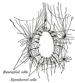
The ependyma is the thin neuroepithelial ( simple columnar ciliated epithelium) lining of the ventricular system of the brain and the central canal of the spinal cord. The ependyma is one of the four types of neuroglia in the central nervous system (CNS). It is involved in the production of cerebrospinal fluid (CSF), and is shown to serve as a reservoir for neuroregeneration. Structure The ependyma is made up of ependymal cells called ependymocytes, a type of glial cell. These cells line the ventricles in the brain and the central canal of the spinal cord, which become filled with cerebrospinal fluid. These are nervous tissue cells with simple columnar shape, much like that of some mucosal epithelial cells. Early monociliated ependymal cells are differentiated to multiciliated ependymal cells for their function in circulating cerebrospinal fluid. The basal membranes of these cells are characterized by tentacle-like extensions that attach to astrocytes. The apical side i ... [...More Info...] [...Related Items...] OR: [Wikipedia] [Google] [Baidu] |
Glioma
A glioma is a type of primary tumor that starts in the glial cells of the brain or spinal cord. They are malignant but some are extremely slow to develop. Gliomas comprise about 30% of all brain and central nervous system tumors and 80% of all malignant brain tumors. They are a few common types that include astrocytoma (cancer of astrocytes), glioblastoma (an aggressive form of astrocytoma), oligodendroglioma (cancer of oligodendrocytes), and ependymoma (cancer of ependymal cells). Signs and symptoms Symptoms of gliomas depend on the part of the central nervous system (CNS) that is affected. A brain glioma can cause headaches, vomiting, memory loss, seizures, vision problems, speech difficulties, and cranial nerve disorders as a result of increased intracranial pressure. Cognitive impairments such as vision loss arise in glioma patients when a tumor arises in or around their optic nerve. Spinal cord gliomas can cause pain, weakness, or numbness in the extremities ... [...More Info...] [...Related Items...] OR: [Wikipedia] [Google] [Baidu] |
Micrograph
A micrograph is an image, captured photographically or digitally, taken through a microscope or similar device to show a magnify, magnified image of an object. This is opposed to a macrograph or photomacrograph, an image which is also taken on a microscope but is only slightly magnified, usually less than 10 times. Micrography is the practice or art of using microscopes to make photographs. A photographic micrograph is a photomicrograph, and one taken with an electron microscope is an electron micrograph. A micrograph contains extensive details of microstructure. A wealth of information can be obtained from a simple micrograph like behavior of the material under different conditions, the phases found in the system, failure analysis, grain size estimation, elemental analysis and so on. Micrographs are widely used in all fields of microscopy. Types Photomicrograph A light micrograph or photomicrograph is a micrograph prepared using an optical microscope, a process referred to ... [...More Info...] [...Related Items...] OR: [Wikipedia] [Google] [Baidu] |
Central Canal
The central canal (also known as spinal foramen or ependymal canal) is the cerebrospinal fluid-filled space that runs through the spinal cord. The central canal lies below and is connected to the ventricular system of the brain, from which it receives cerebrospinal fluid, and shares the same ependymal lining. The central canal helps to transport nutrients to the spinal cord as well as protect it by cushioning the impact of a force when the spine is affected. The central canal represents the adult remainder of the central cavity of the neural tube. It generally occludes (closes off) with age. Structure The central canal below at the ventricular system of the brain, beginning at a region called the obex where the fourth ventricle, a cavity present in the brainstem, narrows. The central canal is located in the third of the spinal cord in the cervical vertebrae, cervical and thoracic spine, thoracic regions. In the lumbar spine it enlarges and is located more centrally. At the c ... [...More Info...] [...Related Items...] OR: [Wikipedia] [Google] [Baidu] |
HPS Stain
In histology, the HPS stain, or hematoxylin phloxine saffron stain, is a way of marking tissues. HPS is similar to H&E, the standard bearer in histology. However, it differentiates between the most common connective tissue (collagen) and muscle and cytoplasm by staining the former yellow and the latter two pink, . polysciences.com. Accessed 6 December 2009. unlike an H&E stain, which stains all three pink. HPS stained sections are more expensive than H&E stained sections, primarily due to the cost of saffron. See also *Histopathology
Histopathology (compound of three Greek words: 'tissue', 'suffering', and '' -logia'' 'study ...
[...More Info...] [...Related Items...] OR: [Wikipedia] [Google] [Baidu] |
Ventricular System
In neuroanatomy, the ventricular system is a set of four interconnected cavities known as cerebral ventricles in the brain. Within each ventricle is a region of choroid plexus which produces the circulating cerebrospinal fluid (CSF). The ventricular system is continuous with the central canal of the spinal cord from the fourth ventricle, allowing for the flow of CSF to circulate. All of the ventricular system and the central canal of the spinal cord are lined with ependyma, a specialised form of epithelium connected by tight junctions that make up the blood–cerebrospinal fluid barrier. Structure The system comprises four ventricles: * lateral ventricles right and left (one for each hemisphere) * third ventricle * fourth ventricle There are several foramina, openings acting as channels, that connect the ventricles. The interventricular foramina (also called the foramina of Monro) connect the lateral ventricles to the third ventricle through which the cerebrospinal ... [...More Info...] [...Related Items...] OR: [Wikipedia] [Google] [Baidu] |
Anaplastic
Anaplasia () is a condition of cells with poor cellular differentiation, losing the morphological characteristics of mature cells and their orientation with respect to each other and to endothelial cells. The term also refers to a group of morphological changes in a cell (nuclear pleomorphism, altered nuclear-cytoplasmic ratio, presence of nucleoli, high proliferation index) that point to a possible malignant transformation. Such loss of structural differentiation is especially seen in most, but not all, malignant neoplasms. Sometimes, the term also includes an increased capacity for multiplication. Lack of differentiation is considered a hallmark of aggressive malignancies (for example, it differentiates leiomyosarcomas from leiomyomas). The term ''anaplasia'' literally means "to form backward". It implies dedifferentiation, or loss of structural and functional differentiation of normal cells. It is now known, however, that at least some cancers arise from stem cells in t ... [...More Info...] [...Related Items...] OR: [Wikipedia] [Google] [Baidu] |
Benign
Malignancy () is the tendency of a medical condition to become progressively worse; the term is most familiar as a characterization of cancer. A ''malignant'' tumor contrasts with a non-cancerous benign tumor, ''benign'' tumor in that a malignancy is not self-limited in its growth, is capable of invading into adjacent tissues, and may be capable of spreading to distant tissues. A benign tumor has none of those properties, but may still be harmful to health. The term benign in more general medical use characterizes a condition or growth that is not cancerous, i.e. does not spread to other parts of the body or invade nearby tissue. Sometimes the term is used to suggest that a condition is not dangerous or serious. Malignancy in cancers is characterized by anaplasia, invasiveness, and metastasis. Malignant tumors are also characterized by genome instability, so that cancers, as assessed by whole genome sequencing, frequently have between 10,000 and 100,000 mutations in their ent ... [...More Info...] [...Related Items...] OR: [Wikipedia] [Google] [Baidu] |
Ependymal Canal
The central canal (also known as spinal foramen or ependymal canal) is the cerebrospinal fluid-filled space that runs through the spinal cord. The central canal lies below and is connected to the ventricular system of the brain, from which it receives cerebrospinal fluid, and shares the same ependymal lining. The central canal helps to transport nutrients to the spinal cord as well as protect it by cushioning the impact of a force when the spine is affected. The central canal represents the adult remainder of the central cavity of the neural tube. It generally occludes (closes off) with age. Structure The central canal below at the ventricular system of the brain, beginning at a region called the obex where the fourth ventricle, a cavity present in the brainstem, narrows. The central canal is located in the third of the spinal cord in the cervical vertebrae, cervical and thoracic spine, thoracic regions. In the lumbar spine it enlarges and is located more centrally. At the c ... [...More Info...] [...Related Items...] OR: [Wikipedia] [Google] [Baidu] |


