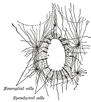
Ependymal Canal on:
[Wikipedia]
[Google]
[Amazon]
The central canal (also known as spinal foramen or ependymal canal) is the
 The central canal below at the
cerebrospinal fluid
Cerebrospinal fluid (CSF) is a clear, colorless Extracellular fluid#Transcellular fluid, transcellular body fluid found within the meninges, meningeal tissue that surrounds the vertebrate brain and spinal cord, and in the ventricular system, ven ...
-filled space that runs through the spinal cord
The spinal cord is a long, thin, tubular structure made up of nervous tissue that extends from the medulla oblongata in the lower brainstem to the lumbar region of the vertebral column (backbone) of vertebrate animals. The center of the spinal c ...
. The central canal lies below and is connected to the ventricular system
In neuroanatomy, the ventricular system is a set of four interconnected cavities known as cerebral ventricles in the brain. Within each ventricle is a region of choroid plexus which produces the circulating cerebrospinal fluid (CSF). The ventric ...
of the brain
The brain is an organ (biology), organ that serves as the center of the nervous system in all vertebrate and most invertebrate animals. It consists of nervous tissue and is typically located in the head (cephalization), usually near organs for ...
, from which it receives cerebrospinal fluid, and shares the same ependyma
The ependyma is the thin neuroepithelial ( simple columnar ciliated epithelium) lining of the ventricular system of the brain and the central canal of the spinal cord. The ependyma is one of the four types of neuroglia in the central nervous s ...
l lining. The central canal helps to transport nutrients to the spinal cord as well as protect it by cushioning the impact of a force when the spine is affected.
The central canal represents the adult remainder of the central cavity of the neural tube
In the developing chordate (including vertebrates), the neural tube is the embryonic precursor to the central nervous system, which is made up of the brain and spinal cord. The neural groove gradually deepens as the neural folds become elevated, ...
. It generally occludes (closes off) with age.
Structure
 The central canal below at the
 ventricular system
In neuroanatomy, the ventricular system is a set of four interconnected cavities known as cerebral ventricles in the brain. Within each ventricle is a region of choroid plexus which produces the circulating cerebrospinal fluid (CSF). The ventric ...
of the brain
The brain is an organ (biology), organ that serves as the center of the nervous system in all vertebrate and most invertebrate animals. It consists of nervous tissue and is typically located in the head (cephalization), usually near organs for ...
, beginning at a region called the obex
The obex () is the point in the human brain at which the fourth ventricle narrows to become the central canal of the spinal cord. Cerebrospinal fluid can flow from the fourth ventricle into the obex. In anatomical studies, the obex has been fo ...
where the fourth ventricle
The fourth ventricle is one of the four connected fluid-filled cavities within the human brain. These cavities, known collectively as the ventricular system, consist of the left and right lateral ventricles, the third ventricle, and the fourth ...
, a cavity present in the brainstem, narrows.
The central canal is located in the third of the spinal cord in the cervical and thoracic
The thorax (: thoraces or thoraxes) or chest is a part of the anatomy of mammals and other tetrapod animals located between the neck and the abdomen.
In insects, crustaceans, and the extinct trilobites, the thorax is one of the three main ...
regions. In the lumbar spine
The lumbar vertebrae are located between the thoracic vertebrae and pelvis. They form the lower part of the back in humans, and the tail end of the back in quadrupeds. In humans, there are five lumbar vertebrae. The term is used to describe t ...
it enlarges and is located more centrally. At the conus medullaris
The conus medullaris (Latin for "medullary cone") or conus terminalis is the tapered, lower end of the spinal cord. It occurs near lumbar vertebral levels 1 (L1) and 2 (L2), occasionally lower. The upper end of the conus medullaris is usually no ...
, where the spinal cord tapers, it is located more .
Terminal ventricle
The terminal ventricle (ventriculus terminalis, fifth ventricle or ampulla caudalis) is the widest part of the central canal of thespinal cord
The spinal cord is a long, thin, tubular structure made up of nervous tissue that extends from the medulla oblongata in the lower brainstem to the lumbar region of the vertebral column (backbone) of vertebrate animals. The center of the spinal c ...
that is located at or near the conus medullaris
The conus medullaris (Latin for "medullary cone") or conus terminalis is the tapered, lower end of the spinal cord. It occurs near lumbar vertebral levels 1 (L1) and 2 (L2), occasionally lower. The upper end of the conus medullaris is usually no ...
. It was described by Stilling in 1859 and Krause in 1875. Krause introduced the term fifth ventricle after observation of normal ependymal cells
The ependyma is the thin neuroepithelial ( simple columnar ciliated epithelium) lining of the ventricular system of the brain and the central canal of the spinal cord. The ependyma is one of the four types of neuroglia in the central nervous sys ...
. The central canal expands as a fusiform terminal ventricle, and approximately 8–10 mm in length in the conus medullaris (or conus terminalis). Although the terminal ventricle is visible in the fetus and children, it is usually absent in adults.
Sometimes, the terminal ventricle is observed by MRI
Magnetic resonance imaging (MRI) is a medical imaging technique used in radiology to generate pictures of the anatomy and the physiological processes inside the body. MRI scanners use strong magnetic fields, magnetic field gradients, and rad ...
or ultrasound
Ultrasound is sound with frequency, frequencies greater than 20 Hertz, kilohertz. This frequency is the approximate upper audible hearing range, limit of human hearing in healthy young adults. The physical principles of acoustic waves apply ...
in children less than 5 years old.
Microanatomy
The central canal shares the sameependyma
The ependyma is the thin neuroepithelial ( simple columnar ciliated epithelium) lining of the ventricular system of the brain and the central canal of the spinal cord. The ependyma is one of the four types of neuroglia in the central nervous s ...
l lining as the ventricular system of the brain.
The canal is lined by cilia
The cilium (: cilia; ; in Medieval Latin and in anatomy, ''cilium'') is a short hair-like membrane protrusion from many types of eukaryotic cell. (Cilia are absent in bacteria and archaea.) The cilium has the shape of a slender threadlike proj ...
ted, column-shaped cells, outside of which is a band of gelatinous substance, called the substantia gelatinosa of Rolando
The apex of the posterior grey column, one of the three grey columns of the spinal cord, is capped by a V-shaped or crescentic mass of translucent, gelatinous neuroglia, termed the substantia gelatinosa of Rolando (or SGR) (or gelatinous substan ...
also substantia gelatinosa centralis or central gelatinous substance of spinal cord. This gelatinous substance consists mainly of neuroglia
Glia, also called glial cells (gliocytes) or neuroglia, are non- neuronal cells in the central nervous system (the brain and the spinal cord) and in the peripheral nervous system that do not produce electrical impulses. The neuroglia make up ...
, but contains a few nerve cells and fibers; it is traversed by processes from the deep ends of the columnar ciliated cells which line the central canal.
Development
The central canal represents the adult remainder of the central cavity of theneural tube
In the developing chordate (including vertebrates), the neural tube is the embryonic precursor to the central nervous system, which is made up of the brain and spinal cord. The neural groove gradually deepens as the neural folds become elevated, ...
. It generally occludes (closes off) with age.
Function
The central canal carriescerebrospinal fluid
Cerebrospinal fluid (CSF) is a clear, colorless Extracellular fluid#Transcellular fluid, transcellular body fluid found within the meninges, meningeal tissue that surrounds the vertebrate brain and spinal cord, and in the ventricular system, ven ...
(CSF), which it receives from the ventricular system
In neuroanatomy, the ventricular system is a set of four interconnected cavities known as cerebral ventricles in the brain. Within each ventricle is a region of choroid plexus which produces the circulating cerebrospinal fluid (CSF). The ventric ...
of the brain. The central canal helps to transport nutrients to the spinal cord as well as protect it by cushioning the impact of a force when the spine is affected.
Clinical significance
Syringomyelia
Syringomyelia is a generic term referring to a disorder in which a cyst or cavity forms within the spinal cord. Often, syringomyelia is used as a generic term before an etiology is determined. This cyst, called a syrinx, can expand and elongate ...
is a disease caused by the blockage of the central canal. Blockage of the central canal usually occurs at the lower cervical and upper thoracic levels. This typically damages white matter
White matter refers to areas of the central nervous system that are mainly made up of myelinated axons, also called Nerve tract, tracts. Long thought to be passive tissue, white matter affects learning and brain functions, modulating the distr ...
fibers that cross in anterior white commissure
The anterior white commissure (ventral white commissure) is a bundle of nerve fibers which cross the midline of the spinal cord just anterior (in front of) to the gray commissure ( Rexed lamina X). A delta fibers (Aδ fibers) and C fibers carr ...
, leading to the loss of temperature, pain, and motor function at the affected levels on side opposite to the damage.
Other relevant conditions include:
* Spina bifida
Spina bifida (SB; ; Latin for 'split spine') is a birth defect in which there is incomplete closing of the vertebral column, spine and the meninges, membranes around the spinal cord during embryonic development, early development in pregnancy. T ...
* Arnold-Chiari syndrome
In neurology, the Chiari malformation ( ; CM) is a structural defect in the cerebellum, characterized by a downward displacement of one or both cerebellar tonsils through the foramen magnum (the opening at the base of the skull).
CMs can cau ...
* Spinal tumor
Spinal tumors are neoplasms located in either the vertebral column or the spinal cord. There are three main types of spinal tumors classified based on their location: extradural and intradural (intradural-intramedullary and intradural-extramedulla ...
* Myelomeningocele
Spina bifida (SB; ; Latin for 'split spine') is a birth defect in which there is incomplete closing of the spine and the membranes around the spinal cord during early development in pregnancy. There are three main types: spina bifida occulta, ...
* Syringomyelia
Syringomyelia is a generic term referring to a disorder in which a cyst or cavity forms within the spinal cord. Often, syringomyelia is used as a generic term before an etiology is determined. This cyst, called a syrinx, can expand and elongate ...
* Hydromyelia
Syringomyelia is a generic term referring to a disorder in which a cyst or cavity forms within the spinal cord. Often, syringomyelia is used as a generic term before an etiology is determined. This cyst, called a syrinx, can expand and elongate o ...
. In hydromyelia, a dilation of the central canal of the spinal cord is caused by an increase of cerebrospinal fluid.
* Syringohydromyelia
Syringomyelia is a generic term referring to a disorder in which a cyst or cavity forms within the spinal cord. Often, syringomyelia is used as a generic term before an etiology is determined. This cyst, called a Syrinx (medicine), syrinx, can ex ...
(i.e., both Syringomyelia and Hydromyelia)
* Tethered cord
Tethered cord syndrome (TCS) refers to a group of neurological disorders that relate to malformations of the spinal cord.Spinal cord
Ventricular system
Tomsick T, Peak E, Wang L: Fluid-Signal Structures in the Cervical Spinal Cord on MRI: Anterior Median Fissure vs. Central Canal. AJNR 2017; 38:840–45
Tomsick T, Wang L, Zuccarello M, Ringer AJ. Fluid-signal structures in the cervical spinal cord on MRI in Chiari patients: Central canal or anterior median fissure? AJNR Am J Neuroradiol. 2021 Apr;42(4):801-806. doi: 10.3174/ajnr.A7046. Epub 2021 Mar 11.PMID: 33707286