|
Scanning Laser Ophthalmoscopy
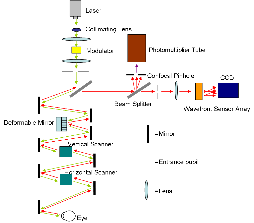
Scanning laser ophthalmoscopy (SLO) is a method of examination of the eye. It uses the technique of confocal laser scanning microscopy for diagnostic imaging of the retina or cornea of the human eye. As a method used to image the retina with a high degree of spatial sensitivity, it is helpful in the diagnosis of glaucoma, macular degeneration, and other retinal disorders. It has further been combined with adaptive optics technology to provide sharper images of the retina."Roorda Lab" — (last accessed: 9 December 2006) Published on October 25, 2006—(last accessed: 9 December 2006) Scanning laser ophthalmoscopy SLO utilizes horizontal and vertical scanning mirrors to scan a specific region of ...[...More Info...] [...Related Items...] OR: [Wikipedia] [Google] [Baidu] |
Confocal Laser Scanning Microscopy
Confocal microscopy, most frequently confocal laser scanning microscopy (CLSM) or laser scanning confocal microscopy (LSCM), is an optical imaging technique for increasing optical resolution and contrast of a micrograph by means of using a spatial pinhole to block out-of-focus light in image formation. Capturing multiple two-dimensional images at different depths in a sample enables the reconstruction of three-dimensional structures (a process known as optical sectioning) within an object. This technique is used extensively in the scientific and industrial communities and typical applications are in life sciences, semiconductor inspection and materials science. Light travels through the sample under a conventional microscope as far into the specimen as it can penetrate, while a confocal microscope only focuses a smaller beam of light at one narrow depth level at a time. The CLSM achieves a controlled and highly limited depth of field. Basic concept The principle of c ... [...More Info...] [...Related Items...] OR: [Wikipedia] [Google] [Baidu] |
Charge-coupled Device
A charge-coupled device (CCD) is an integrated circuit containing an array of linked, or coupled, capacitors. Under the control of an external circuit, each capacitor can transfer its electric charge to a neighboring capacitor. CCD sensors are a major technology used in digital imaging. Overview In a CCD image sensor, pixels are represented by Doping (semiconductor), p-doped metal–oxide–semiconductor (MOS) capacitors. These MOS capacitors, the basic building blocks of a CCD, are biased above the threshold for inversion when image acquisition begins, allowing the conversion of incoming photons into electron charges at the semiconductor-oxide interface; the CCD is then used to read out these charges. Although CCDs are not the only technology to allow for light detection, CCD image sensors are widely used in professional, medical, and scientific applications where high-quality image data are required. In applications with less exacting quality demands, such as consumer and pr ... [...More Info...] [...Related Items...] OR: [Wikipedia] [Google] [Baidu] |
Ophthalmoscopy
Ophthalmoscopy, also called funduscopy, is a test that allows a health professional to see inside the fundus of the eye and other structures using an ophthalmoscope (or funduscope). It is done as part of an eye examination and may be done as part of a routine physical examination. It is crucial in determining the health of the retina, optic disc, and vitreous humor. The pupil is a hole through which the eye's interior can be viewed. For better viewing, the pupil can be opened wider (dilated; mydriasis) before ophthalmoscopy using medicated eye drops ( dilated fundus examination). However, undilated examination is more convenient (albeit not as comprehensive), and is the most common type in primary care. An alternative or complement to ophthalmoscopy is to perform a fundus photography, where the image can be analysed later by a professional. Types There are two major types of ophthalmoscopy: * direct ophthalmoscopy, which produces an upright (unreversed) image of approxima ... [...More Info...] [...Related Items...] OR: [Wikipedia] [Google] [Baidu] |
Fundus Photography
Fundus photography involves photographing the rear of an eye, also known as the fundus (eye), fundus. Specialized fundus cameras consisting of an intricate microscope attached to a flash (photography), flash enabled camera are used in fundus photography. The main structures that can be visualized on a fundus photo are the central and peripheral retina, optic disc and Macula of retina, macula. Fundus photography can be performed with colored filters, or with specialized dyes including Fluorescein angiography, fluorescein and indocyanine green. The models and technology of fundus photography have advanced and evolved rapidly over the last century. History The concept of fundus photography was first introduced in the mid 19th century, after the introduction of photography in 1839. In 1851, Hermann von Helmholtz introduced the Ophthalmoscopy, Ophthalmoscope, and James Clerk Maxwell presented a colour photography method in 1861. In the early 1860s, Henry Noyes and Abner Mulhollan ... [...More Info...] [...Related Items...] OR: [Wikipedia] [Google] [Baidu] |
Optical Coherence Tomography
Optical coherence tomography (OCT) is a high-resolution imaging technique with most of its applications in medicine and biology. OCT uses coherent near-infrared light to obtain micrometer-level depth resolved images of biological tissue or other scattering media. It uses interferometry techniques to detect the amplitude and time-of-flight of reflected light. OCT uses transverse sample scanning of the light beam to obtain two- and three-dimensional images. Short-coherence-length light can be obtained using a superluminescent diode (SLD) with a broad spectral bandwidth or a broadly tunable laser with narrow linewidth. The first demonstration of OCT imaging (in vitro) was published by a team from MIT and Harvard Medical School in a 1991 article in the journal ''Science (journal), Science''. The article introduced the term "OCT" to credit its derivation from optical coherence-domain reflectometry, in which the axial resolution is based on temporal coherence. The first demonstrat ... [...More Info...] [...Related Items...] OR: [Wikipedia] [Google] [Baidu] |
Fluorescein Angiography
Fluorescein angiography (FA), fluorescent angiography (FAG), or fundus fluorescein angiography (FFA) is a technique for examining the circulation of the retina and choroid (parts of the fundus) using a fluorescent dye and a specialized camera. Sodium fluorescein is added into the systemic circulation, the retina is illuminated with blue-green light at a wavelength of 490 nanometers, and an angiogram is obtained by photographing the fluorescent green light that is emitted by the dye. The fluorescein is administered intravenously in intravenous fluorescein angiography (IVFA) and orally in oral fluorescein angiography (OFA). The test is a dye tracing method. The fluorescein dye also reappears in the patient urine, causing the urine to appear darker, and sometimes orange. It can also cause discolouration of the saliva. Fluorescein angiography is one of several health care applications of this dye, all of which have a risk of severe adverse effects. See fluorescein safety in h ... [...More Info...] [...Related Items...] OR: [Wikipedia] [Google] [Baidu] |
Macula
The macula (/ˈmakjʊlə/) or macula lutea is an oval-shaped pigmented area in the center of the retina of the human eye and in other animals. The macula in humans has a diameter of around and is subdivided into the umbo, foveola, foveal avascular zone, fovea, parafovea, and perifovea areas. The anatomical macula at a size of is much larger than the clinical macula which, at a size of , corresponds to the anatomical fovea. The macula is responsible for the central, high-resolution, color vision that is possible in good light. This kind of vision is impaired if the macula is damaged, as in macular degeneration. The clinical macula is seen when viewed from the pupil, as in ophthalmoscopy or retinal photography. The term macula lutea comes from Latin ''macula'', "spot", and ''lutea'', "yellow". Structure The macula is an oval-shaped pigmented area in the center of the retina of the human eye and other animal eyes. Its center is shifted slightly away from the optical axi ... [...More Info...] [...Related Items...] OR: [Wikipedia] [Google] [Baidu] |
Rhesus Macaque
The rhesus macaque (''Macaca mulatta''), colloquially rhesus monkey, is a species of Old World monkey. There are between six and nine recognised subspecies split between two groups, the Chinese-derived and the Indian-derived. Generally brown or grey in colour, it is in length with a tail and weighs . It is native to South Asia, South, Central Asia, Central, and Southeast Asia and has the widest geographic range of all non-human primates, occupying a great diversity of altitudes and habitats. The rhesus macaque is diurnality, diurnal, arboreal, and terrestrial. It is mostly herbivorous, feeding mainly on fruit, but also eating seeds, roots, buds, Bark (botany), bark, and cereals. Rhesus macaques living in cities also eat human food and trash. They are gregarious, with troops comprising 20–200 individuals. The social groups are matrilineal. Individuals communicate with a variety of facial expressions, vocalisations, body postures, and gestures. As a result of the rhesus macaq ... [...More Info...] [...Related Items...] OR: [Wikipedia] [Google] [Baidu] |
Lipofuscin
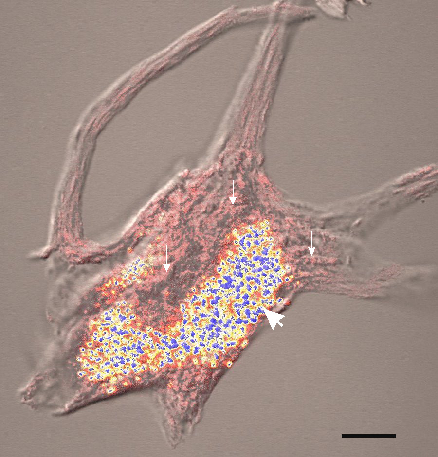
Lipofuscin is the name given to fine yellow-brown pigment Granule (cell biology), granules composed of lipid-containing residues of Lysosome, lysosomal digestion. It is considered to be one of the aging or "wear-and-tear" pigments, found in the liver, kidney, heart muscle, retina, adrenals, nerve cells, and ganglion cells. Formation and turnover Lipofuscin appears to be the product of the oxidation of unsaturated fatty acids and may be symptomatic of membrane damage, or damage to mitochondria and lysosomes. Aside from a large lipid content, lipofuscin is known to contain sugars and metals, including mercury (element), mercury, aluminium, iron, copper and zinc.Chris Gaugler,Lipofuscin", ''Stanislaus Journal of Biochemical Reviews'' May 1997 Lipofuscin is also accepted as consisting of oxidized proteins (30–70%) as well as lipids (20–50%). It is a type of lipochrome and is specifically arranged around the nucleus. The accumulation of lipofuscin-like material may be the result ... [...More Info...] [...Related Items...] OR: [Wikipedia] [Google] [Baidu] |
Retinal Pigment Epithelium
The pigmented layer of retina or retinal pigment epithelium (RPE) is the pigment A pigment is a powder used to add or alter color or change visual appearance. Pigments are completely or nearly solubility, insoluble and reactivity (chemistry), chemically unreactive in water or another medium; in contrast, dyes are colored sub ...ed cell layer just outside the neurosensory retina that nourishes retinal visual cells, and is firmly attached to the underlying choroid and overlying retinal visual cells. History The RPE was known in the 18th and 19th centuries as the pigmentum nigrum, referring to the observation that the RPE is dark (black in many animals, brown in humans); and as the tapetum nigrum, referring to the observation that in animals with a tapetum lucidum, in the region of the tapetum lucidum the RPE is not pigmented. Anatomy The RPE is composed of a single layer of hexagonal cells that are densely packed with pigment granules. When viewed from the outer surface, ... [...More Info...] [...Related Items...] OR: [Wikipedia] [Google] [Baidu] |
Retinitis Pigmentosa
Retinitis pigmentosa (RP) is a member of a group of genetic disorders called inherited retinal dystrophy (IRD) that cause loss of vision. Symptoms include trouble seeing at night and decreasing peripheral vision (side and upper or lower visual field). As peripheral vision worsens, people may experience " tunnel vision". Complete blindness is uncommon. Onset of symptoms is generally gradual and often begins in childhood. Retinitis pigmentosa is generally inherited from one or both parents. It is caused by genetic variants in nearly 100 genes. The underlying mechanism involves the progressive loss of rod photoreceptor cells that line the retina of the eyeball. The rod cells secrete a neuroprotective substance (rod-derived cone viability factor, RdCVF) that protects the cone cells from apoptosis. When these rod cells die, this substance is no longer provided. This is generally followed by the loss of cone photoreceptor cells. Diagnosis is through eye examination of the retina ... [...More Info...] [...Related Items...] OR: [Wikipedia] [Google] [Baidu] |
Cone Dystrophy
A cone dystrophy is an inherited ocular disorder characterized by the loss of cone cells, the photoreceptors responsible for both central and color vision. Presentation The most common symptoms of cone dystrophy are vision loss (age of onset ranging from the late teens to the sixties), sensitivity to bright lights, and poor color vision. Therefore, patients see better at dusk. Visual acuity usually deteriorates gradually, but it can deteriorate rapidly to 20/200; later, in more severe cases, it drops to "counting fingers" vision. Color vision testing using color test plates (HRR series) reveals many errors on both red-green and blue-yellow plates. Dystrophy of the rods and cones Dystrophy of the light-sensing cells of the eye may also occur in the rods as well, or in both the cones and the rods. A type of rod-cone dystrophy—where rod function decline is typically earlier or more pronounced than cone dystrophy—has been identified as a relatively common characteris ... [...More Info...] [...Related Items...] OR: [Wikipedia] [Google] [Baidu] |


