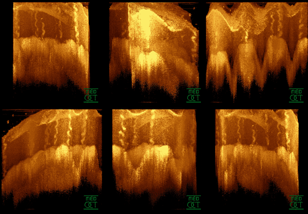
Optical Coherence Tomography on:
[Wikipedia]
[Google]
[Amazon]
 Optical coherence tomography (OCT) is a high-resolution imaging technique with most of its applications in medicine and biology. OCT uses coherent near-infrared light to obtain micrometer-level depth resolved images of biological tissue or other
 Interferometric reflectometry of biological tissue, especially of the human eye using short-coherence-length light (also referred to as partially-coherent, low-coherence, or broadband, broad-spectrum, or white light) was investigated in parallel by multiple groups worldwide since 1980s. Lending ideas from


 Optical coherence tomography (OCT) is a technique for obtaining sub-surface images of translucent or opaque materials at a resolution equivalent to a low-power microscope. It is effectively "optical ultrasound", imaging reflections from within tissue to provide cross-sectional images.
OCT has attracted interest among the medical community because it provides tissue morphology imagery at much higher resolution (less than 10 μm axially and less than 20 μm laterally ) than other imaging modalities such as MRI or ultrasound.
The key benefits of OCT are:
* Live sub-surface images at near-microscopic resolution
* Instant, direct imaging of tissue morphology
* No preparation of the sample or subject, no contact
* No ionizing radiation
OCT delivers high resolution because it is based on light, rather than sound or radio frequency. An optical beam is directed at the tissue, and the small portion of this light that reflects directly back from sub-surface features is collected. Note that most light scatters off at large angles. In conventional imaging, this diffusely scattered light contributes background that obscures an image. However, in OCT, a technique called interferometry is used to record the optical path length of received photons, allowing rejection of most photons that scatter multiple times before detection. Thus OCT can build up clear 3D images of thick samples by rejecting background signal while collecting light directly reflected from surfaces of interest.
Within the range of noninvasive three-dimensional imaging techniques that have been introduced to the medical research community, OCT as an echo technique is similar to
 Fourier-domain (or Frequency-domain) OCT (FD-OCT) has speed and signal-to-noise ratio (SNR) advantages over time-domain OCT (TD-OCT) and has become the standard in the industry since 2006. The idea of using frequency modulation and coherent detection to obtain ranging information was already demonstrated in optical frequency domain reflectometry and laser radar in the 1980s, though the distance resolution and range were much longer than OCT. There are two types of FD-OCT – swept-source OCT (SS-OCT) and spectral-domain OCT (SD-OCT) – both of which acquire spectral interferograms which are then
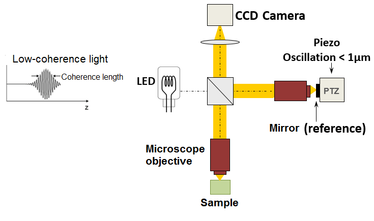
 An imaging approach to temporal OCT was developed by Claude Boccara's team in 1998, with an acquisition of the images without beam scanning. In this technique called full-field OCT (FF-OCT), unlike other OCT techniques that acquire cross-sections of the sample, the images are here "en-face" i.e. like images of classical microscopy: orthogonal to the light beam of illumination.
More precisely, interferometric images are created by a Michelson interferometer where the path length difference is varied by a fast electric component (usually a piezo mirror in the reference arm). These images acquired by a CCD camera are combined in post-treatment (or online) by the phase shift interferometry method, where usually 2 or 4 images per modulation period are acquired, depending on the algorithm used. More recently, approaches that allow rapid single-shot imaging were developed to simultaneously capture multiple phase-shifted images required for reconstruction, using single camera. Single-shot time-domain OCM is limited only by the camera frame rate and available illumination.
The "en-face" tomographic images are thus produced by a wide-field illumination, ensured by the Linnik configuration of the Michelson interferometer where a microscope objective is used in both arms. Furthermore, while the temporal coherence of the source must remain low as in classical OCT (i.e. a broad spectrum), the spatial coherence must also be low to avoid parasitical interferences (i.e. a source with a large size).
Creative Commons Attribution 3.0 Unported (CC BY 3.0)
license. Other promising areas of application include the imaging of lesions where excisions are hazardous or impossible and the guidance of surgical interventions through identification of tumor margins.
 Optical coherence tomography (OCT) is a high-resolution imaging technique with most of its applications in medicine and biology. OCT uses coherent near-infrared light to obtain micrometer-level depth resolved images of biological tissue or other
 scattering
In physics, scattering is a wide range of physical processes where moving particles or radiation of some form, such as light or sound, are forced to deviate from a straight trajectory by localized non-uniformities (including particles and radiat ...
media. It uses interferometry
Interferometry is a technique which uses the ''interference (wave propagation), interference'' of Superposition principle, superimposed waves to extract information. Interferometry typically uses electromagnetic waves and is an important inves ...
techniques to detect the amplitude and time-of-flight of reflected light.
OCT uses transverse sample scanning of the light beam to obtain two- and three-dimensional images. Short-coherence-length light can be obtained using a superluminescent diode (SLD) with a broad spectral bandwidth or a broadly tunable laser with narrow linewidth
A spectral line is a weaker or stronger region in an otherwise uniform and continuous spectrum. It may result from emission or absorption of light in a narrow frequency range, compared with the nearby frequencies. Spectral lines are often used ...
. The first demonstration of OCT imaging (in vitro) was published by a team from MIT and Harvard Medical School in a 1991 article in the journal ''Science
Science is a systematic discipline that builds and organises knowledge in the form of testable hypotheses and predictions about the universe. Modern science is typically divided into twoor threemajor branches: the natural sciences, which stu ...
''. The article introduced the term "OCT" to credit its derivation from optical coherence-domain reflectometry
Reflectometry is a general term for the use of the reflection of waves or pulses at surfaces and interfaces to detect or characterize objects, sometimes to detect anomalies as in fault detection and medical diagnosis.
There are many different ...
, in which the axial resolution is based on temporal coherence
Coherence expresses the potential for two waves to Wave interference, interfere. Two Monochromatic radiation, monochromatic beams from a single source always interfere. Wave sources are not strictly monochromatic: they may be ''partly coherent''. ...
. The first demonstrations of in vivo OCT imaging quickly followed.
The first US patents on OCT by the MIT/Harvard group described a time-domain
In mathematics and signal processing, the time domain is a representation of how a signal, function, or data set varies with time. It is used for the analysis of function (mathematics), mathematical functions, physical signal (information theory), ...
OCT (TD-OCT) system. These patents were licensed by Zeiss and formed the basis of the first generations of OCT products until 2006.
In the decade preceding the invention of OCT, interferometry with short-coherence-length light had been investigated for a variety of applications. The potential to use interferometry for imaging was proposed, and measurement of retina
The retina (; or retinas) is the innermost, photosensitivity, light-sensitive layer of tissue (biology), tissue of the eye of most vertebrates and some Mollusca, molluscs. The optics of the eye create a focus (optics), focused two-dimensional ...
l elevation profile and thickness had been demonstrated.
The initial commercial clinical OCT systems were based on point-scanning TD-OCT technology, which primarily produced cross-sectional
In statistics and econometrics, cross-sectional data is a type of data collected by observing many subjects (such as individuals, firms, countries, or regions) at a single point or period of time. Analysis of cross-sectional data usually consists ...
images due to the speed limitation (tens to thousands of axial scans per second). Fourier-domain OCT became available clinically 2006, enabling much greater image acquisition rate (tens of thousands to hundreds of thousands axial scans per second) without sacrificing signal strength. The higher speed allowed for three-dimensional imaging, which can be visualized in both en face and cross-sectional views. Novel contrasts such as angiography
Angiography or arteriography is a medical imaging technique used to visualize the inside, or lumen, of blood vessels and organs of the body, with particular interest in the arteries, veins, and the heart chambers. Modern angiography is perfo ...
, elastography, and optoretinography also became possible by detecting signal change over time. Over the past three decades, the speed of commercial clinical OCT systems has increased more than 1000-fold, doubling every three years and rivaling Moore's law
Moore's law is the observation that the Transistor count, number of transistors in an integrated circuit (IC) doubles about every two years. Moore's law is an observation and Forecasting, projection of a historical trend. Rather than a law of ...
of computer chip performance. Development of parallel image acquisition approaches such as line-field and full-field technology may allow the performance improvement trend to continue.
OCT is most widely used in ophthalmology
Ophthalmology (, ) is the branch of medicine that deals with the diagnosis, treatment, and surgery of eye diseases and disorders.
An ophthalmologist is a physician who undergoes subspecialty training in medical and surgical eye care. Following a ...
, in which it has transformed the diagnosis and monitoring of retinal diseases, optic nerve
In neuroanatomy, the optic nerve, also known as the second cranial nerve, cranial nerve II, or simply CN II, is a paired cranial nerve that transmits visual system, visual information from the retina to the brain. In humans, the optic nerve i ...
diseases, and cornea
The cornea is the transparency (optics), transparent front part of the eyeball which covers the Iris (anatomy), iris, pupil, and Anterior chamber of eyeball, anterior chamber. Along with the anterior chamber and Lens (anatomy), lens, the cornea ...
l diseases. It has greatly improved the management of the top three causes of blindness – macular degeneration
Macular degeneration, also known as age-related macular degeneration (AMD or ARMD), is a medical condition which may result in blurred vision, blurred or vision loss, no vision in the center of the visual field. Early on there are often no sym ...
, diabetic retinopathy
Diabetic retinopathy (also known as diabetic eye disease) is a medical condition in which damage occurs to the retina due to diabetes. It is a leading cause of blindness in developed countries and one of the lead causes of sight loss in the wor ...
, and glaucoma
Glaucoma is a group of eye diseases that can lead to damage of the optic nerve. The optic nerve transmits visual information from the eye to the brain. Glaucoma may cause vision loss if left untreated. It has been called the "silent thief of ...
– thereby preventing vision loss in many patients. By 2016 OCT was estimated to be used in more than 30 million imaging procedures per year worldwide.
Intravascular OCT imaging is used in the intravascular evaluation of coronary artery
The coronary arteries are the arterial blood vessels of coronary circulation, which transport oxygenated blood to the heart muscle. The heart requires a continuous supply of oxygen to function and survive, much like any other tissue or organ of ...
plaques and to guide stent
In medicine, a stent is a tube usually constructed of a metallic alloy or a polymer. It is inserted into the Lumen (anatomy), lumen (hollow space) of an anatomic vessel or duct to keep the passageway open.
Stenting refers to the placement of ...
placement. Beyond ophthalmology and cardiology, applications are also developing in other medical specialties such as dermatology
Dermatology is the branch of medicine dealing with the Human skin, skin.''Random House Webster's Unabridged Dictionary.'' Random House, Inc. 2001. Page 537. . It is a speciality with both medical and surgical aspects. A List of dermatologists, ...
, gastroenterology
Gastroenterology (from the Greek gastḗr- "belly", -énteron "intestine", and -logía "study of") is the branch of medicine focused on the digestive system and its disorders. The digestive system consists of the gastrointestinal tract, sometime ...
, neurology
Neurology (from , "string, nerve" and the suffix wikt:-logia, -logia, "study of") is the branch of specialty (medicine) , medicine dealing with the diagnosis and treatment of all categories of conditions and disease involving the nervous syst ...
and neurovascular imaging, oncology
Oncology is a branch of medicine that deals with the study, treatment, diagnosis, and prevention of cancer. A medical professional who practices oncology is an ''oncologist''. The name's Etymology, etymological origin is the Greek word ὄγ ...
, and dentistry
Dentistry, also known as dental medicine and oral medicine, is the branch of medicine focused on the Human tooth, teeth, gums, and Human mouth, mouth. It consists of the study, diagnosis, prevention, management, and treatment of diseases, dis ...
.
Introduction
 Interferometric reflectometry of biological tissue, especially of the human eye using short-coherence-length light (also referred to as partially-coherent, low-coherence, or broadband, broad-spectrum, or white light) was investigated in parallel by multiple groups worldwide since 1980s. Lending ideas from
 ultrasound imaging
Medical ultrasound includes diagnostic techniques (mainly imaging) using ultrasound, as well as therapeutic applications of ultrasound. In diagnosis, it is used to create an image of internal body structures such as tendons, muscles, join ...
and merging the time-of-flight detection with optical interferometry to detect optical delays in the pico- and femtosecond range as known from the autocorrelator in the 1960's, the technique's development was and is tightly associated with the availability of novel electronic, mechanical and photonic abilities. Stemming from single lateral point low-coherence interferometry the addition of a wide range of technologies enabled key milestones in this computational imaging technique. High-speed axial and lateral scanners, ultra-broad spectrum or ultra-fast spectrally tunable lasers or other high brightness radiation sources, increasingly sensitive detectors, like high resolution and high speed cameras or fast A/D-converters that picked up from and drove ideas in the rapidly developing photonics field, together with the increasing availability of computing power were essential for its birth and success. In 1991, David Huang, then a student in James Fujimoto laboratory at Massachusetts Institute of Technology
The Massachusetts Institute of Technology (MIT) is a Private university, private research university in Cambridge, Massachusetts, United States. Established in 1861, MIT has played a significant role in the development of many areas of moder ...
, working with Eric Swanson at the MIT Lincoln Laboratory and colleagues at the Harvard Medical School, successfully demonstrated imaging and called the new imaging modality "optical coherence tomography". Since then, OCT with micrometer axial resolution and below and cross-sectional imaging capabilities has become a prominent biomedical imaging technique that has continually improved in technical performance and range of applications. The improvement in image acquisition rate is particularly spectacular, starting with the original 0.8 Hz axial scan repetition rate to the current commercial clinical OCT systems operating at several hundred kHz and laboratory prototypes at multiple MHz. The range of applications has expanded from ophthalmology to cardiology and other medical specialties. For their roles in the invention of OCT, Fujimoto, Huang, and Swanson received the 2023 Lasker-DeBakey Clinical Medical Research Award and the National Medal of Technology and Innovation. These developments have been reviewed in articles written for the general scientific and medical readership.
It is particularly suited to ophthalmic applications and other tissue imaging requiring micrometer resolution and millimeter penetration depth. OCT has also been used for various art conservation
conservation and restoration of cultural property focuses on protection and care of cultural property (tangible cultural heritage), including artworks, architecture, archaeology, and museum collections. Conservation activities include preve ...
projects, where it is used to analyze different layers in a painting. OCT has interesting advantages over other medical imaging systems. Medical ultrasonography
Medical ultrasound includes Medical diagnosis, diagnostic techniques (mainly medical imaging, imaging) using ultrasound, as well as therapeutic ultrasound, therapeutic applications of ultrasound. In diagnosis, it is used to create an image of ...
, magnetic resonance imaging
Magnetic resonance imaging (MRI) is a medical imaging technique used in radiology to generate pictures of the anatomy and the physiological processes inside the body. MRI scanners use strong magnetic fields, magnetic field gradients, and ...
(MRI), confocal microscopy, and OCT are differently suited to morphological tissue imaging: while the first two have whole body but low resolution imaging capability (typically a fraction of a millimeter), the third one can provide images with resolutions well below 1 micrometer (i.e. sub-cellular), between 0 and 100 micrometers in depth, and the fourth can probe as deep as 500 micrometers, but with a lower (i.e. architectural) resolution (around 10 micrometers in lateral and a few micrometers in depth in ophthalmology, for instance, and 20 micrometers in lateral in endoscopy).
OCT is based on low-coherence interferometry. In conventional interferometry with long coherence length (i.e., laser interferometry), interference of light occurs over a distance of meters. In OCT, this interference is shortened to a distance of micrometers, owing to the use of broad-bandwidth light sources (i.e., sources that emit light over a broad range of frequencies). Light with broad bandwidths can be generated by using superluminescent diodes or lasers with extremely short pulses ( femtosecond lasers). White light is an example of a broadband source with lower power.
Light in an OCT system is broken into two arms – a sample arm (containing the item of interest) and a reference arm (usually a mirror). The combination of reflected light from the sample arm and reference light from the reference arm gives rise to an interference pattern, but only if light from both arms have traveled the "same" optical distance ("same" meaning a difference of less than a coherence length). By scanning the mirror in the reference arm, a reflectivity profile of the sample can be obtained (this is time domain OCT). Areas of the sample that reflect back a lot of light will create greater interference than areas that don't. Any light that is outside the short coherence length will not interfere. This reflectivity profile, called an A-scan, contains information about the spatial dimensions and location of structures within the item of interest. A cross-sectional tomogram ( B-scan) may be achieved by laterally combining a series of these axial depth scans (A-scan). En face imaging at an acquired depth is possible depending on the imaging engine used.
Layperson's explanation


 Optical coherence tomography (OCT) is a technique for obtaining sub-surface images of translucent or opaque materials at a resolution equivalent to a low-power microscope. It is effectively "optical ultrasound", imaging reflections from within tissue to provide cross-sectional images.
OCT has attracted interest among the medical community because it provides tissue morphology imagery at much higher resolution (less than 10 μm axially and less than 20 μm laterally ) than other imaging modalities such as MRI or ultrasound.
The key benefits of OCT are:
* Live sub-surface images at near-microscopic resolution
* Instant, direct imaging of tissue morphology
* No preparation of the sample or subject, no contact
* No ionizing radiation
OCT delivers high resolution because it is based on light, rather than sound or radio frequency. An optical beam is directed at the tissue, and the small portion of this light that reflects directly back from sub-surface features is collected. Note that most light scatters off at large angles. In conventional imaging, this diffusely scattered light contributes background that obscures an image. However, in OCT, a technique called interferometry is used to record the optical path length of received photons, allowing rejection of most photons that scatter multiple times before detection. Thus OCT can build up clear 3D images of thick samples by rejecting background signal while collecting light directly reflected from surfaces of interest.
Within the range of noninvasive three-dimensional imaging techniques that have been introduced to the medical research community, OCT as an echo technique is similar to
 ultrasound imaging
Medical ultrasound includes diagnostic techniques (mainly imaging) using ultrasound, as well as therapeutic applications of ultrasound. In diagnosis, it is used to create an image of internal body structures such as tendons, muscles, join ...
. Other medical imaging techniques such as computerized axial tomography, magnetic resonance imaging, or positron emission tomography do not use the echo-location principle.
The technique is limited to imaging 1 to 2 mm below the surface in biological tissue, because at greater depths the proportion of light that escapes without scattering is too small to be detected. No special preparation of a biological specimen is required, and images can be obtained "non-contact" or through a transparent window or membrane.
The laser output from the instruments used is loweye-safe near-infrared or visible-lightand no damage to the sample is therefore likely.
Theory
The principle of OCT is white light, or low coherence, interferometry. The optical setup typically consists of an interferometer (Fig. 1, typically Michelson type) with a low coherence, broad bandwidth light source. Light is split into and recombined from reference and sample arms, respectively.Time domain
: In time domain OCT the path length of the reference arm is varied in time (the reference mirror is translated longitudinally). A property of low coherence interferometry is that interference, i.e. the series of dark and bright fringes, is only achieved when the path difference lies within the coherence length of the light source. This interference is called autocorrelation in a symmetric interferometer (both arms have the same reflectivity), or cross-correlation in the common case. The envelope of this modulation changes as path length difference is varied, where the peak of the envelope corresponds to path length matching. The interference of two partially coherent light beams can be expressed in terms of the source intensity, , as : where represents the interferometer beam splitting ratio, and is called the complex degree of coherence, i.e. the interference envelope and carrier dependent on reference arm scan or time delay , and whose recovery is of interest in OCT. Due to the coherence gating effect of OCT the complex degree of coherence is represented as a Gaussian function expressed as : where represents the spectral width of the source in the optical frequency domain, and is the centre optical frequency of the source. In equation (2), the Gaussian envelope is amplitude modulated by an optical carrier. The peak of this envelope represents the location of the microstructure of the sample under test, with an amplitude dependent on the reflectivity of the surface. The optical carrier is due to theDoppler effect
The Doppler effect (also Doppler shift) is the change in the frequency of a wave in relation to an observer who is moving relative to the source of the wave. The ''Doppler effect'' is named after the physicist Christian Doppler, who described ...
resulting from scanning one arm of the interferometer, and the frequency of this modulation is controlled by the speed of scanning. Therefore, translating one arm of the interferometer has two functions; depth scanning and a Doppler-shifted optical carrier are accomplished by pathlength variation. In OCT, the Doppler-shifted optical carrier has a frequency expressed as
:
where is the central optical frequency of the source, is the scanning velocity of the pathlength variation, and is the speed of light.
The axial and lateral resolutions of OCT are decoupled from one another; the former being an equivalent to the coherence length of the light source and the latter being a function of the optics. The axial resolution of OCT is defined as
:
where and are respectively the central wavelength and the spectral width of the light source.
Fourier domain
Fourier transform
In mathematics, the Fourier transform (FT) is an integral transform that takes a function as input then outputs another function that describes the extent to which various frequencies are present in the original function. The output of the tr ...
ed to obtain an axial scan of reflectance amplitude versus depth. In SS-OCT, the spectral interferogram is acquired sequentially by tuning the wavelength of a laser light source. SD-OCT acquires spectral interferogram simultaneously in a spectrometer. An implementation of SS-OCT was described by the MIT group as early as 1994. A group based in the University of Vienna described measurement of intraocular distance using both tunable laser and spectrometer-based interferometry as early as 1995. SD-OCT imaging was first demonstrated both in vitro and in vivo by a collaboration between the Vienna group and a group based in the Nicholas Copernicus University in a series of articles between 2000 and 2002. The SNR advantage of FD-OCT over TD-OCT was first demonstrated in eye imaging and further analyzed by multiple groups of researchers in 2003.
Spectral-domain OCT
Spectral-domain OCT (spatially encoded frequency domain OCT) extracts spectral information by distributing different optical frequencies onto a detector stripe (line-array CCD or CMOS) via a dispersive element (see Fig. 4). Thereby the information of the full depth scan can be acquired within a single exposure. However, the large signal-to-noise advantage of FD-OCT is reduced due to the lower dynamic range of stripe detectors with respect to single photosensitive diodes, resulting in an SNR advantage of ~10 dB at much higher speeds. This is not much of a problem when working at 1300 nm, however, since dynamic range is not a serious problem at this wavelength range. The drawbacks of this technology are found in a strong fall-off of the SNR, which is proportional to the distance from the zero delay and a sinc-type reduction of the depth-dependent sensitivity because of limited detection linewidth. (One pixel detects a quasi-rectangular portion of an optical frequency range instead of a single frequency, the Fourier transform leads to the sinc(z) behavior). Additionally, the dispersive elements in the spectroscopic detector usually do not distribute the light equally spaced in frequency on the detector, but mostly have an inverse dependence. Therefore, the signal has to be resampled before processing, which cannot take care of the difference in local (pixelwise) bandwidth, which results in further reduction of the signal quality. However, the fall-off is not a serious problem with the development of new generation CCD orphotodiode
A photodiode is a semiconductor diode sensitive to photon radiation, such as visible light, infrared or ultraviolet radiation, X-rays and gamma rays. It produces an electrical current when it absorbs photons. This can be used for detection and me ...
array with a larger number of pixels.
Synthetic array heterodyne detection offers another approach to this problem without the need for high dispersion.
Swept-source OCT
Swept-source OCT (Time-encoded frequency domain OCT) tries to combine some of the advantages of standard TD and spectral domain OCT. Here the spectral components are not encoded by spatial separation, but they are encoded in time. The spectrum is either filtered or generated in single successive frequency steps and reconstructed before Fourier transformation. By accommodation of a frequency scanning light source (i.e. frequency scanning laser) the optical setup (see Fig. 3) becomes simpler than spectral domain OCT, but the problem of scanning is essentially translated from the TD-OCT reference arm into the swept source OCT light source. Here the advantage lies in the proven high SNR detection technology, while swept laser sources achieve very small instantaneous bandwidths (linewidths) at very high frequencies (20–200 kHz). Drawbacks are the nonlinearities in the wavelength (especially at high scanning frequencies), the broadening of the linewidth at high frequencies and a high sensitivity to movements of the scanning geometry or the sample (below the range of nanometers within successive frequency steps).Scanning schemes
Focusing the light beam to a point on the surface of the sample under test, and recombining the reflected light with the reference will yield an interferogram with sample information corresponding to a single A-scan (Z axis only). Scanning of the sample can be accomplished by either scanning the light on the sample, or by moving the sample under test. A linear scan will yield a two-dimensional data set corresponding to a cross-sectional image (X-Z axes scan), whereas an area scan achieves a three-dimensional data set corresponding to a volumetric image (X-Y-Z axes scan).Single point
Systems based on single point, confocal, or flying-spot time domain OCT, must scan the sample in two lateral dimensions and reconstruct a three-dimensional image using depth information obtained by coherence-gating through an axially scanning reference arm (Fig. 2). Two-dimensional lateral scanning has been electromechanically implemented by moving the sample using a translation stage, and using a novel micro-electro-mechanical system scanner.Line-field OCT
Line-field confocal optical coherence tomography (LC-OCT) is an imaging technique based on the principle of time-domain OCT with line illumination using a broadband laser and line detection using a line-scan camera. LC-OCT produces B-scans in real-time from multiple A-scans acquired in parallel. En face as well as three-dimensional images can also be obtained by scanning the illumination line laterally. The focus is continuously adjusted during the scan of the sample depth, using a high numerical aperture (NA) microscope objective to image with high lateral resolution. By using a supercontinuum laser as a light source, a quasi-isotropic spatial resolution of ~ 1 μm is achieved at a central wavelength of ~ 800 nm. On the other hand, line illumination and detection, combined with the use of a high NA microscope objective, produce a confocal gate that prevents most scattered light that does not contribute to the signal from being detected by the camera. This confocal gate, which is absent in the full-field OCT technique, gives LC-OCT an advantage in terms of detection sensitivity and penetration in highly scattering media such as skin tissues. So far this technique has been used mainly for skin imaging in the fields of dermatology and cosmetology.Full-field OCT
 An imaging approach to temporal OCT was developed by Claude Boccara's team in 1998, with an acquisition of the images without beam scanning. In this technique called full-field OCT (FF-OCT), unlike other OCT techniques that acquire cross-sections of the sample, the images are here "en-face" i.e. like images of classical microscopy: orthogonal to the light beam of illumination.
More precisely, interferometric images are created by a Michelson interferometer where the path length difference is varied by a fast electric component (usually a piezo mirror in the reference arm). These images acquired by a CCD camera are combined in post-treatment (or online) by the phase shift interferometry method, where usually 2 or 4 images per modulation period are acquired, depending on the algorithm used. More recently, approaches that allow rapid single-shot imaging were developed to simultaneously capture multiple phase-shifted images required for reconstruction, using single camera. Single-shot time-domain OCM is limited only by the camera frame rate and available illumination.
The "en-face" tomographic images are thus produced by a wide-field illumination, ensured by the Linnik configuration of the Michelson interferometer where a microscope objective is used in both arms. Furthermore, while the temporal coherence of the source must remain low as in classical OCT (i.e. a broad spectrum), the spatial coherence must also be low to avoid parasitical interferences (i.e. a source with a large size).
Selected applications
Optical coherence tomography is an established medical imaging technique and is used across several medical specialties including ophthalmology and cardiology and is widely used in basic science research applications.Ophthalmology
Ocular (or ophthalmic) OCT is used heavily byophthalmologists
Ophthalmology (, ) is the branch of medicine that deals with the diagnosis, treatment, and surgery of eye diseases and disorders.
An ophthalmologist is a physician who undergoes subspecialty training in medical and surgical eye care. Following a ...
and optometrists to obtain high-resolution images of the retina
The retina (; or retinas) is the innermost, photosensitivity, light-sensitive layer of tissue (biology), tissue of the eye of most vertebrates and some Mollusca, molluscs. The optics of the eye create a focus (optics), focused two-dimensional ...
and anterior segment
The anterior segment or anterior cavity is the front third of the eye that includes the structures in front of the vitreous humour: the cornea, iris, ciliary body, and lens
A lens is a transmissive optical device that focuses or dispers ...
. Owing to OCT's capability to show cross-sections of tissue layers with micrometer resolution, OCT provides a straightforward method of assessing cellular organization, photoreceptor integrity, and axon
An axon (from Greek ἄξων ''áxōn'', axis) or nerve fiber (or nerve fibre: see American and British English spelling differences#-re, -er, spelling differences) is a long, slender cellular extensions, projection of a nerve cell, or neuron, ...
al thickness in glaucoma
Glaucoma is a group of eye diseases that can lead to damage of the optic nerve. The optic nerve transmits visual information from the eye to the brain. Glaucoma may cause vision loss if left untreated. It has been called the "silent thief of ...
, macular degeneration
Macular degeneration, also known as age-related macular degeneration (AMD or ARMD), is a medical condition which may result in blurred vision, blurred or vision loss, no vision in the center of the visual field. Early on there are often no sym ...
, diabetic macular edema, multiple sclerosis
Multiple sclerosis (MS) is an autoimmune disease resulting in damage to myelinthe insulating covers of nerve cellsin the brain and spinal cord. As a demyelinating disease, MS disrupts the nervous system's ability to Action potential, transmit ...
, optic neuritis, and other eye disease
This is a partial list of human eye diseases and disorders.
The World Health Organization (WHO) publishes a classification of known diseases and injuries, the International Statistical Classification of Diseases and Related Health Problems, or ...
s or systemic pathologies which have ocular signs. Additionally, ophthalmologists leverage OCT to assess the vascular health of the retina via a technique called OCT angiography (OCTA). In ophthalmological surgery, especially retinal surgery, an OCT can be mounted on the microscope. Such a system is called an ''intraoperative OCT'' (iOCT) and provides support during the surgery with clinical benefits. Polarization-sensitive OCT was recently applied in the human retina to determine optical polarization properties of vessel walls near the optic nerve.
Retinal imaging with PS-OCT demonstrated how the thickness and birefringence of blood vessel wall tissue of healthy subjects could be quantified, in vivo. PS-OCT was subsequently applied to patients with diabetes and age-matched healthy subjects, and showed an almost 100% increase in vessel wall birefringence due to diabetes, without a significant change in vessel wall thickness. In patients with hypertension however, the retinal vessel wall thickness increased by 60% while the vessel wall birefringence dropped by 20%, on average. The large differences measured in healthy subjects and patients suggest that retinal measurements with PS-OCT could be used as a screening tool for hypertension and diabetes.
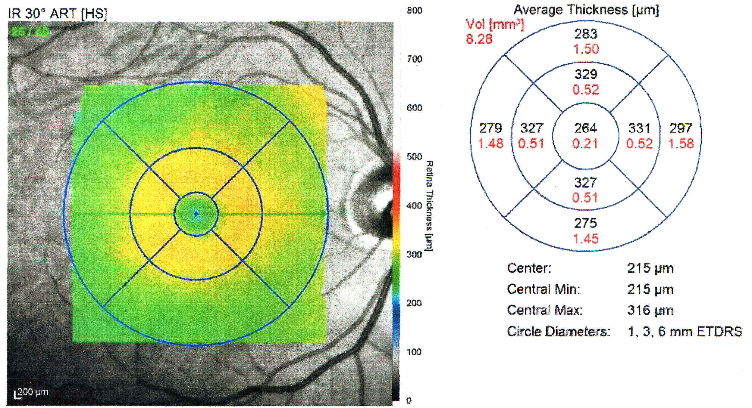
OCT can used to measure the thickness of the Retinal nerve fiber layer (RNFL).
Cardiology
In the settings of cardiology, OCT is used to imagecoronary arteries
The coronary arteries are the arteries, arterial blood vessels of coronary circulation, which transport oxygenated blood to the Cardiac muscle, heart muscle. The heart requires a continuous supply of oxygen to function and survive, much like any ...
to visualize vessel wall lumen morphology and microstructure at a resolution ~10 times higher than other existing modalities such as intravascular ultrasound
Intravascular ultrasound (IVUS) or intravascular echocardiography is a medical imaging methodology using a specially designed catheter with a miniaturized ultrasound probe attached to the distal end of the catheter. The proximal end of the cathe ...
s, and x-ray angiography ( intracoronary optical coherence tomography). For this type of application, 1 mm in diameter or smaller fiber-optics catheters are used to access artery lumen through semi-invasive interventions such as percutaneous coronary intervention
Percutaneous coronary intervention (PCI) is a minimally invasive non-surgical procedure used to treat stenosis, narrowing of the coronary artery, coronary arteries of the heart found in coronary artery disease. The procedure is used to place and ...
s.
The first demonstration of endoscopic OCT was reported in 1997, by researchers in Fujimoto's laboratory at Massachusetts Institute of Technology. The first TD-OCT imaging catheter and system was commercialized by LightLab Imaging, Inc., a company based in Massachusetts in 2006. The first FD-OCT imaging study was reported by Massachusetts General Hospital
Massachusetts General Hospital (Mass General or MGH) is a teaching hospital located in the West End neighborhood of Boston, Massachusetts. It is the original and largest clinical education and research facility of Harvard Medical School/Harvar ...
in 2008. Intracoronary FD-OCT was first introduced in the market in 2009 by LightLab Imaging, Inc. followed by Terumo Corporation in 2012 and by Gentuity LLC in 2020. The higher acquisition speed of FD-OCT enabled the widespread adoption of this imaging technology for coronary artery imaging. It is estimated that over 100,000 FD-OCT coronary imaging cases are performed yearly, and that the market is increasing by approximately 20% every year.
Other developments of intracoronary OCT included the combination with other optical imaging modalities for multi-modality imaging. Intravascular OCT has been combined with near-infrared fluorescence molecular imaging (NIRF) to enhance its capability to detect molecular/functional and tissue morphological information simultaneously. In a similar way, combination with near-infrared spectroscopy (NIRS) has been implemented.
Neurovascular
Endoscopic/intravascular OCT has been further developed for use in neurovascular applications including imaging for guiding endovascular treatment of ischemic stroke and brain aneurysms. Initial clinical investigations with existing coronary OCT catheters have been limited to proximal intracranial anatomy of patient with limited tortuosity, as coronary OCT technology was not designed for the tortuous cerebrovasculature encountered in the brain. However, despite these limitations, it showed the potential of OCT for the imaging of neurovascular disease. An intravascular OCT imaging catheter design tailored for use in tortuous neurovascular anatomy has been proposed in 2020. A first-in-human study using endovascular neuro OCT (''n''OCT) has been reported in 2024.Oncology
Endoscopic OCT has been applied to the detection and diagnosis ofcancer
Cancer is a group of diseases involving Cell growth#Disorders, abnormal cell growth with the potential to Invasion (cancer), invade or Metastasis, spread to other parts of the body. These contrast with benign tumors, which do not spread. Po ...
and precancerous lesions
A precancerous condition is a condition, tumor or lesion involving abnormal cells which are associated with an increased risk of developing into cancer. Clinically, precancerous conditions encompass a variety of abnormal tissues with an increas ...
, such as Barrett's esophagus
Barrett's esophagus is a condition in which there is an abnormal ( metaplastic) change in the mucosal cells that line the lower part of the esophagus. The cells change from stratified squamous epithelium to simple columnar epithelium, intersper ...
and esophageal dysplasia
Dysplasia is any of various types of abnormal growth or development of cells (microscopic scale) or organs (macroscopic scale), and the abnormal histology or anatomical structure(s) resulting from such growth. Dysplasias on a mainly microscopic ...
.
Dermatology
The first use of OCT in dermatology dates back to 1997. Since then, OCT has been applied to the diagnosis of various skin lesions including carcinomas. However, the diagnosis of melanoma using conventional OCT is difficult, especially due to insufficient imaging resolution. Emerging high-resolution OCT techniques such as LC-OCT have the potential to improve the clinical diagnostic process, allowing for the early detection of malignant skin tumors – including melanoma – and a reduction in the number of surgical excisions of benign lesions. This article contains quotations from this source, which is available under thCreative Commons Attribution 3.0 Unported (CC BY 3.0)
license. Other promising areas of application include the imaging of lesions where excisions are hazardous or impossible and the guidance of surgical interventions through identification of tumor margins.
Dentistry
Researchers in Tokyo medical and Dental University were able to detect enamel white spot lesions around and beneath the orthodontic brackets using swept source OCT.Research applications
Researchers have used OCT to produce detailed images of mice brains, through a "window" made of zirconia that has been modified to be transparent and implanted in the skull. Optical coherence tomography is also applicable and increasingly used in industrial applications, such asnondestructive testing
Nondestructive testing (NDT) is any of a wide group of analysis techniques used in science and technology industry to evaluate the properties of a material, component or system without causing damage.
The terms nondestructive examination (NDE), n ...
(NDT), material thickness measurements, and in particular thin silicon wafers and compound semiconductor wafers thickness measurements surface roughness characterization, surface and cross-section imaging and volume loss measurements. OCT systems with feedback can be used to control manufacturing processes. With high speed data acquisition, and sub-micron resolution, OCT is adaptable to perform both inline and off-line. Due to the high volume of produced pills, an interesting field of application is in the pharmaceutical industry to control the coating of tablets. Fiber-based OCT systems are particularly adaptable to industrial environments. These can access and scan interiors of hard-to-reach spaces, and are able to operate in hostile environments—whether radioactive, cryogenic, or very hot. Novel optical biomedical diagnostic and imaging technologies are currently being developed to solve problems in biology and medicine. As of 2014, attempts have been made to use optical coherence tomography to identify root canals in teeth, specifically canal in the maxillary molar, however, there is no difference with the current methods of dental operatory microscope. Research conducted in 2015 was successful in utilizing a smartphone as an OCT platform, although much work remains to be done before such a platform would be commercially viable. Photonic integrated circuits may be a promising option to miniaturized OCT. Similarly to integrated circuits
An integrated circuit (IC), also known as a microchip or simply chip, is a set of electronic circuits, consisting of various electronic components (such as transistors, resistors, and capacitors) and their interconnections. These components a ...
silicon-based fabrication techniques can be used to produce miniaturized photonic systems. First in vivo human retinal imaging has been reported recently. In 3D microfabrication, OCT enables non-destructive testing and real-time inspection during additive manufacturing. Its high-resolution imaging detects defects, characterizes material properties and ensures the integrity of internal geometries without damaging the part.
See also
*Angle-resolved low-coherence interferometry
Angle-resolved low-coherence interferometry (a/LCI) is an emerging biomedical imaging technology which uses the properties of scattered light to measure the average size of cell structures, including cell nuclei. The technology shows promise as ...
* Ballistic photon
Ballistic light, also known as ballistic photons, is photons of light that have traveled through a scattering (turbid) medium in a straight line.
When pulses of laser light pass through a turbid medium such as fog or body tissue, most of the pho ...
* Confocal microscopy
Confocal microscopy, most frequently confocal laser scanning microscopy (CLSM) or laser scanning confocal microscopy (LSCM), is an optical imaging technique for increasing optical resolution and contrast (vision), contrast of a micrograph by me ...
* Dual-axis optical coherence tomography
* Interferometry
Interferometry is a technique which uses the ''interference (wave propagation), interference'' of Superposition principle, superimposed waves to extract information. Interferometry typically uses electromagnetic waves and is an important inves ...
* Intracoronary optical coherence tomography
* Leica Microsystems
Leica Microsystems GmbH is a German microscope manufacturing company. It is a manufacturer of optical microscopes, equipment for the preparation of microscopic specimens and related products. There are ten plants in eight countries with distri ...
* Medical imaging
Medical imaging is the technique and process of imaging the interior of a body for clinical analysis and medical intervention, as well as visual representation of the function of some organs or tissues (physiology). Medical imaging seeks to revea ...
* Novacam Technologies
Novacam Technologies Inc. specializes in designing and manufacturing advanced metrology and imaging systems for industrial and bio-medical applications. Novacam's fiber-based optical profilometers and Optical Coherence Tomography (OCT) systems ar ...
* Optical heterodyne detection
Optical heterodyne detection is a method of extracting information encoded as modulation of the phase, frequency or both of electromagnetic radiation in the wavelength band of visible or infrared light. The light signal is compared with standard o ...
* Optical projection tomography
* Spectroscopic optical coherence tomography
* Terahertz tomography
* Tomography
Tomography is imaging by sections or sectioning that uses any kind of penetrating wave. The method is used in radiology, archaeology, biology, atmospheric science, geophysics, oceanography, plasma physics, materials science, cosmochemistry, ast ...
References
{{Lasers Eye procedures Laser medicine Medical equipment Optical imaging