|
Ophthalmoscopy
Ophthalmoscopy, also called funduscopy, is a test that allows a health professional to see inside the fundus of the eye and other structures using an ophthalmoscope (or funduscope). It is done as part of an eye examination and may be done as part of a routine physical examination. It is crucial in determining the health of the retina, optic disc, and vitreous humor. The pupil is a hole through which the eye's interior can be viewed. For better viewing, the pupil can be opened wider (dilated; mydriasis) before ophthalmoscopy using medicated eye drops ( dilated fundus examination). However, undilated examination is more convenient (albeit not as comprehensive), and is the most common type in primary care. An alternative or complement to ophthalmoscopy is to perform a fundus photography, where the image can be analysed later by a professional. Types There are two major types of ophthalmoscopy: * direct ophthalmoscopy, which produces an upright (unreversed) image of approxima ... [...More Info...] [...Related Items...] OR: [Wikipedia] [Google] [Baidu] |
Fundus Photographs
Fundus photography involves photographing the rear of an eye, also known as the fundus (eye), fundus. Specialized fundus cameras consisting of an intricate microscope attached to a flash (photography), flash enabled camera are used in fundus photography. The main structures that can be visualized on a fundus photo are the central and peripheral retina, optic disc and Macula of retina, macula. Fundus photography can be performed with colored filters, or with specialized dyes including Fluorescein angiography, fluorescein and indocyanine green. The models and technology of fundus photography have advanced and evolved rapidly over the last century. History The concept of fundus photography was first introduced in the mid 19th century, after the introduction of photography in 1839. In 1851, Hermann von Helmholtz introduced the Ophthalmoscopy, Ophthalmoscope, and James Clerk Maxwell presented a colour photography method in 1861. In the early 1860s, Henry Noyes and Abner Mulhollan ... [...More Info...] [...Related Items...] OR: [Wikipedia] [Google] [Baidu] |
Ophthalmologists
Ophthalmology (, ) is the branch of medicine that deals with the diagnosis, treatment, and surgery of eye diseases and disorders. An ophthalmologist is a physician who undergoes subspecialty training in medical and surgical eye care. Following a medical degree, a doctor specialising in ophthalmology must pursue additional postgraduate residency training specific to that field. In the United States, following graduation from medical school, one must complete a four-year residency in ophthalmology to become an ophthalmologist. Following residency, additional specialty training (or fellowship) may be sought in a particular aspect of eye pathology. Ophthalmologists prescribe medications to treat ailments, such as eye diseases, implement laser therapy, and perform surgery when needed. Ophthalmologists provide both primary and specialty eye care—medical and surgical. Most ophthalmologists participate in academic research on eye diseases at some point in their training and many incl ... [...More Info...] [...Related Items...] OR: [Wikipedia] [Google] [Baidu] |
Retina
The retina (; or retinas) is the innermost, photosensitivity, light-sensitive layer of tissue (biology), tissue of the eye of most vertebrates and some Mollusca, molluscs. The optics of the eye create a focus (optics), focused two-dimensional image of the visual world on the retina, which then processes that image within the retina and sends nerve impulses along the optic nerve to the visual cortex to create visual perception. The retina serves a function which is in many ways analogous to that of the photographic film, film or image sensor in a camera. The neural retina consists of several layers of neurons interconnected by Chemical synapse, synapses and is supported by an outer layer of pigmented epithelial cells. The primary light-sensing cells in the retina are the photoreceptor cells, which are of two types: rod cell, rods and cone cell, cones. Rods function mainly in dim light and provide monochromatic vision. Cones function in well-lit conditions and are responsible fo ... [...More Info...] [...Related Items...] OR: [Wikipedia] [Google] [Baidu] |
Optic Disc
The optic disc or optic nerve head is the point of exit for ganglion cell axons leaving the eye. Because there are no rods or cones overlying the optic disc, it corresponds to a small blind spot in each eye. The ganglion cell axons form the optic nerve after they leave the eye. The optic disc represents the beginning of the optic nerve and is the point where the axons of retinal ganglion cells come together. The optic disc in a normal human eye carries 1–1.2 million afferent nerve fibers from the eye toward the brain. The optic disc is also the entry point for the major arteries that supply the retina with blood, and the exit point for the veins from the retina. Structure The optic disc is located 3 to 4 mm to the nasal side of the fovea. It is a vertical oval, with average dimensions of 1.76mm horizontally by 1.92mm vertically. There is a central depression, of variable size, called the optic cup. This depression can be a variety of shapes from a shallow indent ... [...More Info...] [...Related Items...] OR: [Wikipedia] [Google] [Baidu] |
Etymology And Pronunciation
Etymology ( ) is the study of the origin and evolution of words—including their constituent units of sound and meaning—across time. In the 21st century a subfield within linguistics, etymology has become a more rigorously scientific study. Most directly tied to historical linguistics, philology, and semiotics, it additionally draws upon comparative semantics, morphology, pragmatics, and phonetics in order to attempt a comprehensive and chronological catalogue of all meanings and changes that a word (and its related parts) carries throughout its history. The origin of any particular word is also known as its ''etymology''. For languages with a long written history, etymologists make use of texts, particularly texts about the language itself, to gather knowledge about how words were used during earlier periods, how they developed in meaning and form, or when and how they entered the language. Etymologists also apply the methods of comparative linguistics to reconstruct inform ... [...More Info...] [...Related Items...] OR: [Wikipedia] [Google] [Baidu] |
Slit Lamp
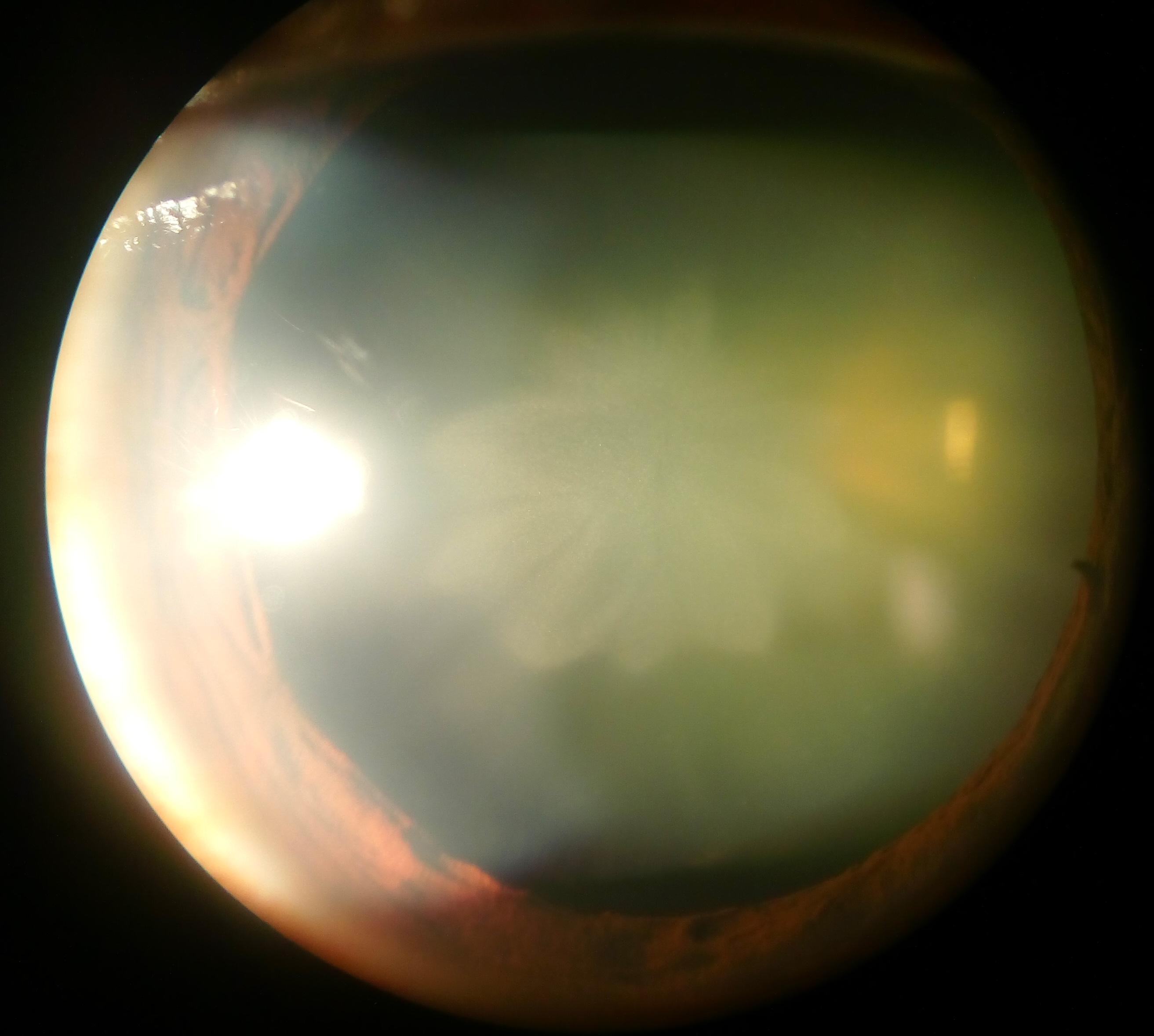
In ophthalmology and optometry, a slit lamp is an instrument consisting of a high-intensity light source that can be focused to shine a thin sheet of light into the eye. It is used in conjunction with a biomicroscope. The lamp facilitates an examination of the anterior segment and posterior segment of the human eye, which includes the eyelid, sclera, conjunctiva, iris (anatomy), iris, natural crystalline lens, and cornea. The binocular slit-lamp examination provides a stereoscopic magnified view of the eye structures in detail, enabling anatomical diagnoses to be made for a variety of eye conditions. A second, hand-held lens is used to examine the retina. History Two conflicting trends emerged in the development of the slit lamp. One trend originated from clinical research and aimed to apply the increasingly complex and advanced technology of the time. [...More Info...] [...Related Items...] OR: [Wikipedia] [Google] [Baidu] |
Cataract
A cataract is a cloudy area in the lens (anatomy), lens of the eye that leads to a visual impairment, decrease in vision of the eye. Cataracts often develop slowly and can affect one or both eyes. Symptoms may include faded colours, blurry or double vision, halos around light, trouble with bright lights, and Nyctalopia, difficulty seeing at night. This may result in trouble driving, reading, or recognizing faces. Poor vision caused by cataracts may also result in an increased risk of Falling (accident), falling and Depression (mood), depression. Cataracts cause 51% of all cases of blindness and 33% of visual impairment worldwide. Cataracts are most commonly due to senescence, aging but may also occur due to Trauma (medicine), trauma or radiation exposure, be congenital cataract, present from birth, or occur following eye surgery for other problems. Risk factors include diabetes mellitus, diabetes, longstanding use of corticosteroid medication, smoking tobacco, prolonged exposu ... [...More Info...] [...Related Items...] OR: [Wikipedia] [Google] [Baidu] |
Optometrists
Optometry is the healthcare practice concerned with examining the eyes for visual defects, prescribing corrective lenses, and detecting eye abnormalities. In the United States and Canada, optometrists are those that hold a post-baccalaureate four-year Doctor of Optometry degree. They are trained and licensed to practice medicine for eye related conditions, in addition to providing refractive (optical) eye care. Within their scope of practice, optometrists are considered physicians and bill medical insurance(s) (example: Medicare) accordingly. In the United Kingdom, optometrists may also provide medical care (e.g. prescribe medications and perform various surgeries) for eye-related conditions in addition to providing refractive care. The Doctor of Optometry degree is rarer in the UK. Many optometrists participate in academic research for eye-related conditions and diseases. In addition to prescribing glasses and contact lenses for vision related deficiencies, optometrists are ... [...More Info...] [...Related Items...] OR: [Wikipedia] [Google] [Baidu] |
Retinal Vascular Diseases
Retinal (also known as retinaldehyde) is a polyene chromophore. Retinal, bound to proteins called opsins, is the chemical basis of visual phototransduction, the light-detection stage of visual perception (vision). Some microorganisms use retinal to convert light into metabolic energy. One study suggests that approximately three billion years ago, most living organisms on Earth used retinal, rather than chlorophyll, to convert sunlight into energy. Because retinal absorbs mostly green light and transmits purple light, this gave rise to the Purple Earth hypothesis. Retinal itself is considered to be a form of vitamin A when eaten by an animal. There are many forms of vitamin A, all of which are converted to retinal, which cannot be made without them. The number of different molecules that can be converted to retinal varies from species to species. Retinal was originally called retinene, and was renamed after it was discovered to be vitamin A aldehyde. Vertebrate animals ingest r ... [...More Info...] [...Related Items...] OR: [Wikipedia] [Google] [Baidu] |
Stereopsis
Binocular vision is seeing with two eyes, which increases the size of the Visual field, visual field. If the visual fields of the two eyes overlap, binocular #Depth, depth can be seen. This allows objects to be recognized more quickly, camouflage to be detected, spatial relationships to be perceived more quickly and accurately(#Stereopsis, stereopsis) and perception to be less susceptible to optical illusions, optical illusions. In #Medical, medical attention is paid to the occurrence, defects and sharpness of binocular vision. In #Biological, biological the occurrence of binocular vision in animals is described. Geometric terms When the left eye (LE) and the right eye (RE) observe two objects X and Y, the following concepts are important: Egocentric distance The ''egocentric distance'' to object X is the distance from the observer to X. In the figure: Dx. Metric depth The ''metric depth'' between two objects X and Y is the difference of the egocentric distances to X and Y. In ... [...More Info...] [...Related Items...] OR: [Wikipedia] [Google] [Baidu] |


