|
Venous Return Curve
Venous return is the rate of blood flow back to the heart. It normally limits cardiac output. Superposition of the cardiac function curve and venous return curve is used in one hemodynamic model. __TOC__ Physiology Venous return (VR) is the flow of blood back to the heart. Under steady-state conditions, venous return must equal cardiac output (Q), when averaged over time because the cardiovascular system is essentially a closed loop. Otherwise, blood would accumulate in either the systemic or pulmonary circulations. Although cardiac output and venous return are interdependent, each can be independently regulated. The circulatory system is made up of two circulations (pulmonary and systemic) situated in series between the right ventricle (RV) and left ventricle (LV). Balance is achieved, in large part, by the Frank–Starling mechanism. For example, if systemic venous return is suddenly increased (e.g., changing from upright to supine position), right ventricular preload increas ... [...More Info...] [...Related Items...] OR: [Wikipedia] [Google] [Baidu] |
Cardiac Output
In cardiac physiology, cardiac output (CO), also known as heart output and often denoted by the symbols Q, \dot Q, or \dot Q_ , edited by Catherine E. Williamson, Phillip Bennett is the volumetric flow rate of the heart's pumping output: that is, the volume of blood being pumped by a single Ventricle (heart), ventricle of the heart, per unit time (usually measured per minute). Cardiac output (CO) is the product of the heart rate (HR), i.e. the number of heartbeats per minute (bpm), and the stroke volume (SV), which is the volume of blood pumped from the left ventricle per beat; thus giving the formula: :CO = HR \times SV Values for cardiac output are usually denoted as L/min. For a healthy individual weighing 70 kg, the cardiac output at rest averages about 5 L/min; assuming a heart rate of 70 beats/min, the stroke volume would be approximately 70 mL. Because cardiac output is related to the quantity of blood delivered to various parts of the body, it is an important com ... [...More Info...] [...Related Items...] OR: [Wikipedia] [Google] [Baidu] |
Muscle Contraction
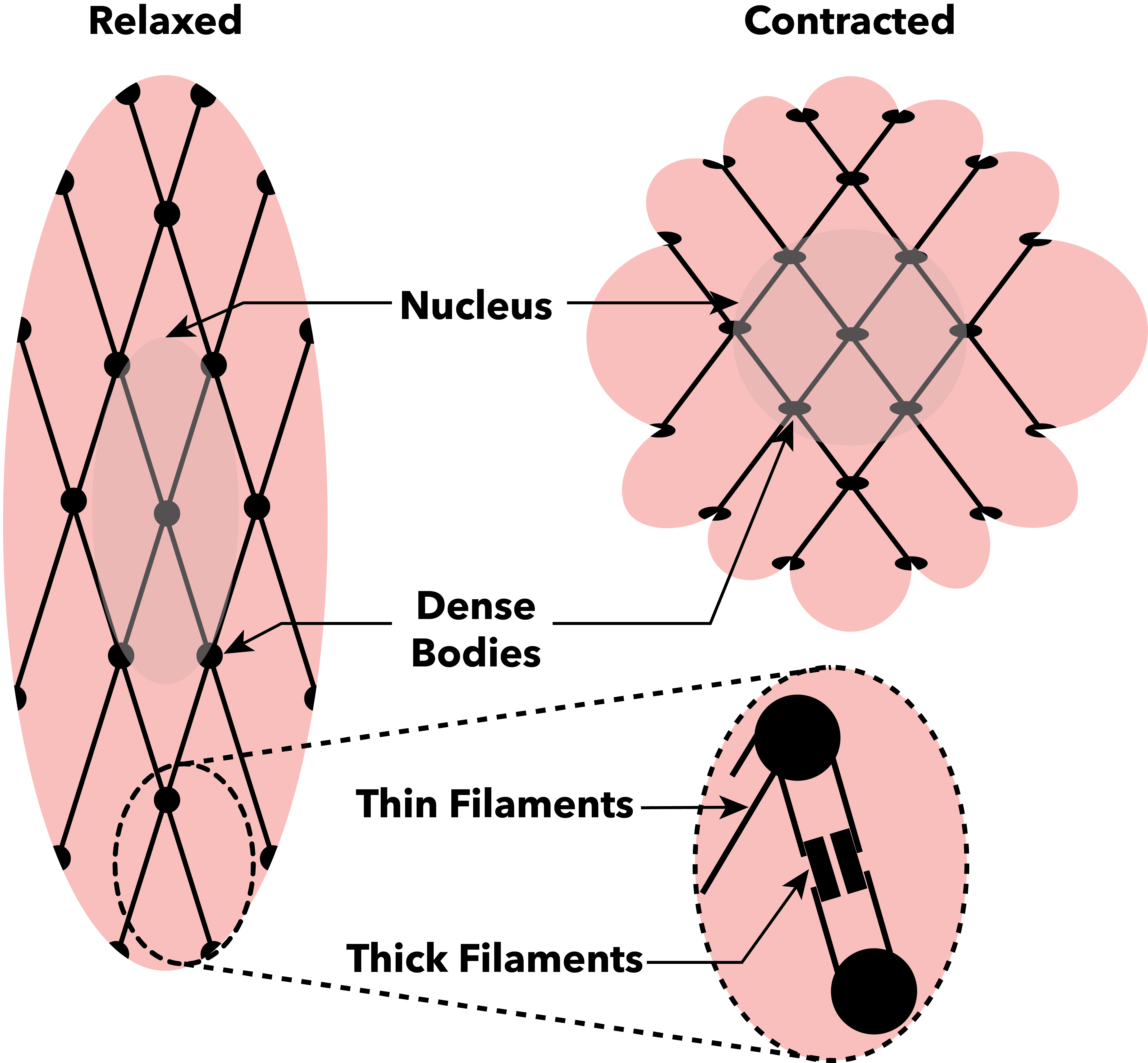
Muscle contraction is the activation of Tension (physics), tension-generating sites within muscle cells. In physiology, muscle contraction does not necessarily mean muscle shortening because muscle tension can be produced without changes in muscle length, such as when holding something heavy in the same position. The termination of muscle contraction is followed by muscle relaxation, which is a return of the muscle fibers to their low tension-generating state. For the contractions to happen, the muscle cells must rely on the change in action of two types of Myofilament, filaments: thin and thick filaments. The major constituent of thin filaments is a chain formed by helical coiling of two strands of actin, and thick filaments dominantly consist of chains of the Motor protein, motor-protein myosin. Together, these two filaments form myofibrils - the basic functional organelles in the skeletal muscle system. In vertebrates, Muscle cell#Muscle contraction in striated muscle, skele ... [...More Info...] [...Related Items...] OR: [Wikipedia] [Google] [Baidu] |
End-diastolic Volume
In cardiovascular physiology, end-diastolic volume (EDV) is the volume of blood in the right or left ventricle at end of filling in diastole which is amount of blood present in ventricle at the end of diastole. Because greater EDVs cause greater distention of the ventricle, ''EDV'' is often used synonymously with '' preload'', which refers to the length of the sarcomeres in cardiac muscle prior to contraction (systole). An increase in EDV increases the preload on the heart and, through the Frank-Starling mechanism of the heart, increases the amount of blood ejected from the ventricle during systole (stroke volume). __TOC__ Sample values The right ventricular end-diastolic volume (RVEDV) ranges between 100 and 160 mL. The right ventricular end-diastolic volume index (RVEDVI) is calculated by RVEDV/ BSA and ranges between 60 and 100 mL/m2. See also * End-systolic volume * Stroke volume In cardiovascular physiology, stroke volume (SV) is the volume of blood pumped from the ventr ... [...More Info...] [...Related Items...] OR: [Wikipedia] [Google] [Baidu] |
Right Atrial Pressure
Right atrial pressure (RAP) is the blood pressure in the right atrium of the heart. RAP reflects the amount of blood returning to the heart and the ability of the heart to pump the blood into the arterial system. RAP is often nearly identical to central venous pressure (CVP), although the two terms are not identical, as a pressure differential can sometimes exist between the venae cavae and the right atrium. CVP and RAP can differ when venous tone (i.e the degree of venous constriction) is altered. This can be graphically depicted as changes in the slope of the venous return plotted against right atrial pressure (where central venous pressure increases, but right atrial pressure stays the same; VR = CVP − RAP). Factors affecting RAP Factors that increase RAP include: * Hypervolemia * Forced exhalation * Tension pneumothorax * Heart failure * Pleural effusion * Decreased cardiac output * Cardiac tamponade * Mechanical ventilation and the application of positive end-expiratory pr ... [...More Info...] [...Related Items...] OR: [Wikipedia] [Google] [Baidu] |
Cardiac Output
In cardiac physiology, cardiac output (CO), also known as heart output and often denoted by the symbols Q, \dot Q, or \dot Q_ , edited by Catherine E. Williamson, Phillip Bennett is the volumetric flow rate of the heart's pumping output: that is, the volume of blood being pumped by a single Ventricle (heart), ventricle of the heart, per unit time (usually measured per minute). Cardiac output (CO) is the product of the heart rate (HR), i.e. the number of heartbeats per minute (bpm), and the stroke volume (SV), which is the volume of blood pumped from the left ventricle per beat; thus giving the formula: :CO = HR \times SV Values for cardiac output are usually denoted as L/min. For a healthy individual weighing 70 kg, the cardiac output at rest averages about 5 L/min; assuming a heart rate of 70 beats/min, the stroke volume would be approximately 70 mL. Because cardiac output is related to the quantity of blood delivered to various parts of the body, it is an important com ... [...More Info...] [...Related Items...] OR: [Wikipedia] [Google] [Baidu] |
Arthur Guyton
Arthur Clifton Guyton (September 8, 1919 – April 3, 2003) was an American physiologist. Guyton is well known for his ''Textbook of Medical Physiology'', which quickly became the standard text on the subject in medical schools. The first edition was published in 1956, the 10th edition in 2000 (the last before Guyton's death), and the 12th edition in 2010. The 14th edition (2020) is the latest version available. It is the world's best-selling medical physiology textbook and has been translated into at least 15 languages. Textbook of medical physiology ''Textbook of Medical Physiology'' is one of the world's best-selling physiology books and has been translated into at least 13 languages (the textbook memoriam states 13, but the online memoriam states at least 15.) From the ninth edition onwards, John E. Hall co-authored the textbook. However, all prior editions were written entirely by Guyton, with the eighth edition published in 1991. Subsequent editions, including the latest, ... [...More Info...] [...Related Items...] OR: [Wikipedia] [Google] [Baidu] |
Valsalva Maneuver
The Valsalva maneuver is performed by a forceful attempt of exhalation against a closed airway, usually done by closing one's mouth and pinching one's nose shut while expelling air, as if blowing up a balloon. Variations of the maneuver can be used either in medicine, medical examination as a test of cardiac function and autonomic nervous system, autonomic nervous control of the heart (because the maneuver raises the pressure in the lungs), or to clear the ears and paranasal sinuses, sinuses (that is, to equalize pressure between them) when ambient pressure changes, as in scuba diving, hyperbaric oxygen therapy, or air travel. A modified version is done by expiring against a closed glottis. This will elicit the cardiovascular responses described below but will not force air into the Eustachian tubes. History The technique is named after Antonio Maria Valsalva, a 17th-century physician and anatomist from Bologna whose principal scientific interest was the human ear. He descri ... [...More Info...] [...Related Items...] OR: [Wikipedia] [Google] [Baidu] |
Vena Cava
In anatomy, the ''venae cavae'' (; ''vena cava'' ; ) are two large veins ( great vessels) that return deoxygenated blood from the body into the heart. In humans they are the superior vena cava and the inferior vena cava, and both empty into the right atrium. They are located slightly off-center, toward the right side of the body. The right atrium receives deoxygenated blood through coronary sinus The coronary sinus () is the largest vein of the heart. It drains over half of the deoxygenated blood from the heart muscle into the right atrium. It begins on the backside of the heart, in between the left atrium, and left ventricle; it begi ... and two large veins called venae cavae. The inferior vena cava (or caudal vena cava in some animals) travels up alongside the abdominal aorta with blood from the lower part of the body. It is the largest vein in the human body. [...More Info...] [...Related Items...] OR: [Wikipedia] [Google] [Baidu] |
Central Venous Pressure
Central venous pressure (CVP) is the blood pressure in the venae cavae, near the right atrium of the heart. CVP reflects the amount of blood returning to the heart and the ability of the heart to pump the blood back into the arterial system. CVP is often a good approximation of right atrial pressure (RAP), although the two terms are not identical, as a pressure differential can sometimes exist between the venae cavae and the right atrium. CVP and RAP can differ when arterial tone is altered. This can be graphically depicted as changes in the slope of the venous return plotted against right atrial pressure (where central venous pressure increases, but right atrial pressure stays the same; VR = CVP − RAP). CVP has been, and often still is, used as a surrogate for preload, and changes in CVP in response to infusions of intravenous fluid have been used to predict volume-responsiveness (i.e. whether more fluid will improve cardiac output). However, there is increasing evidence t ... [...More Info...] [...Related Items...] OR: [Wikipedia] [Google] [Baidu] |
Vasomotor
Vasomotor refers to actions upon a blood vessel which alter its diameter. More specifically, it can refer to vasodilator action and vasoconstrictor action. Control Sympathetic innervation Sympathetic nerve fibers travel around the tunica media of the artery, secrete neurotransmitters such as norepinephrine into the extracellular fluid surrounding the smooth muscle (tunica media) from the terminal knob of the axon. The smooth muscle cell membranes have α and β-adrenergic receptors for these neurotransmitters. Activation of α-adrenergic receptors promotes vasoconstriction, while the activation of β-adrenergic receptors mediates the relaxation of muscle cells, resulting in vasodilation. Normally, α-adrenergic receptors predominate in smooth muscle of resistance vessels.Robert C. Ward et al. (2002). In Foundations for osteopathic medicine'. Lippincott Williams & Wilkins. 2nd edition. p. 98. . Google Book Search. Retrieved on 5 December 2010. Endothelium derived chemicals En ... [...More Info...] [...Related Items...] OR: [Wikipedia] [Google] [Baidu] |
Capacitance Of Blood Vessels
Compliance is the ability of a hollow organ (vessel) to distend and increase volume with increasing transmural pressure or the tendency of a hollow organ to resist recoil toward its original dimensions on application of a distending or compressing force. The reciprocal of compliance is elastance, a measure of the tendency of a hollow organ to recoil toward its original dimensions upon removal of a distending or compressing force. Blood vessels The terms elastance and compliance are of particular significance in cardiovascular physiology and respiratory physiology. In compliance, an increase in volume occurs in a vessel when the pressure in that vessel is increased. The tendency of the arteries and veins to stretch in response to pressure has a large effect on perfusion and blood pressure. This physically means that blood vessels with a higher compliance deform easier than lower compliance blood vessels under the same pressure and volume conditions. Venous compliance is approximately ... [...More Info...] [...Related Items...] OR: [Wikipedia] [Google] [Baidu] |
Muscle
Muscle is a soft tissue, one of the four basic types of animal tissue. There are three types of muscle tissue in vertebrates: skeletal muscle, cardiac muscle, and smooth muscle. Muscle tissue gives skeletal muscles the ability to muscle contraction, contract. Muscle tissue contains special Muscle contraction, contractile proteins called actin and myosin which interact to cause movement. Among many other muscle proteins, present are two regulatory proteins, troponin and tropomyosin. Muscle is formed during embryonic development, in a process known as myogenesis. Skeletal muscle tissue is striated consisting of elongated, multinucleate muscle cells called muscle fibers, and is responsible for movements of the body. Other tissues in skeletal muscle include tendons and perimysium. Smooth and cardiac muscle contract involuntarily, without conscious intervention. These muscle types may be activated both through the interaction of the central nervous system as well as by innervation ... [...More Info...] [...Related Items...] OR: [Wikipedia] [Google] [Baidu] |


