|
End-diastolic Volume
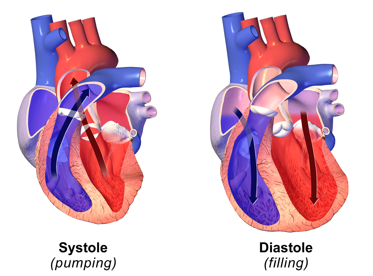
In cardiovascular physiology, end-diastolic volume (EDV) is the volume of blood in the right or left ventricle at end of filling in diastole which is amount of blood present in ventricle at the end of diastole. Because greater EDVs cause greater distention of the ventricle, ''EDV'' is often used synonymously with '' preload'', which refers to the length of the sarcomeres in cardiac muscle prior to contraction (systole). An increase in EDV increases the preload on the heart and, through the Frank-Starling mechanism of the heart, increases the amount of blood ejected from the ventricle during systole (stroke volume). __TOC__ Sample values The right ventricular end-diastolic volume (RVEDV) ranges between 100 and 160 mL. The right ventricular end-diastolic volume index (RVEDVI) is calculated by RVEDV/ BSA and ranges between 60 and 100 mL/m2. See also * End-systolic volume * Stroke volume In cardiovascular physiology, stroke volume (SV) is the volume of blood pumped from the ventr ... [...More Info...] [...Related Items...] OR: [Wikipedia] [Google] [Baidu] |
Cardiovascular Physiology
Cardiovascular physiology is the study of the cardiovascular system, specifically addressing the physiology of the heart ("cardio") and blood vessels ("vascular"). These subjects are sometimes addressed separately, under the names cardiac physiology and circulatory physiology. Although the different aspects of cardiovascular physiology are closely interrelated, the subject is still usually divided into several subtopics. Heart * Cardiac output (= heart rate * stroke volume. Can also be calculated with Fick principle, palpating method.) ** Stroke volume (= end-diastolic volume − end-systolic volume) ** Ejection fraction (= stroke volume / end-diastolic volume) ** Cardiac output is mathematically ` to systole ** Inotropic, chronotropic, and dromotropic states ** Cardiac input (= heart rate * suction volume Can be calculated by inverting terms in Fick principle) ** Suction volume (= end-systolic volume + end-diastolic volume) ** Injection fraction (=suction volum ... [...More Info...] [...Related Items...] OR: [Wikipedia] [Google] [Baidu] |
Diastole
Diastole ( ) is the relaxed phase of the cardiac cycle when the chambers of the heart are refilling with blood. The contrasting phase is systole when the heart chambers are contracting. Atrial diastole is the relaxing of the atria, and ventricular diastole the relaxing of the ventricles. The term originates from the Greek word (''diastolē''), meaning "dilation", from (''diá'', "apart") + (''stéllein'', "to send"). Role in cardiac cycle A typical heart rate is 75 beats per minute (bpm), which means that the cardiac cycle that produces one heartbeat, lasts for less than one second. The cycle requires 0.3 sec in ventricular systole (contraction)—pumping blood to all body systems from the two ventricles; and 0.5 sec in diastole (dilation), re-filling the four chambers of the heart, for a total of 0.8 sec to complete the cycle. Early ventricular diastole During early ventricular diastole, pressure in the two ventricles begins to drop from the peak reached during systole ... [...More Info...] [...Related Items...] OR: [Wikipedia] [Google] [Baidu] |
Preload (cardiology)
In cardiac physiology, preload is the amount of sarcomere stretch experienced by cardiac muscle cells, called cardiomyocytes, at the end of ventricular filling during diastole. Preload is directly related to ventricular filling. As the relaxed ventricle fills during diastole, the walls are stretched and the length of sarcomeres increases. Sarcomere length can be approximated by the volume of the ventricle because each shape has a conserved surface-area-to-volume ratio. This is useful clinically because measuring the sarcomere length is destructive to heart tissue. It requires cutting out a piece of cardiac muscle to look at the sarcomeres under a microscope. It is currently not possible to directly measure preload in the beating heart of a living animal. Preload is estimated from end-diastolic ventricular pressure and is measured in millimeters of mercury (mmHg). Estimating preload Though not exactly equivalent to the strict definition of ''preload,'' end-diastolic volume ... [...More Info...] [...Related Items...] OR: [Wikipedia] [Google] [Baidu] |
Sarcomere
A sarcomere (Greek σάρξ ''sarx'' "flesh", μέρος ''meros'' "part") is the smallest functional unit of striated muscle tissue. It is the repeating unit between two Z-lines. Skeletal striated muscle, Skeletal muscles are composed of tubular muscle cells (called muscle fibers or myofibers) which are formed during embryonic development, embryonic myogenesis. Muscle fibers contain numerous tubular myofibrils. Myofibrils are composed of repeating sections of sarcomeres, which appear under the microscope as alternating dark and light bands. Sarcomeres are composed of long, fibrous proteins as filaments that slide past each other when a muscle contracts or relaxes. The costamere is a different component that connects the sarcomere to the sarcolemma. Two of the important proteins are myosin, which forms the thick filament, and actin, which forms the thin filament. Myosin has a long fibrous tail and a globular head that binds to actin. The myosin head also binds to Adenosine triphos ... [...More Info...] [...Related Items...] OR: [Wikipedia] [Google] [Baidu] |
Cardiac Muscle
Cardiac muscle (also called heart muscle or myocardium) is one of three types of vertebrate muscle tissues, the others being skeletal muscle and smooth muscle. It is an involuntary, striated muscle that constitutes the main tissue of the wall of the heart. The cardiac muscle (myocardium) forms a thick middle layer between the outer layer of the heart wall (the pericardium) and the inner layer (the endocardium), with blood supplied via the coronary circulation. It is composed of individual cardiac muscle cells joined by intercalated discs, and encased by collagen fibers and other substances that form the extracellular matrix. Cardiac muscle contracts in a similar manner to skeletal muscle, although with some important differences. Electrical stimulation in the form of a cardiac action potential triggers the release of calcium from the cell's internal calcium store, the sarcoplasmic reticulum. The rise in calcium causes the cell's myofilaments to slide past each other i ... [...More Info...] [...Related Items...] OR: [Wikipedia] [Google] [Baidu] |
Systole
Systole ( ) is the part of the cardiac cycle during which some chambers of the heart contract after refilling with blood. Its contrasting phase is diastole, the relaxed phase of the cardiac cycle when the chambers of the heart are refilling with blood. Etymology The term originates, via Neo-Latin, from Ancient Greek (''sustolē''), from (''sustéllein'' 'to contract'; from ''sun'' 'together' + ''stéllein'' 'to send'), and is similar to the use of the English term ''to squeeze''. Terminology, general explanation The mammalian heart has four chambers: the left atrium above the left ventricle (lighter pink, see graphic), which two are connected through the mitral (or bicuspid) valve; and the right atrium above the right ventricle (lighter blue), connected through the tricuspid valve. The atria are the receiving blood chambers for the circulation of blood and the ventricles are the discharging chambers. In late ventricular diastole, the atrial chambers contract and ... [...More Info...] [...Related Items...] OR: [Wikipedia] [Google] [Baidu] |
Frank–Starling Law
The Frank–Starling law of the heart (also known as Starling's law and the Frank–Starling mechanism) represents the relationship between stroke volume and end diastolic volume.Widmaier, E. P., Hershel, R., & Strang, K. T. (2016).''Vander's Human Physiology: The Mechanisms of Body Function''(14th ed.). New York, NY: McGraw-Hill Education. The law states that the stroke volume of the heart increases in response to an increase in the volume of blood in the ventricles, before contraction (the end diastolic volume), when all other factors remain constant. As a larger volume of blood flows into the ventricle, the blood stretches cardiac muscle, leading to an increase in the force of contraction. The Frank-Starling mechanism allows the cardiac output to be synchronized with the venous return, arterial blood supply and humoral length, without depending upon external regulation to make alterations. The physiological importance of the mechanism lies mainly in maintaining left and ri ... [...More Info...] [...Related Items...] OR: [Wikipedia] [Google] [Baidu] |
Stroke Volume
In cardiovascular physiology, stroke volume (SV) is the volume of blood pumped from the ventricle (heart), ventricle per beat. Stroke volume is calculated using measurements of ventricle volumes from an Echocardiography, echocardiogram and subtracting the volume of the blood in the ventricle at the end of a beat (called end-systolic volume) from the volume of blood just prior to the beat (called end-diastolic volume). The term ''stroke volume'' can apply to each of the two ventricles of the heart, although when not explicitly stated it refers to the left ventricle and should therefore be referred to as left stroke volume (LSV). The stroke volumes for each ventricle are generally equal, both being approximately 90 mL in a healthy 70-kg man. Any persistent difference between the two stroke volumes, no matter how small, would inevitably lead to venous congestion of either the systemic or the pulmonary circulation, with a corresponding state of hypotension in the other circulatory sys ... [...More Info...] [...Related Items...] OR: [Wikipedia] [Google] [Baidu] |
Body Surface Area
In physiology and medicine, the body surface area (BSA) is the measured or calculated surface area of a human body. For many clinical purposes, BSA is a better indicator of metabolic mass than body weight because it is less affected by abnormal adipose mass. Nevertheless, there have been several important critiques of the use of BSA in determining the dosage of medications with a narrow therapeutic index, such as chemotherapy. Typically there is a 4–10 fold variation in drug clearance between individuals due to differing the activity of drug elimination processes related to genetic and environmental factors. This can lead to significant overdosing and underdosing (and increased risk of disease recurrence). It is also thought to be a distorting factor in Phase I and II trials that may result in potentially helpful medications being prematurely rejected. The trend to personalized medicine is one approach to counter this weakness. Uses Examples of uses of the BSA: * Renal clea ... [...More Info...] [...Related Items...] OR: [Wikipedia] [Google] [Baidu] |
End-systolic Volume
End-systolic volume (ESV) is the volume of blood in a ventricle at the end of contraction, or systole, and the beginning of filling, or diastole. ESV is the lowest volume of blood in the ventricle at any point in the cardiac cycle. The main factors that affect the end-systolic volume are afterload and the contractility of the heart. __TOC__ Uses End systolic volume can be used clinically as a measurement of the adequacy of cardiac emptying, related to systolic function. On an electrocardiogram, or ECG, the end-systolic volume will be seen at the end of the T wave. Clinically, ESV can be measured using two-dimensional echocardiography, MRI (magnetic resonance tomography) or cardiac CT (computed tomography) or SPECT (single photon emission computed tomography). Sample values Along with end-diastolic volume, ESV determines the stroke volume, or output of blood by the heart during a single phase of the cardiac cycle. The stroke volume In cardiovascular physiology, stroke volume ... [...More Info...] [...Related Items...] OR: [Wikipedia] [Google] [Baidu] |
Stroke Volume
In cardiovascular physiology, stroke volume (SV) is the volume of blood pumped from the ventricle (heart), ventricle per beat. Stroke volume is calculated using measurements of ventricle volumes from an Echocardiography, echocardiogram and subtracting the volume of the blood in the ventricle at the end of a beat (called end-systolic volume) from the volume of blood just prior to the beat (called end-diastolic volume). The term ''stroke volume'' can apply to each of the two ventricles of the heart, although when not explicitly stated it refers to the left ventricle and should therefore be referred to as left stroke volume (LSV). The stroke volumes for each ventricle are generally equal, both being approximately 90 mL in a healthy 70-kg man. Any persistent difference between the two stroke volumes, no matter how small, would inevitably lead to venous congestion of either the systemic or the pulmonary circulation, with a corresponding state of hypotension in the other circulatory sys ... [...More Info...] [...Related Items...] OR: [Wikipedia] [Google] [Baidu] |


