|
TNXB
Tenascin X (TN-X), also known as flexillin or hexabrachion-like protein, is a 450kDa glycoprotein, a member of the tenascin family, that is expressed in connective tissues. In humans it is encoded by the ''TNXB'' gene. The TN-X protein is expressed in many parts of the human body, including the skin, muscles, kidneys, blood vessels, and digestive tract. Deficiencies in the TN-X protein due to mutations or not enough of it being produced (haploinsufficiency) can lead to a rare condition called classical-like Ehlers-Danlos syndrome (EDS). People with EDS may have loose joints and weak tissues because their bodies make defective collagen. Structure TN-X possesses a modular structure composed, from the N- to the C-terminal part by a Tenascin assembly domain (TAD), a series of 18.5 repeats of epidermal growth factor (EGF)-like motif, a high number of Fibronectin type III (FNIII) module, and a fibrinogen (FBG)-like globular domain. Gene TNXB (functional gene) The ''TNXB'' gene ... [...More Info...] [...Related Items...] OR: [Wikipedia] [Google] [Baidu] |
RCCX
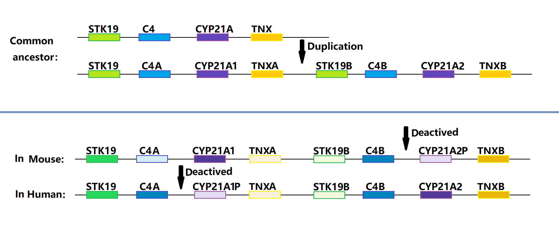
RCCX is a complex, multiallelic, and tandem copy number variation (CNV) human DNA locus on chromosome 6p21.3, a cluster located in the major histocompatibility complex (MHC) class III region. CNVs are segments of DNA that vary in copy number compared to a reference genome and play a significant role in human phenotypic variation and disease development. The RCCX cluster consists of one or more modules each having a series of genes close to each other: serine/threonine kinase 19 (''STK19''), complement 4 (''C4''), steroid 21-hydroxylase (''CYP21''), and tenascin-X (''TNX''). Name The RCCX abbreviation is composed of the names of the genes RP (a former name for ''STK19'' serine/threonine kinase 19), '' C4'', '' CYP21'' and '' TNX''). The RCCX abbreviation was first mentioned in a 1994 article published in Immunogenetics, an academic journal, for a study by Dangel et al. Structure The number of RCCX segments varies between one and four in a chromosome, with the prevalence of ap ... [...More Info...] [...Related Items...] OR: [Wikipedia] [Google] [Baidu] |
Ehlers–Danlos Syndrome
Ehlers–Danlos syndromes (EDS) is a group of 14 genetic connective-tissue disorders. Symptoms often include loose joints, joint pain, stretchy velvety skin, and abnormal scar formation. These may be noticed at birth or in early childhood. Complications may include aortic dissection, joint dislocations, scoliosis, chronic pain, or early osteoarthritis. The existing classification was last updated in 2017, when a number of rarer forms of EDS were added. EDS occurs due to mutations in one or more particular genes—there are 19 genes that can contribute to the condition. The specific gene affected determines the type of EDS, though the genetic causes of hypermobile Ehlers–Danlos syndrome are still unknown. Some cases result from a new variation occurring during early development, while others are inherited in an autosomal dominant or recessive manner. Typically, these variations result in defects in the structure or processing of the protein collagen or tenascin. Diagnos ... [...More Info...] [...Related Items...] OR: [Wikipedia] [Google] [Baidu] |
21-Hydroxylase
Steroid 21-hydroxylase is a protein that in humans is encoded by the ''CYP21A2'' gene. The protein is an enzyme that hydroxylates steroids at the C21 position on the molecule. Naming conventions for enzymes are based on the substrate acted upon and the chemical process performed. Biochemically, this enzyme is involved in the biosynthesis of the adrenal gland hormones aldosterone and cortisol, which are important in blood pressure regulation, sodium homeostasis and blood sugar control. The enzyme converts progesterone and 17α-hydroxyprogesterone into 11-deoxycorticosterone and 11-deoxycortisol, respectively, within metabolic pathways which in humans ultimately lead to aldosterone and cortisol creation—deficiency in the enzyme may cause congenital adrenal hyperplasia. Steroid 21-hydroxylase is a member of the cytochrome P450 family of monooxygenase enzymes that use an iron-containing heme cofactor to oxidize substrates. In humans, the enzyme is localized in endoplasmic ... [...More Info...] [...Related Items...] OR: [Wikipedia] [Google] [Baidu] |
EGF-like Domain
The EGF-like domain is an evolutionary conserved protein domain, which derives its name from the epidermal growth factor where it was first described. It comprises about 30 to 40 amino-acid residues and has been found in a large number of mostly animal proteins. Most occurrences of the EGF-like domain are found in the extracellular domain of membrane-bound proteins or in proteins known to be secreted. An exception to this is the prostaglandin-endoperoxide synthase. The EGF-like domain includes 6 cysteine residues which in the epidermal growth factor have been shown to form 3 disulfide bonds. The structures of 4-disulfide EGF-domains have been solved from the laminin and integrin proteins. The main structure of EGF-like domains is a two-stranded β-sheet followed by a loop to a short C-terminal, two-stranded β-sheet. These two β-sheets are usually denoted as the major (N-terminal) and minor (C-terminal) sheets. EGF-like domains frequently occur in numerous tandem copies in p ... [...More Info...] [...Related Items...] OR: [Wikipedia] [Google] [Baidu] |
Fibronectin Type III Domain
The Fibronectin type III domain is an evolutionarily conserved protein domain that is widely found in animal proteins. The fibronectin protein in which this domain was first identified contains 16 copies of this domain. The domain is about 100 amino acids long and possesses a beta sandwich structure. Of the three fibronectin-type domains, type III is the only one without disulfide bonding present. Fibronectin domains are found in a wide variety of extracellular proteins. They are widely distributed in animal species, but also found sporadically in yeast, plant and bacterial proteins. Human proteins containing this domain ABI3BP; ANKFN1; ASTN2; AXL; BOC; BZRAP1; C20orf75; CDON; CHL1; CMYA5; CNTFR; CNTN1; CNTN2; CNTN3; CNTN4; CNTN5; CNTN6; COL12A1; COL14A1; COL20A1; COL7A1; CRLF1; CRLF3; CSF2RB; CSF3R; DCC; DSCAM; DSCAML1; EBI3; EGFLAM; EPHA1; EPH ... [...More Info...] [...Related Items...] OR: [Wikipedia] [Google] [Baidu] |
Ovary
The ovary () is a gonad in the female reproductive system that produces ova; when released, an ovum travels through the fallopian tube/ oviduct into the uterus. There is an ovary on the left and the right side of the body. The ovaries are endocrine glands, secreting various hormones that play a role in the menstrual cycle and fertility. The ovary progresses through many stages beginning in the prenatal period through menopause. Structure Each ovary is whitish in color and located alongside the lateral wall of the uterus in a region called the ovarian fossa. The ovarian fossa is the region that is bounded by the external iliac artery and in front of the ureter and the internal iliac artery. This area is about 4 cm x 3 cm x 2 cm in size.Daftary, Shirish; Chakravarti, Sudip (2011). Manual of Obstetrics, 3rd Edition. Elsevier. pp. 1-16. . The ovaries are surrounded by a capsule, and have an outer cortex and an inner medulla. The capsule is of dense connect ... [...More Info...] [...Related Items...] OR: [Wikipedia] [Google] [Baidu] |
Optic Nerve
In neuroanatomy, the optic nerve, also known as the second cranial nerve, cranial nerve II, or simply CN II, is a paired cranial nerve that transmits visual system, visual information from the retina to the brain. In humans, the optic nerve is derived from optic stalks during the seventh week of development and is composed of retinal ganglion cell axons and glial cells; it extends from the optic disc to the optic chiasma and continues as the optic tract to the lateral geniculate nucleus, Pretectal area, pretectal nuclei, and superior colliculus. Structure The optic nerve has been classified as the second of twelve paired cranial nerves, but it is technically a myelinated tract of the central nervous system, rather than a classical nerve of the peripheral nervous system because it is derived from an out-pouching of the diencephalon (optic stalks) during embryonic development. As a consequence, the fibers of the optic nerve are covered with myelin produced by oligodendrocytes, r ... [...More Info...] [...Related Items...] OR: [Wikipedia] [Google] [Baidu] |
Adrenal Gland
The adrenal glands (also known as suprarenal glands) are endocrine glands that produce a variety of hormones including adrenaline and the steroids aldosterone and cortisol. They are found above the kidneys. Each gland has an outer adrenal cortex, cortex which produces steroid hormones and an inner Adrenal medulla, medulla. The adrenal cortex itself is divided into three main zones: the zona glomerulosa, the zona fasciculata and the zona reticularis. The adrenal cortex produces three main types of steroid hormones: mineralocorticoids, glucocorticoids, and androgens. Mineralocorticoids (such as aldosterone) produced in the zona glomerulosa help in the regulation of blood pressure and osmoregulation, electrolyte balance. The glucocorticoids cortisol and cortisone are synthesized in the zona fasciculata; their functions include the regulation of metabolism and immune system suppression. The innermost layer of the cortex, the zona reticularis, produces androgens that are converted to ... [...More Info...] [...Related Items...] OR: [Wikipedia] [Google] [Baidu] |
Blood Vessel
Blood vessels are the tubular structures of a circulatory system that transport blood throughout many Animal, animals’ bodies. Blood vessels transport blood cells, nutrients, and oxygen to most of the Tissue (biology), tissues of a Body (biology), body. They also take waste and carbon dioxide away from the tissues. Some tissues such as cartilage, epithelium, and the lens (anatomy), lens and cornea of the eye are not supplied with blood vessels and are termed ''avascular''. There are five types of blood vessels: the arteries, which carry the blood away from the heart; the arterioles; the capillaries, where the exchange of water and chemicals between the blood and the tissues occurs; the venules; and the veins, which carry blood from the capillaries back towards the heart. The word ''vascular'', is derived from the Latin ''vas'', meaning ''vessel'', and is mostly used in relation to blood vessels. Etymology * artery – late Middle English; from Latin ''arteria'', from Gree ... [...More Info...] [...Related Items...] OR: [Wikipedia] [Google] [Baidu] |
Testicle
A testicle or testis ( testes) is the gonad in all male bilaterians, including humans, and is Homology (biology), homologous to the ovary in females. Its primary functions are the production of sperm and the secretion of Androgen, androgens, primarily testosterone. The release of testosterone is regulated by luteinizing hormone (LH) from the anterior pituitary gland. Sperm production is controlled by follicle-stimulating hormone (FSH) from the anterior pituitary gland and by testosterone produced within the gonads. Structure Appearance Males have two testicles of similar size contained within the scrotum, which is an extension of the abdominal wall. Scrotal asymmetry, in which one testicle extends farther down into the scrotum than the other, is common. This is because of the differences in the vasculature's anatomy. For 85% of men, the right testis hangs lower than the left one. Measurement and volume The volume of the testicle can be estimated by palpating it and compari ... [...More Info...] [...Related Items...] OR: [Wikipedia] [Google] [Baidu] |
Glycoprotein
Glycoproteins are proteins which contain oligosaccharide (sugar) chains covalently attached to amino acid side-chains. The carbohydrate is attached to the protein in a cotranslational or posttranslational modification. This process is known as glycosylation. Secreted extracellular proteins are often glycosylated. In proteins that have segments extending extracellularly, the extracellular segments are also often glycosylated. Glycoproteins are also often important integral membrane proteins, where they play a role in cell–cell interactions. It is important to distinguish endoplasmic reticulum-based glycosylation of the secretory system from reversible cytosolic-nuclear glycosylation. Glycoproteins of the cytosol and nucleus can be modified through the reversible addition of a single GlcNAc residue that is considered reciprocal to phosphorylation and the functions of these are likely to be an additional regulatory mechanism that controls phosphorylation-based signalling. In ... [...More Info...] [...Related Items...] OR: [Wikipedia] [Google] [Baidu] |
Gastrointestinal Tract
The gastrointestinal tract (GI tract, digestive tract, alimentary canal) is the tract or passageway of the Digestion, digestive system that leads from the mouth to the anus. The tract is the largest of the body's systems, after the cardiovascular system. The GI tract contains all the major organ (biology), organs of the digestive system, in humans and other animals, including the esophagus, stomach, and intestines. Food taken in through the mouth is digestion, digested to extract nutrients and absorb energy, and the waste expelled at the anus as feces. ''Gastrointestinal'' is an adjective meaning of or pertaining to the stomach and intestines. Nephrozoa, Most animals have a "through-gut" or complete digestive tract. Exceptions are more primitive ones: sponges have small pores (ostium (sponges), ostia) throughout their body for digestion and a larger dorsal pore (osculum) for excretion, comb jellies have both a ventral mouth and dorsal anal pores, while cnidarians and acoels have ... [...More Info...] [...Related Items...] OR: [Wikipedia] [Google] [Baidu] |


