|
Sternohyoid
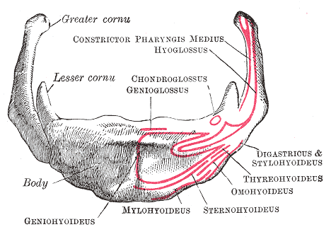
The sternohyoid muscle is a bilaterally paired, long, thin, narrow strap muscle of the anterior neck. It is one of the infrahyoid muscles. It is innervated by the ansa cervicalis. It acts to depress the hyoid bone. The sternohyoid muscle is a flat muscle located on both sides of the neck, part of the infrahyoid muscle group. It originates from the medial edge of the clavicle, sternoclavicular ligament, and posterior side of the manubrium, and ascends to attach to the body of the hyoid bone. The sternohyoid muscle, along with other infrahyoid muscles, functions to depress the hyoid bone, which is important for activities such as speaking, chewing, and swallowing. Additionally, this muscle group contributes to the protection of the trachea, esophagus, blood vessels, and thyroid gland. The sternohyoid muscle also plays a minor role in head movements. Structure The sternohyoid muscle is one of the paired strap muscles of the infrahyoid muscles. The muscle is directed superome ... [...More Info...] [...Related Items...] OR: [Wikipedia] [Google] [Baidu] |
Ansa Cervicalis
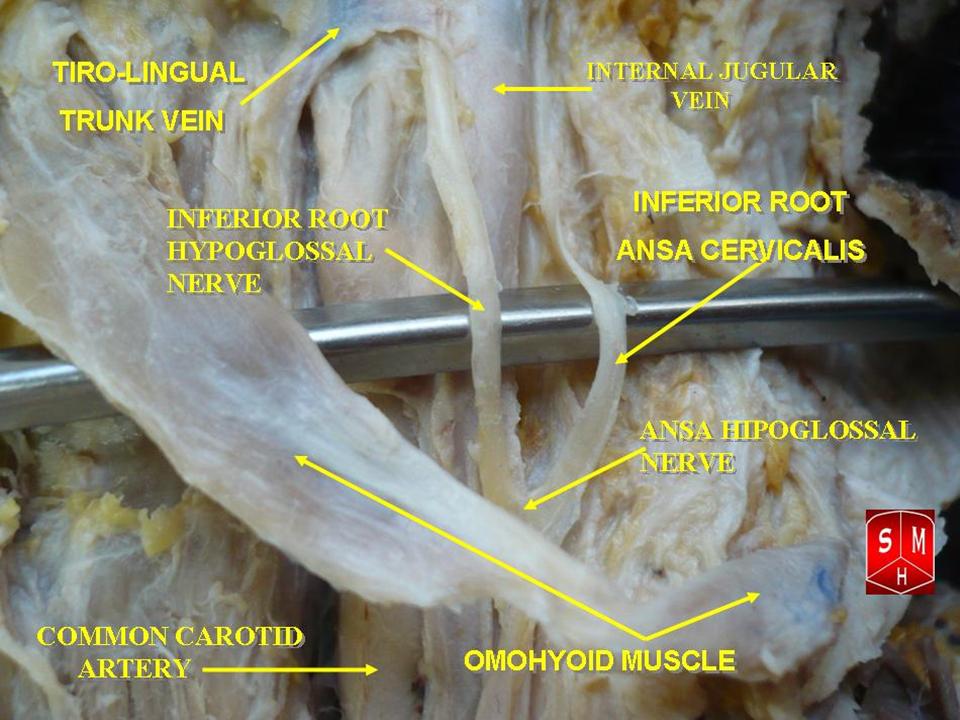
The ansa cervicalis (or ansa hypoglossi in older literature) is a loop formed by muscular branches of the cervical plexus formed by branches of cervical spinal nerves C1-C3. The ansa cervicalis has two roots - a superior root (formed by branch of C1) and an inferior root (formed by union of branches of C2 and C3) - that unite distally, forming a loop. It is situated anterior to the carotid sheath. Branches of the ansa cervicalis innervate three of the four infrahyoid muscles: the sternothyroid, sternohyoid, and omohyoid muscles (note that the thyrohyoid muscle is the one infrahyoid muscle not innervated by the ansa cervicalis - it is instead innervated by cervical spinal nerve 1 via a separate thyrohyoid branch). Its name means "handle of the neck" in Latin. Anatomy The ansa cervicalis is typically embedded within the anterior wall of the carotid sheath anterior to the internal jugular vein. Superior root The superior root of the ansa cervicalis (formerly known as d ... [...More Info...] [...Related Items...] OR: [Wikipedia] [Google] [Baidu] |
Trachea
The trachea (: tracheae or tracheas), also known as the windpipe, is a cartilaginous tube that connects the larynx to the bronchi of the lungs, allowing the passage of air, and so is present in almost all animals' lungs. The trachea extends from the larynx and branches into the two primary bronchi. At the top of the trachea, the cricoid cartilage attaches it to the larynx. The trachea is formed by a number of horseshoe-shaped rings, joined together vertically by overlying annular ligaments of trachea, ligaments, and by the trachealis muscle at their ends. The epiglottis closes the opening to the larynx during swallowing. The trachea begins to form in the second month of embryo development, becoming longer and more fixed in its position over time. Its epithelium is lined with columnar epithelium, column-shaped cells that have hair-like extensions called cilia, with scattered goblet cells that produce protective mucins. The trachea can be affected by inflammation or infection, usua ... [...More Info...] [...Related Items...] OR: [Wikipedia] [Google] [Baidu] |
Hyoid Bone
The hyoid-bone (lingual-bone or tongue-bone) () is a horseshoe-shaped bone situated in the anterior midline of the neck between the chin and the thyroid-cartilage. At rest, it lies between the base of the mandible and the third cervical vertebra. Unlike other bones, the hyoid is only distantly articulated to other bones by muscles or ligaments. It is the only bone in the human body that is not connected to any other bones. The hyoid is anchored by muscles from the anterior, posterior and inferior directions, and aids in tongue movement and swallowing. The hyoid bone provides attachment to the muscles of the floor of the mouth and the tongue above, the larynx below, and the epiglottis and pharynx behind. Its name is derived . Structure The hyoid bone is classed as an irregular bone and consists of a central part called the body, and two pairs of horns, the greater and lesser horns. Body The body of the hyoid bone is the central part of the hyoid bone. *At the fron ... [...More Info...] [...Related Items...] OR: [Wikipedia] [Google] [Baidu] |
Strap Muscles
The infrahyoid muscles, or strap muscles, are a group of four pairs of muscles in the anterior (frontal) part of the neck. The four infrahyoid muscles are the sternohyoid, sternothyroid, thyrohyoid and omohyoid muscles. Excluding the sternothyroid, the infrahyoid muscles either originate from or insert on to the hyoid bone. The term ''infrahyoid'' refers to the region below the hyoid bone, while the term strap muscles refers to the long and flat muscle shapes which resembles a strap. The stylopharyngeus muscle is considered by many to be one of the strap muscles, but is not an infrahyoid muscle. Individual muscles The origin, insertion and innervation of the individual muscles: Nerve supply All of the infrahyoid muscles are innervated by the ansa cervicalis from the cervical plexus ( C1- C3) except the thyrohyoid muscle, which is innervated by fibers only from the first cervical spinal nerve travelling with the hypoglossal nerve. Function The infrahyoid muscles function to ... [...More Info...] [...Related Items...] OR: [Wikipedia] [Google] [Baidu] |
Infrahyoid Muscles
The infrahyoid muscles, or strap muscles, are a group of four pairs of muscles in the anterior (frontal) part of the neck. The four infrahyoid muscles are the sternohyoid, sternothyroid, thyrohyoid and omohyoid muscles. Excluding the sternothyroid, the infrahyoid muscles either originate from or insert on to the hyoid bone. The term ''infrahyoid'' refers to the region below the hyoid bone, while the term strap muscles refers to the long and flat muscle shapes which resembles a strap. The stylopharyngeus muscle is considered by many to be one of the strap muscles, but is not an infrahyoid muscle. Individual muscles The origin, insertion and innervation of the individual muscles: Nerve supply All of the infrahyoid muscles are innervated by the ansa cervicalis from the cervical plexus ( C1- C3) except the thyrohyoid muscle, which is innervated by fibers only from the first cervical spinal nerve travelling with the hypoglossal nerve. Function The infrahyoid muscles ... [...More Info...] [...Related Items...] OR: [Wikipedia] [Google] [Baidu] |
Hyoid Bone
The hyoid-bone (lingual-bone or tongue-bone) () is a horseshoe-shaped bone situated in the anterior midline of the neck between the chin and the thyroid-cartilage. At rest, it lies between the base of the mandible and the third cervical vertebra. Unlike other bones, the hyoid is only distantly articulated to other bones by muscles or ligaments. It is the only bone in the human body that is not connected to any other bones. The hyoid is anchored by muscles from the anterior, posterior and inferior directions, and aids in tongue movement and swallowing. The hyoid bone provides attachment to the muscles of the floor of the mouth and the tongue above, the larynx below, and the epiglottis and pharynx behind. Its name is derived . Structure The hyoid bone is classed as an irregular bone and consists of a central part called the body, and two pairs of horns, the greater and lesser horns. Body The body of the hyoid bone is the central part of the hyoid bone. *At the fron ... [...More Info...] [...Related Items...] OR: [Wikipedia] [Google] [Baidu] |
Neck
The neck is the part of the body in many vertebrates that connects the head to the torso. It supports the weight of the head and protects the nerves that transmit sensory and motor information between the brain and the rest of the body. Additionally, the neck is highly flexible, allowing the head to turn and move in all directions. Anatomically, the human neck is divided into four compartments: vertebral, visceral, and two vascular compartments. Within these compartments, the neck houses the cervical vertebrae, the cervical portion of the spinal cord, upper parts of the respiratory and digestive tracts, endocrine glands, nerves, arteries and veins. The muscles of the neck, which are separate from the compartments, form the boundaries of the neck triangles. In anatomy, the neck is also referred to as the or . However, when the term ''cervix'' is used alone, it often refers to the uterine cervix, the neck of the uterus. Therefore, the adjective ''cervical'' ... [...More Info...] [...Related Items...] OR: [Wikipedia] [Google] [Baidu] |
Cervical Spinal Nerve
A spinal nerve is a mixed nerve, which carries motor, sensory, and autonomic signals between the spinal cord and the body. In the human body there are 31 pairs of spinal nerves, one on each side of the vertebral column. These are grouped into the corresponding cervical, thoracic, lumbar, sacral and coccygeal regions of the spine. There are eight pairs of cervical nerves, twelve pairs of thoracic nerves, five pairs of lumbar nerves, five pairs of sacral nerves, and one pair of coccygeal nerves. The spinal nerves are part of the peripheral nervous system. Structure Each spinal nerve is a mixed nerve, formed from the combination of nerve root fibers from its dorsal and ventral roots. The dorsal root is the afferent sensory root and carries sensory information to the brain. The ventral root is the efferent motor root and carries motor information from the brain. The spinal nerve emerges from the spinal column through an opening (intervertebral foramen) between adjacent ... [...More Info...] [...Related Items...] OR: [Wikipedia] [Google] [Baidu] |
Superior Thyroid Artery
The superior thyroid artery arises from the external carotid artery just below the level of the greater cornu of the hyoid bone and ends in the thyroid gland. Structure From its origin under the anterior border of the sternocleidomastoid the superior thyroid artery runs upward and forward for a short distance in the carotid triangle, where it is covered by the skin, platysma, and fascia; it then arches downward beneath the omohyoid, sternohyoid, and sternothyroid muscles. To its medial side are the inferior pharyngeal constrictor muscle and the external branch of the superior laryngeal nerve. Branches It distributes twigs to the adjacent muscles, and numerous branches to the thyroid gland, connecting with its fellow of the opposite side, and with the inferior thyroid arteries. The branches to the gland are generally two in number. One, the larger, supplies principally the anterior surface; on the isthmus of the gland it connects with the corresponding artery of the opposite s ... [...More Info...] [...Related Items...] OR: [Wikipedia] [Google] [Baidu] |
Manubrium
The sternum (: sternums or sterna) or breastbone is a long flat bone located in the central part of the chest. It connects to the ribs via cartilage and forms the front of the rib cage, thus helping to protect the heart, human lung, lungs, and major blood vessels from injury. Shaped roughly like a necktie, it is one of the largest and longest flat bones of the body. Its three regions are the manubrium, the body, and the xiphoid process. The word ''sternum'' originates from Ancient Greek στέρνον (''stérnon'') 'chest'. Structure The sternum is a narrow, flat bone, forming the middle portion of the front of the chest. The top of the sternum supports the clavicles (collarbones) and its edges join with the costal cartilages of the first two pairs of ribs. The inner surface of the sternum is also the attachment of the sternopericardial ligaments. Its top is also connected to the sternocleidomastoid muscle. The sternum consists of three main parts, listed from the top: * Manubr ... [...More Info...] [...Related Items...] OR: [Wikipedia] [Google] [Baidu] |
Head
A head is the part of an organism which usually includes the ears, brain, forehead, cheeks, chin, eyes, nose, and mouth, each of which aid in various sensory functions such as sight, hearing, smell, and taste. Some very simple animals may not have a head, but many bilaterally symmetric forms do, regardless of size. Heads develop in animals by an evolutionary trend known as cephalization. In bilaterally symmetrical animals, nervous tissue concentrate at the anterior region, forming structures responsible for information processing. Through biological evolution, sense organs and feeding structures also concentrate into the anterior region; these collectively form the head. Human head The human head is an anatomical unit that consists of the skull, hyoid bone and cervical vertebrae. The skull consists of the brain case which encloses the cranial cavity, and the facial skeleton, which includes the mandible. There are eight bones in the brain case ... [...More Info...] [...Related Items...] OR: [Wikipedia] [Google] [Baidu] |


