|
Infrahyoid Muscles
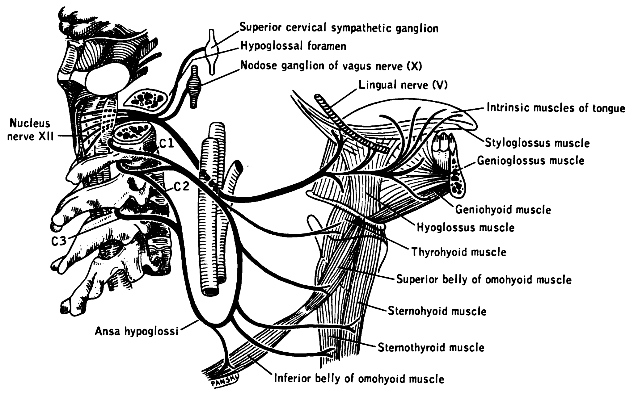
The infrahyoid muscles, or strap muscles, are a group of four pairs of muscles in the anterior (frontal) part of the neck. The four infrahyoid muscles are the sternohyoid, sternothyroid, thyrohyoid and omohyoid muscles. Excluding the sternothyroid, the infrahyoid muscles either originate from or insert on to the hyoid bone. The term ''infrahyoid'' refers to the region below the hyoid bone, while the term strap muscles refers to the long and flat muscle shapes which resembles a strap. The stylopharyngeus muscle is considered by many to be one of the strap muscles, but is not an infrahyoid muscle. Individual muscles The origin, insertion and innervation of the individual muscles: Nerve supply All of the infrahyoid muscles are innervated by the ansa cervicalis from the cervical plexus ( C1- C3) except the thyrohyoid muscle, which is innervated by fibers only from the first cervical spinal nerve travelling with the hypoglossal nerve. Function The infrahyoid muscles ... [...More Info...] [...Related Items...] OR: [Wikipedia] [Google] [Baidu] |
Neck
The neck is the part of the body in many vertebrates that connects the head to the torso. It supports the weight of the head and protects the nerves that transmit sensory and motor information between the brain and the rest of the body. Additionally, the neck is highly flexible, allowing the head to turn and move in all directions. Anatomically, the human neck is divided into four compartments: vertebral, visceral, and two vascular compartments. Within these compartments, the neck houses the cervical vertebrae, the cervical portion of the spinal cord, upper parts of the respiratory and digestive tracts, endocrine glands, nerves, arteries and veins. The muscles of the neck, which are separate from the compartments, form the boundaries of the neck triangles. In anatomy, the neck is also referred to as the or . However, when the term ''cervix'' is used alone, it often refers to the uterine cervix, the neck of the uterus. Therefore, the adjective ''cervical'' ca ... [...More Info...] [...Related Items...] OR: [Wikipedia] [Google] [Baidu] |
Clavicle
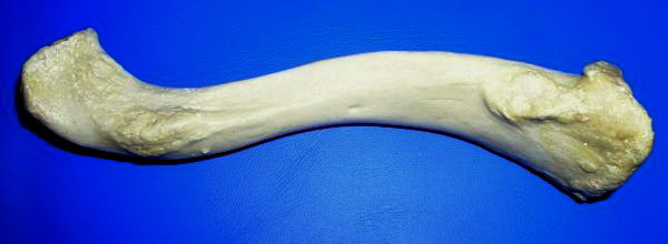
The clavicle, collarbone, or keybone is a slender, S-shaped long bone approximately long that serves as a strut between the scapula, shoulder blade and the sternum (breastbone). There are two clavicles, one on each side of the body. The clavicle is the only long bone in the body that lies horizontally. Together with the shoulder blade, it makes up the shoulder girdle. It is a palpable bone and, in people who have less fat in this region, the location of the bone is clearly visible. It receives its name from Latin ''clavicula'' 'little key' because the bone rotates along its axis like a key when the shoulder is Abduction (kinesiology), abducted. The clavicle is the most commonly fractured bone. It can easily be fractured by impacts to the shoulder from the force of falling on outstretched arms or by a direct hit. Structure The collarbone is a thin doubly curved long bone that connects the human arm, arm to the torso, trunk of the body. Located directly above the first rib, it ac ... [...More Info...] [...Related Items...] OR: [Wikipedia] [Google] [Baidu] |
Cervical Spinal Nerve
A spinal nerve is a mixed nerve, which carries motor, sensory, and autonomic signals between the spinal cord and the body. In the human body there are 31 pairs of spinal nerves, one on each side of the vertebral column. These are grouped into the corresponding cervical, thoracic, lumbar, sacral and coccygeal regions of the spine. There are eight pairs of cervical nerves, twelve pairs of thoracic nerves, five pairs of lumbar nerves, five pairs of sacral nerves, and one pair of coccygeal nerves. The spinal nerves are part of the peripheral nervous system. Structure Each spinal nerve is a mixed nerve, formed from the combination of nerve root fibers from its dorsal and ventral roots. The dorsal root is the afferent sensory root and carries sensory information to the brain. The ventral root is the efferent motor root and carries motor information from the brain. The spinal nerve emerges from the spinal column through an opening (intervertebral foramen) between adjacent ... [...More Info...] [...Related Items...] OR: [Wikipedia] [Google] [Baidu] |
Cervical Plexus
The cervical plexus is a nerve plexus of the anterior rami of the first (i.e. upper-most) four cervical spinal nerves C1-C4. The cervical plexus provides motor innervation to some muscles of the neck, and the diaphragm; it provides sensory innervation to parts of the head, neck, and chest. Anatomy They are located laterally to the transverse processes between prevertebral muscles from the medial side and vertebral (m. scalenus, m. levator scapulae, m. splenius cervicis) from lateral side. There is anastomosis with accessory nerve, hypoglossal nerve and sympathetic trunk. It is located in the neck, deep to the sternocleidomastoid muscle. The branches of the cervical plexus emerge from the posterior triangle at the nerve point, a point which lies midway on the posterior border of the sternocleidomastoid. Relations The cervical plexus is situated deep to the sternocleidomastoid muscle, internal jugular vein, and deep cervical fascia. It is situated anterior to the mid ... [...More Info...] [...Related Items...] OR: [Wikipedia] [Google] [Baidu] |
Ansa Cervicalis
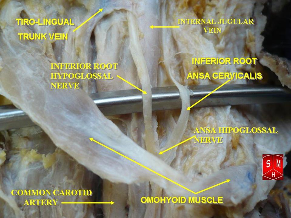
The ansa cervicalis (or ansa hypoglossi in older literature) is a loop formed by muscular branches of the cervical plexus formed by branches of cervical spinal nerves C1-C3. The ansa cervicalis has two roots - a superior root (formed by branch of C1) and an inferior root (formed by union of branches of C2 and C3) - that unite distally, forming a loop. It is situated anterior to the carotid sheath. Branches of the ansa cervicalis innervate three of the four infrahyoid muscles: the sternothyroid, sternohyoid, and omohyoid muscles (note that the thyrohyoid muscle is the one infrahyoid muscle not innervated by the ansa cervicalis - it is instead innervated by cervical spinal nerve 1 via a separate thyrohyoid branch). Its name means "handle of the neck" in Latin. Anatomy The ansa cervicalis is typically embedded within the anterior wall of the carotid sheath anterior to the internal jugular vein. Superior root The superior root of the ansa cervicalis (formerly known as d ... [...More Info...] [...Related Items...] OR: [Wikipedia] [Google] [Baidu] |
Cervical Spinal Nerve 3
The cervical spinal nerve 3 (C3) is a spinal nerve of the cervical segment The spinal cord is a long, thin, tubular structure made up of nervous tissue that extends from the medulla oblongata in the lower brainstem to the lumbar region of the vertebral column (backbone) of vertebrate animals. The center of the spinal c .... Nervous System -- Groups of Nerves It originates from the spinal column from above the cervical vertebra 3 (C3). References Spinal nerves {{neuroanatomy-stub ...[...More Info...] [...Related Items...] OR: [Wikipedia] [Google] [Baidu] |
Superior Border Of Scapula
The scapula (: scapulae or scapulas), also known as the shoulder blade, is the bone that connects the humerus (upper arm bone) with the clavicle (collar bone). Like their connected bones, the scapulae are paired, with each scapula on either side of the body being roughly a mirror image of the other. The name derives from the Classical Latin word for trowel or small shovel, which it was thought to resemble. In compound terms, the prefix omo- is used for the shoulder blade in medical terminology. This prefix is derived from ὦμος (ōmos), the Ancient Greek word for shoulder, and is cognate with the Latin , which in Latin signifies either the shoulder or the upper arm bone. The scapula forms the back of the shoulder girdle. In humans, it is a flat bone, roughly triangular in shape, placed on a posterolateral aspect of the thoracic cage. Structure The scapula is a thick, flat bone lying on the thoracic wall that provides an attachment for three groups of muscles: intrinsic, e ... [...More Info...] [...Related Items...] OR: [Wikipedia] [Google] [Baidu] |
Superior Root Of Ansa Cervicalis
The ansa cervicalis (or ansa hypoglossi in older literature) is a loop formed by muscular branches of the cervical plexus formed by branches of cervical spinal nerves C1-C3. The ansa cervicalis has two roots - a superior root (formed by branch of C1) and an inferior root (formed by union of branches of C2 and C3) - that unite distally, forming a loop. It is situated anterior to the carotid sheath. Branches of the ansa cervicalis innervate three of the four infrahyoid muscles: the sternothyroid, sternohyoid, and omohyoid muscles (note that the thyrohyoid muscle is the one infrahyoid muscle not innervated by the ansa cervicalis - it is instead innervated by cervical spinal nerve 1 via a separate thyrohyoid branch). Its name means "handle of the neck" in Latin. Anatomy The ansa cervicalis is typically embedded within the anterior wall of the carotid sheath anterior to the internal jugular vein. Superior root The superior root of the ansa cervicalis (formerly known as desc ... [...More Info...] [...Related Items...] OR: [Wikipedia] [Google] [Baidu] |
Hyoid Bone
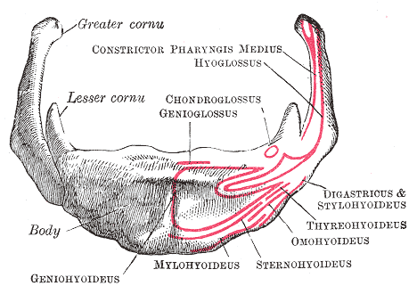
The hyoid-bone (lingual-bone or tongue-bone) () is a horseshoe-shaped bone situated in the anterior midline of the neck between the chin and the thyroid-cartilage. At rest, it lies between the base of the mandible and the third cervical vertebra. Unlike other bones, the hyoid is only distantly articulated to other bones by muscles or ligaments. It is the only bone in the human body that is not connected to any other bones. The hyoid is anchored by muscles from the anterior, posterior and inferior directions, and aids in tongue movement and swallowing. The hyoid bone provides attachment to the muscles of the floor of the mouth and the tongue above, the larynx below, and the epiglottis and pharynx behind. Its name is derived . Structure The hyoid bone is classed as an irregular bone and consists of a central part called the body, and two pairs of horns, the greater and lesser horns. Body The body of the hyoid bone is the central part of the hyoid bone. *At the fron ... [...More Info...] [...Related Items...] OR: [Wikipedia] [Google] [Baidu] |
Hypoglossal Nerve
The hypoglossal nerve, also known as the twelfth cranial nerve, cranial nerve XII, or simply CN XII, is a cranial nerve that innervates all the extrinsic and intrinsic muscles of the tongue except for the palatoglossus, which is innervated by the vagus nerve. CN XII is a nerve with a sole motor function. The nerve arises from the hypoglossal nucleus in the medulla as a number of small rootlets, pass through the hypoglossal canal and down through the neck, and eventually passes up again over the tongue muscles it supplies into the tongue. The nerve is involved in controlling tongue movements required for speech and swallowing, including sticking out the tongue and moving it from side to side. Damage to the nerve or the neural pathways which control it can affect the ability of the tongue to move and its appearance, with the most common sources of damage being injury from trauma or surgery, and motor neuron disease. The first recorded description of the nerve was by Her ... [...More Info...] [...Related Items...] OR: [Wikipedia] [Google] [Baidu] |
Cervical Spinal Nerve 1
The cervical spinal nerve 1 (C1) is a spinal nerve of the cervical segment. from spinalcordinjuryzone.com. Published February 23, 2004 Archived Dec 23, 2011. Retrieved June 12, 2018. C1 carries predominantly motor fibres, but also a small meningeal branch that supplies sensation to parts of the dura around the foramen magnum (via dorsal rami). It originates from the spinal column from above the (C1). [...More Info...] [...Related Items...] OR: [Wikipedia] [Google] [Baidu] |
Greater Cornu
The hyoid-bone (lingual-bone or tongue-bone) () is a horseshoe-shaped bone situated in the anterior midline of the neck between the chin and the thyroid-cartilage. At rest, it lies between the base of the mandible and the third cervical vertebra. Unlike other bones, the hyoid is only distantly articulated to other bones by muscles or ligaments. It is the only bone in the human body that is not connected to any other bones. The hyoid is anchored by muscles from the anterior, posterior and inferior directions, and aids in tongue movement and swallowing. The hyoid bone provides attachment to the muscles of the floor of the mouth and the tongue above, the larynx below, and the epiglottis and pharynx behind. Its name is derived . Structure The hyoid bone is classed as an irregular bone and consists of a central part called the body, and two pairs of horns, the greater and lesser horns. Body The body of the hyoid bone is the central part of the hyoid bone. *At the front, ... [...More Info...] [...Related Items...] OR: [Wikipedia] [Google] [Baidu] |


