|
Posterior Perforated Substance
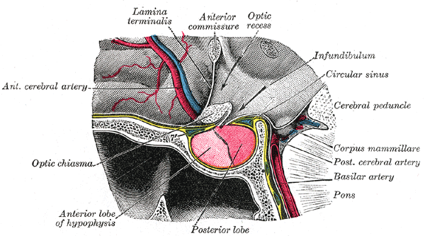
The posterior perforated substance (PPS) is a layer of gray matter which is pierced by small apertures for the transmission of blood vessels. Its inferior part lies on the ventral aspect of the medial portions of the tegmenta and contains the interpeduncular nucleus; its superior part forms part of the floor of the third ventricle. The PPS is situated between the two cerebral peduncles in the midbrain. and posterior to the two mammillary bodies. It is perforated by the posteromedial central arteries – branches of the posterior cerebral arteries en route to the thalamus and basal ganglia. See also * Anterior perforated substance The anterior perforated substance is a part of the brain. It is bilateral. It is irregular and quadrilateral. It lies in front of the optic tract and behind the olfactory trigone. Structure The anterior perforated substance is bilateral. It ... Additional images File:Human brainstem anterior view 2 description.JPG, Human brainstem anterior v ... [...More Info...] [...Related Items...] OR: [Wikipedia] [Google] [Baidu] |
Brain
The brain is an organ (biology), organ that serves as the center of the nervous system in all vertebrate and most invertebrate animals. It consists of nervous tissue and is typically located in the head (cephalization), usually near organs for special senses such as visual perception, vision, hearing, and olfaction. Being the most specialized organ, it is responsible for receiving information from the sensory nervous system, processing that information (thought, cognition, and intelligence) and the coordination of motor control (muscle activity and endocrine system). While invertebrate brains arise from paired segmental ganglia (each of which is only responsible for the respective segmentation (biology), body segment) of the ventral nerve cord, vertebrate brains develop axially from the midline dorsal nerve cord as a brain vesicle, vesicular enlargement at the rostral (anatomical term), rostral end of the neural tube, with centralized control over all body segments. All vertebr ... [...More Info...] [...Related Items...] OR: [Wikipedia] [Google] [Baidu] |
Pons
The pons (from Latin , "bridge") is part of the brainstem that in humans and other mammals, lies inferior to the midbrain, superior to the medulla oblongata and anterior to the cerebellum. The pons is also called the pons Varolii ("bridge of Varolius"), after the Italian anatomist and surgeon Costanzo Varolio (1543–75). This region of the brainstem includes neural pathways and tracts that conduct signals from the brain down to the cerebellum and medulla, and tracts that carry the sensory signals up into the thalamus. Structure The pons in humans measures about in length. It is the part of the brainstem situated between the midbrain and the medulla oblongata. The horizontal ''medullopontine sulcus'' demarcates the boundary between the pons and medulla oblongata on the ventral aspect of the brainstem, and the roots of cranial nerves VI/VII/VIII emerge from the brainstem along this groove. The junction of pons, medulla oblongata, and cerebellum forms the cerebellopontine ... [...More Info...] [...Related Items...] OR: [Wikipedia] [Google] [Baidu] |
Blood Vessels
Blood vessels are the tubular structures of a circulatory system that transport blood throughout many animals’ bodies. Blood vessels transport blood cells, nutrients, and oxygen to most of the tissues of a body. They also take waste and carbon dioxide away from the tissues. Some tissues such as cartilage, epithelium, and the lens and cornea of the eye are not supplied with blood vessels and are termed ''avascular''. There are five types of blood vessels: the arteries, which carry the blood away from the heart; the arterioles; the capillaries, where the exchange of water and chemicals between the blood and the tissues occurs; the venules; and the veins, which carry blood from the capillaries back towards the heart. The word ''vascular'', is derived from the Latin ''vas'', meaning ''vessel'', and is mostly used in relation to blood vessels. Etymology * artery – late Middle English; from Latin ''arteria'', from Greek ''artēria'', probably from ''airein'' ("raise ... [...More Info...] [...Related Items...] OR: [Wikipedia] [Google] [Baidu] |
Ventral
Standard anatomical terms of location are used to describe unambiguously the anatomy of humans and other animals. The terms, typically derived from Latin or Greek roots, describe something in its standard anatomical position. This position provides a definition of what is at the front ("anterior"), behind ("posterior") and so on. As part of defining and describing terms, the body is described through the use of anatomical planes and axes. The meaning of terms that are used can change depending on whether a vertebrate is a biped or a quadruped, due to the difference in the neuraxis, or if an invertebrate is a non-bilaterian. A non-bilaterian has no anterior or posterior surface for example but can still have a descriptor used such as proximal or distal in relation to a body part that is nearest to, or furthest from its middle. International organisations have determined vocabularies that are often used as standards for subdisciplines of anatomy. For example, '' Terminolog ... [...More Info...] [...Related Items...] OR: [Wikipedia] [Google] [Baidu] |
Tegmenta
The midbrain is anatomically delineated into the tectum (roof) and the tegmentum (floor). The midbrain tegmentum extends from the substantia nigra to the cerebral aqueduct in a horizontal section of the midbrain. It forms the floor of the midbrain that surrounds below the cerebral aqueduct as well as the floor of the fourth ventricle while the midbrain tectum forms the roof of the fourth ventricle. The tegmentum contains a collection of tracts and nuclei with movement-related, species-specific, and pain-perception functions. The general structures of midbrain tegmentum include red nucleus and the periaqueductal grey matter. Unlike the midbrain tectum (which is a sensory structure located posteriorly), the midbrain tegmentum, which locates anteriorly, is related to a number of motor functions. Within the tegmentum, the red nucleus is in charge of motor coordination (specifically for limb movements) and the periaqueductal gray matter (PAG) contains critical circuits for modulating ... [...More Info...] [...Related Items...] OR: [Wikipedia] [Google] [Baidu] |
Interpeduncular Nucleus
The interpeduncular nucleus (IPN) is an unpaired, ovoid group of neurons at the base of the midbrain tegmentum. In the midbrain it lies below the interpeduncular fossa. As the name suggests, the interpeduncular nucleus lies ''in between'' the cerebral peduncles. Composition The Interpeduncular nucleus is primarily GABAergic and contains at least two neuron clusters of different morphologies. The region is divided into 7 paired and unpaired subnuclei Subdivisions The presence of non-homologous subdivisions of the Interpeduncular nucleus was first noticed by Santiago Ramón y Cajal, Cajal over a hundred years ago. The currently recognized standard subdivision notation was mostly established by Hammill and Lenn in 1984 by combining the work and notations of four groups. Although most of their proposed convention stuck, at some point the proposed "rostral lateral" sub-nucleus was renamed "dorsomedial" and became immortalized in brain atlases. * Apical sub-nucleus (IPA) Unpaired sub-nu ... [...More Info...] [...Related Items...] OR: [Wikipedia] [Google] [Baidu] |
Third Ventricle
The third ventricle is one of the four connected cerebral ventricles of the ventricular system within the mammalian brain. It is a slit-like cavity formed in the diencephalon between the two thalami, in the midline between the right and left lateral ventricles, and is filled with cerebrospinal fluid (CSF). Running through the third ventricle is the interthalamic adhesion, which contains thalamic neurons and fibers that may connect the two thalami. Structure The third ventricle is a narrow, laterally flattened, vaguely rectangular region, filled with cerebrospinal fluid, and lined by ependyma. It is connected at the superior anterior corner to the lateral ventricles, by the interventricular foramina, and becomes the cerebral aqueduct (''aqueduct of Sylvius'') at the posterior caudal corner. Since the interventricular foramina are on the lateral edge, the corner of the third ventricle itself forms a bulb, known as the ''anterior recess'' (it is also known as the ''bulb ... [...More Info...] [...Related Items...] OR: [Wikipedia] [Google] [Baidu] |
Cerebral Peduncle
The cerebral peduncles (In Latin, ''ped-'' means 'foot'.) are the two stalks that attach the cerebrum to the brainstem. They are structures at the front of the midbrain which arise from the ventral pons and contain the large ascending (sensory) and descending (motor) tracts that run to and from the cerebrum from the pons. Mainly, the three common areas that give rise to the cerebral peduncles are the cerebral cortex, the spinal cord and the cerebellum. The region includes the tegmentum, crus cerebri and pretectum. By this definition, the cerebral peduncles are also known as the basis pedunculi, while the large ventral bundle of efferent fibers is referred to as the cerebral crus (''crus'' means ‘leg’ in Latin.) or the pes pedunculi (''pes'' means 'foot' in Latin.). The cerebral peduncles are located on either side of the midbrain and are the frontmost part of the midbrain, and act as the connectors between the rest of the midbrain and the thalamic nuclei and thus the cer ... [...More Info...] [...Related Items...] OR: [Wikipedia] [Google] [Baidu] |
Midbrain
The midbrain or mesencephalon is the uppermost portion of the brainstem connecting the diencephalon and cerebrum with the pons. It consists of the cerebral peduncles, tegmentum, and tectum. It is functionally associated with vision, hearing, motor control, sleep and wakefulness, arousal (alertness), and temperature regulation.Breedlove, Watson, & Rosenzweig. Biological Psychology, 6th Edition, 2010, pp. 45-46 The name ''mesencephalon'' comes from the Greek ''mesos'', "middle", and ''enkephalos'', "brain". Structure The midbrain is the shortest segment of the brainstem, measuring less than 2cm in length. It is situated mostly in the posterior cranial fossa, with its superior part extending above the tentorial notch. The principal regions of the midbrain are the tectum, the cerebral aqueduct, tegmentum, and the cerebral peduncles. Rostral and caudal, Rostrally the midbrain adjoins the diencephalon (thalamus, hypothalamus, etc.), while Rostral and caudal, cau ... [...More Info...] [...Related Items...] OR: [Wikipedia] [Google] [Baidu] |
Mammillary Bodies
The mammillary bodies also mamillary bodies, are a pair of small round brainstem nuclei. They are located on the undersurface of the brain that, as part of the diencephalon, form part of the limbic system. They are located at the ends of the anterior arches of the fornix. They consist of two groups of nuclei, the medial mammillary nuclei and the lateral mammillary nuclei. Neuroanatomists have often categorized the mammillary bodies as part of the posterior part of hypothalamus. Structure Connections They are connected to other parts of the brain (as shown in the schematic, below left), and act as a relay for impulses coming from the amygdalae and hippocampi, via the mamillothalamic tract to the thalamus. The lateral mammillary nucleus has bidirectional connections with the dorsal tegmental nucleus. The medial mammillary nucleus connects with the ventral tegmental nucleus. Function File:Slide5dd.JPG, Mammillary body Mammillary bodies, and their projections to th ... [...More Info...] [...Related Items...] OR: [Wikipedia] [Google] [Baidu] |
Posteromedial Central Arteries
Central arteries (or perforating or ganglionic arteries) of the brain are numerous small arteries branching from the Circle of Willis, and adjacent arteries that often enter the substance of the brain through the anterior and posterior perforated substances. They supply structures of the base of the brain and internal structures of the cerebral hemispheres. They are separated into four principal groups: anteromedial central arteries; anterolateral central arteries (lenticulostriate arteries); posteromedial central arteries (paramedian arteries); and posterolateral central arteries. Anteromedial central arteries Anteromedial central arteries (also anteromedial perforating arteries, or anteromedial ganglionic arteries) are arteries that arise from the anterior cerebral artery and anterior communicating artery, and pass into the substance of the cerebral hemispheres through the (medial portion of) the anterior perforated substance to supply the optic chiasm, ( anterior nucleus, preop ... [...More Info...] [...Related Items...] OR: [Wikipedia] [Google] [Baidu] |
Posterior Cerebral Arteries
The posterior cerebral artery (PCA) is one of a pair of cerebral arteries that supply oxygenated blood to the occipital lobe, as well as the medial and inferior aspects of the temporal lobe of the human brain. The two arteries originate from the distal end of the basilar artery, where it bifurcates into the left and right posterior cerebral arteries. These anastomose with the middle cerebral arteries and internal carotid arteries via the posterior communicating arteries. Structure The posterior cerebral artery is subdivided into 4 segments: P1: pre-communicating segment * Originated at the termination of the basilar artery * May give rise to the artery of Percheron if present P2: post-communicating segment * From the PCOM around the midbrain * Terminates as it enters the quadrigeminal ganglion * Gives rise to the choroidal branches (medial and lateral posterior choroidal arteries) P3: quadrigeminal segment * Courses posteromedially through the quadrigeminal cistern * ... [...More Info...] [...Related Items...] OR: [Wikipedia] [Google] [Baidu] |


