|
Mandibular Incisive Canal
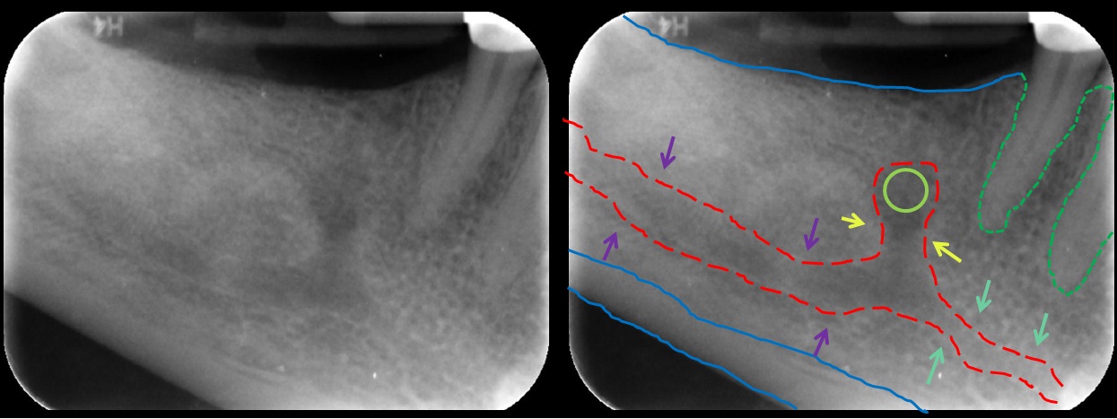
The mandibular incisive canal is a bony canal within the anterior mandible that runs bilaterally from the mental foramina usually to the region of the ipsilateral lateral incisor teeth. After branching into the mental nerve that exits the foramen of the same name, the inferior alveolar nerve continues anteriorly within the mandibular incisive canal as the incisive nerve, providing innervation to the mandibular first premolar, canine Canine may refer to: Zoology and anatomy * a dog-like Canid animal in the subfamily Caninae ** '' Canis'', a genus including dogs, wolves, coyotes, and jackals ** Dog, the domestic dog * Canine tooth, in mammalian oral anatomy People with the ... and lateral and central incisors.Greenstein, G; Cavallaro, J; Tarnow, D. "Practical Application of Anatomy for the Dental Implant Surgeon," ''J Perio'' 2008;79:1833-1846 The mandibular incisive nerve either terminates as nerve endings within the anterior teeth or adjacent bone, or may join nerve endi ... [...More Info...] [...Related Items...] OR: [Wikipedia] [Google] [Baidu] |
Mandibular Canal
In human anatomy, the mandibular canal is a canal within the mandible that contains the inferior alveolar nerve, inferior alveolar artery, and inferior alveolar vein. It runs obliquely downward and forward in the ramus, and then horizontally forward in the body, where it is placed under the alveoli and communicates with them by small openings. On arriving at the incisor teeth, it turns back to communicate with the mental foramen, giving off a small canal known as the mandibular incisive canal, which run to the cavities containing the incisor teeth. It carries branches of the inferior alveolar nerve and artery. It is continuous with the mental foramen (which opens onto front of mandible) and mandibular foramen (on medial aspect of ramus). Variations The mandibular canal is fairly close to the apices of the second molar in 50% of the radiographs. In 40%, canal is away from the root apices, and in only 10% of the radiographs the root apices appeared to penetrate the ca ... [...More Info...] [...Related Items...] OR: [Wikipedia] [Google] [Baidu] |
Mental Foramen
The mental foramen is one of two foramina (openings) located on the anterior surface of the mandible. It is part of the mandibular canal. It transmits the terminal branches of the inferior alveolar nerve and the mental vessels. Structure The mental foramen is located on the anterior surface of the mandible. It is directly below the commisure of the lips, and the tendon of depressor labii inferioris muscle. It is at the end of the mandibular canal, which begins at the mandibular foramen on the posterior surface of the mandible. It transmits the terminal branches of the inferior alveolar nerve (the mental nerve), the mental artery, and the mental vein. Variation The mental foramen descends slightly in toothless individuals. The mental foramen is in line with the longitudinal axis of the 2nd premolar in 63% of people. It generally lies at the level of the vestibular fornix and about a finger's breadth above the inferior border of the mandible. In the general populat ... [...More Info...] [...Related Items...] OR: [Wikipedia] [Google] [Baidu] |
Bone
A bone is a rigid organ that constitutes part of the skeleton in most vertebrate animals. Bones protect the various other organs of the body, produce red and white blood cells, store minerals, provide structure and support for the body, and enable mobility. Bones come in a variety of shapes and sizes and have complex internal and external structures. They are lightweight yet strong and hard and serve multiple functions. Bone tissue (osseous tissue), which is also called bone in the uncountable sense of that word, is hard tissue, a type of specialized connective tissue. It has a honeycomb-like matrix internally, which helps to give the bone rigidity. Bone tissue is made up of different types of bone cells. Osteoblasts and osteocytes are involved in the formation and mineralization of bone; osteoclasts are involved in the resorption of bone tissue. Modified (flattened) osteoblasts become the lining cells that form a protective layer on the bone surface. The mineralize ... [...More Info...] [...Related Items...] OR: [Wikipedia] [Google] [Baidu] |
Human Mandible
In anatomy, the mandible, lower jaw or jawbone is the largest, strongest and lowest bone in the human facial skeleton. It forms the lower jaw and holds the lower teeth in place. The mandible sits beneath the maxilla. It is the only movable bone of the skull (discounting the ossicles of the middle ear). It is connected to the temporal bones by the temporomandibular joints. The bone is formed in the fetus from a fusion of the left and right mandibular prominences, and the point where these sides join, the mandibular symphysis, is still visible as a faint ridge in the midline. Like other symphyses in the body, this is a midline articulation where the bones are joined by fibrocartilage, but this articulation fuses together in early childhood.Illustrated Anatomy of the Head and Neck, Fehrenbach and Herring, Elsevier, 2012, p. 59 The word "mandible" derives from the Latin word ''mandibula'', "jawbone" (literally "one used for chewing"), from '' mandere'' "to chew" and ''-bula' ... [...More Info...] [...Related Items...] OR: [Wikipedia] [Google] [Baidu] |
Bilateral Symmetry
Symmetry in biology refers to the symmetry observed in organisms, including plants, animals, fungi A fungus (plural, : fungi or funguses) is any member of the group of Eukaryote, eukaryotic organisms that includes microorganisms such as yeasts and Mold (fungus), molds, as well as the more familiar mushrooms. These organisms are classified ..., and bacteria. External symmetry can be easily seen by just looking at an organism. For example, take the face of a human being which has a plane of symmetry down its centre, or a pine cone with a clear symmetrical spiral pattern. Internal features can also show symmetry, for example the tubes in the human body (responsible for transporting gases, nutrients, and waste products) which are cylindrical and have several planes of symmetry. Biological symmetry can be thought of as a balanced distribution of duplicate body parts or shapes within the body of an organism. Importantly, unlike in mathematics, symmetry in biology is always ... [...More Info...] [...Related Items...] OR: [Wikipedia] [Google] [Baidu] |
Ipsilateral
Standard anatomical terms of location are used to unambiguously describe the anatomy of animals, including humans. The terms, typically derived from Latin or Greek roots, describe something in its standard anatomical position. This position provides a definition of what is at the front ("anterior"), behind ("posterior") and so on. As part of defining and describing terms, the body is described through the use of anatomical planes and anatomical axes. The meaning of terms that are used can change depending on whether an organism is bipedal or quadrupedal. Additionally, for some animals such as invertebrates, some terms may not have any meaning at all; for example, an animal that is radially symmetrical will have no anterior surface, but can still have a description that a part is close to the middle ("proximal") or further from the middle ("distal"). International organisations have determined vocabularies that are often used as standard vocabularies for subdisciplines of anatom ... [...More Info...] [...Related Items...] OR: [Wikipedia] [Google] [Baidu] |
Lateral Incisor
Incisors (from Latin ''incidere'', "to cut") are the front teeth present in most mammals. They are located in the premaxilla above and on the mandible below. Humans have a total of eight (two on each side, top and bottom). Opossums have 18, whereas armadillos have none. Structure Adult humans normally have eight incisors, two of each type. The types of incisor are: * maxillary central incisor (upper jaw, closest to the center of the lips) * maxillary lateral incisor (upper jaw, beside the maxillary central incisor) * mandibular central incisor (lower jaw, closest to the center of the lips) * mandibular lateral incisor (lower jaw, beside the mandibular central incisor) Children with a full set of deciduous teeth (primary teeth) also have eight incisors, named the same way as in permanent teeth. Young children may have from zero to eight incisors depending on the stage of their tooth eruption and tooth development. Typically, the mandibular central incisors erupt first ... [...More Info...] [...Related Items...] OR: [Wikipedia] [Google] [Baidu] |
Mental Nerve
The mental nerve is a sensory nerve of the face. It is a branch of the posterior trunk of the inferior alveolar nerve, itself a branch of the mandibular nerve (CN V3), itself a branch of the trigeminal nerve (CN V). It provides sensation to the front of the chin and the lower lip, as well as the gums of the anterior mandibular (lower) teeth. It can be blocked with local anaesthesia for procedures on the chin, lower lip, and mucous membrane of the inner cheek. Problems with the nerve cause chin numbness. Structure The mental nerve is a branch of the posterior trunk of the inferior alveolar nerve. This is a branch of the mandibular nerve (CN V3), itself a branch of the trigeminal nerve (CN V). It emerges from the mental foramen in the mandible. It divides into three branches beneath the depressor anguli oris muscle. One branch descends to the skin of the chin. Two branches ascend to the skin and mucous membrane of the lower lip. These branches communicate freely with the faci ... [...More Info...] [...Related Items...] OR: [Wikipedia] [Google] [Baidu] |
Inferior Alveolar Nerve
The inferior alveolar nerve (IAN) (also the inferior dental nerve) is a branch of the mandibular nerve, which is itself the third branch of the trigeminal nerve. The inferior alveolar nerves supply sensation to the lower teeth. Structure The inferior alveolar nerve is a branch of the mandibular nerve. After branching from the mandibular nerve, the inferior alveolar nerve travels behind the lateral pterygoid muscle. It gives off a branch, the mylohyoid nerve, and then enters the mandibular foramen. While in the mandibular canal within the mandible, it supplies the lower teeth (molars and second premolar) with sensory branches that form into the inferior dental plexus and give off small gingival and dental nerves to the teeth. Anteriorly, the nerve gives off the mental nerve at about the level of the mandibular 2nd premolars, which exits the mandible via the mental foramen and supplies sensory branches to the chin and lower lip. The inferior alveolar nerve continues anterior ... [...More Info...] [...Related Items...] OR: [Wikipedia] [Google] [Baidu] |
Premolar
The premolars, also called premolar teeth, or bicuspids, are transitional teeth located between the canine and molar teeth. In humans, there are two premolars per quadrant in the permanent set of teeth, making eight premolars total in the mouth. They have at least two cusps. Premolars can be considered transitional teeth during chewing, or mastication. They have properties of both the canines, that lie anterior and molars that lie posterior, and so food can be transferred from the canines to the premolars and finally to the molars for grinding, instead of directly from the canines to the molars. Human anatomy The premolars in humans are the maxillary first premolar, maxillary second premolar, mandibular first premolar, and the mandibular second premolar. Premolar teeth by definition are permanent teeth distal to the canines, preceded by deciduous molars. Morphology There is always one large buccal cusp, especially so in the mandibular first premolar. The lower sec ... [...More Info...] [...Related Items...] OR: [Wikipedia] [Google] [Baidu] |
Canine Tooth
In mammalian oral anatomy, the canine teeth, also called cuspids, dog teeth, or (in the context of the upper jaw) fangs, eye teeth, vampire teeth, or vampire fangs, are the relatively long, pointed teeth. They can appear more flattened however, causing them to resemble incisors and leading them to be called ''incisiform''. They developed and are used primarily for firmly holding food in order to tear it apart, and occasionally as weapons. They are often the largest teeth in a mammal's mouth. Individuals of most species that develop them normally have four, two in the upper jaw and two in the lower, separated within each jaw by incisors; humans and dogs are examples. In most species, canines are the anterior-most teeth in the maxillary bone. The four canines in humans are the two maxillary canines and the two mandibular canines. Details There are generally four canine teeth: two in the upper (maxillary) and two in the lower (mandibular) arch. A canine is placed laterall ... [...More Info...] [...Related Items...] OR: [Wikipedia] [Google] [Baidu] |
Central Incisor
Incisors (from Latin ''incidere'', "to cut") are the front teeth present in most mammals. They are located in the premaxilla above and on the mandible below. Humans have a total of eight (two on each side, top and bottom). Opossums have 18, whereas armadillos have none. Structure Adult humans normally have eight incisors, two of each type. The types of incisor are: * maxillary central incisor (upper jaw, closest to the center of the lips) * maxillary lateral incisor (upper jaw, beside the maxillary central incisor) * mandibular central incisor (lower jaw, closest to the center of the lips) * mandibular lateral incisor (lower jaw, beside the mandibular central incisor) Children with a full set of deciduous teeth (primary teeth) also have eight incisors, named the same way as in permanent teeth. Young children may have from zero to eight incisors depending on the stage of their tooth eruption and tooth development. Typically, the mandibular central incisors erupt first, followe ... [...More Info...] [...Related Items...] OR: [Wikipedia] [Google] [Baidu] |


