|
Inferior Alveolar Nerve
The inferior alveolar nerve (IAN) (also the inferior dental nerve) is a sensory branch of the mandibular nerve (CN V3) (which is itself the third branch of the trigeminal nerve (CN V)). The nerve provides sensory innervation to the lower/mandibular teeth and their corresponding gingiva as well as a small area of the face (via its mental nerve). Structure Origin The inferior alveolar nerve arises from the mandibular nerve. Course After branching from the mandibular nerve, the inferior alveolar nerve passes posterior to the lateral pterygoid muscle. It issues a branch (the mylohyoid nerve) before entering the mandibular foramen to come to pass in the mandibular canal within the mandible. Passing through the canal, it issues sensory branches for the molar and second premolar teeth; the branches first form the inferior dental plexus which then gives off small gingival and dental nerves to these teeth themselves. The nerve terminates distally/anteriorly (near the second ... [...More Info...] [...Related Items...] OR: [Wikipedia] [Google] [Baidu] |
Maxillary Nerve
In neuroanatomy, the maxillary nerve (V) is one of the three branches or divisions of the trigeminal nerve, the fifth (CN V) cranial nerve. It comprises the principal functions of Sense, sensation from the maxilla, nasal cavity, Sinus (anatomy), sinuses, the palate and subsequently that of the mid-face, and is intermediate, both in position and size, between the ophthalmic nerve and the mandibular nerve.Illustrated Anatomy of the Head and Neck, Fehrenbach and Herring, Elsevier, 2012, page 180 Structure It begins at the middle of the trigeminal ganglion as a flattened plexiform band then it passes through the lateral wall of the cavernous sinus. It leaves the skull through the foramen rotundum, where it becomes more cylindrical in form, and firmer in texture. After leaving foramen rotundum it gives two branches to the pterygopalatine ganglion. It then crosses the pterygopalatine fossa, inclines lateralward on the back of the maxilla, and enters the orbit through the inferior orb ... [...More Info...] [...Related Items...] OR: [Wikipedia] [Google] [Baidu] |
Mandibular Incisive Canal
The mandibular incisive canal is a bilaterally paired bony canal within the anterior portion of the mandible that extends from the mental foramen (usually) to near the ipsilateral lateral incisor teeth. The inferior alveolar nerve splits into its two terminal branches within the mandibular canal: the mental nerve (which exits the mandible through the mental foramen), and the incisive nerve which represents an anterior continuation of the inferior alveolar nerve and continues to course within the mandible in the mandibular incisive canal. The incisive nerve provides innervation to the mandibular first premolar The premolars, also called premolar Tooth (human), teeth, or bicuspids, are transitional teeth located between the Canine tooth, canine and Molar (tooth), molar teeth. In humans, there are two premolars per dental terminology#Quadrant, quadrant in ..., canine and lateral and central incisors.Greenstein, G; Cavallaro, J; Tarnow, D. "Practical Application of Anatomy f ... [...More Info...] [...Related Items...] OR: [Wikipedia] [Google] [Baidu] |
Nonsteroidal Anti-inflammatory Drug
Non-steroidal anti-inflammatory drugs (NSAID) are members of a Indication (medicine), therapeutic drug class which Analgesic, reduces pain, Anti-inflammatory, decreases inflammation, Antipyretic, decreases fever, and Antithrombotic, prevents blood clots. Side effects depend on the specific drug, its dose and duration of use, but largely include an increased risk of Stomach ulcers, gastrointestinal ulcers and bleeds, heart attack, and kidney disease. The term ''non-steroidal'', common from around 1960, distinguishes these drugs from corticosteroids, another class of anti-inflammatory drugs, which during the 1950s had acquired a bad reputation due to overuse and side-effect problems after their introduction in 1948. NSAIDs work by inhibiting the activity of cyclooxygenase enzymes (the COX-1 and COX-2 isozyme, isoenzymes). In cells, these enzymes are involved in the synthesis of key biological mediators, namely prostaglandins, which are involved in inflammation, and thromboxanes, ... [...More Info...] [...Related Items...] OR: [Wikipedia] [Google] [Baidu] |
Lower Lip
The lips are a horizontal pair of soft appendages attached to the jaws and are the most visible part of the mouth of many animals, including humans. Mammal lips are soft, movable and serve to facilitate the ingestion of food (e.g. suckling and gulping) and the articulation of sound and speech. Human lips are also a somatosensory organ, and can be an erogenous zone when used in kissing and other acts of intimacy. Structure The upper and lower lips are referred to as the ''labium superius oris'' and ''labium inferius oris'', respectively. The juncture where the lips meet the surrounding skin of the mouth area is the vermilion border, and the typically reddish area within the borders is called the vermilion zone. The vermilion border of the upper lip is known as the Cupid's bow. The fleshy protuberance located in the center of the upper lip is a tubercle known by various terms including the procheilon (also spelled ''prochilon''), the "tuberculum labii superioris", a ... [...More Info...] [...Related Items...] OR: [Wikipedia] [Google] [Baidu] |
Teeth
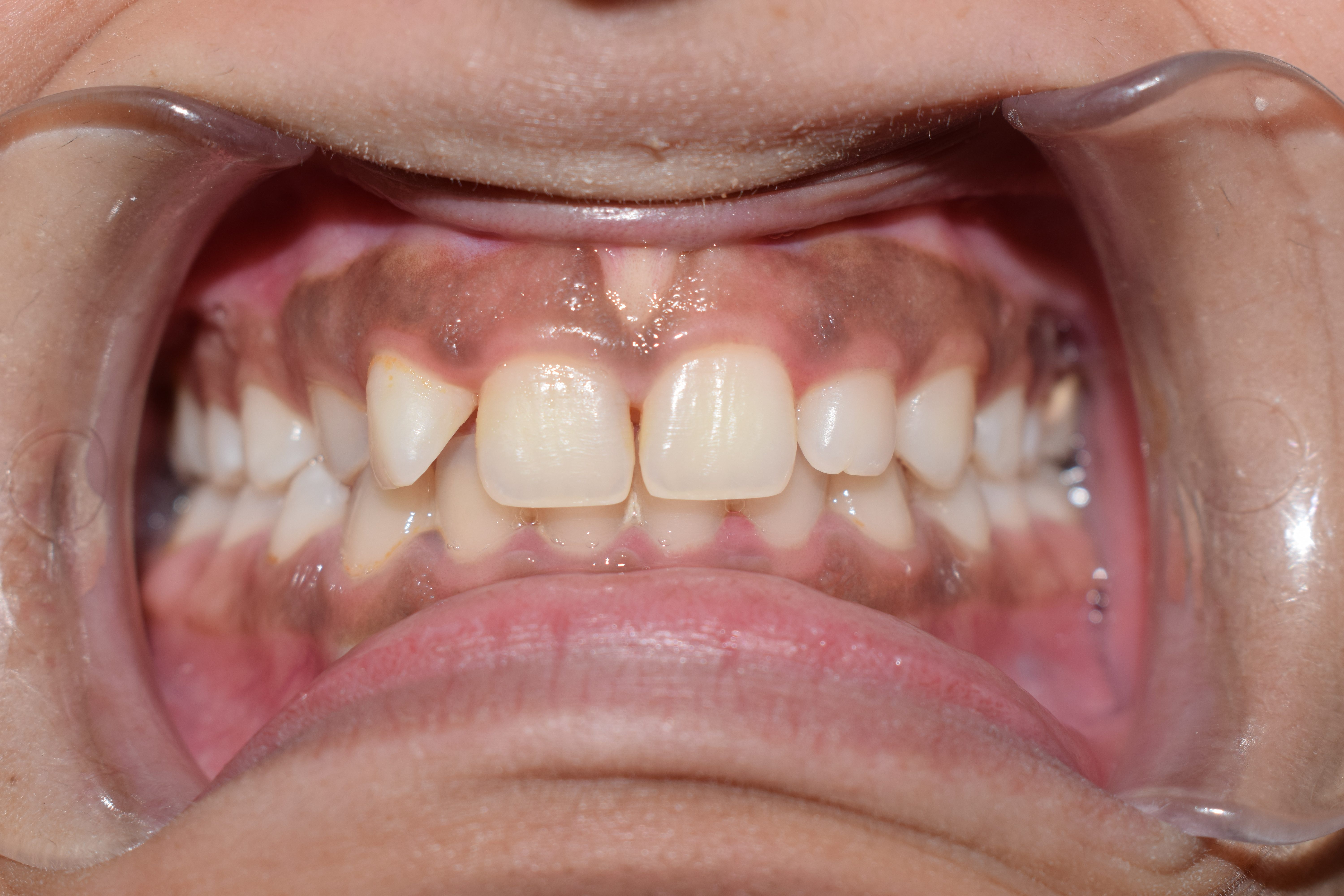
A tooth (: teeth) is a hard, calcified structure found in the jaws (or mouths) of many vertebrates and used to break down food. Some animals, particularly carnivores and omnivores, also use teeth to help with capturing or wounding prey, tearing food, for defensive purposes, to intimidate other animals often including their own, or to carry prey or their young. The roots of teeth are covered by gums. Teeth are not made of bone, but rather of multiple tissues of varying density and hardness that originate from the outermost embryonic germ layer, the ectoderm. The general structure of teeth is similar across the vertebrates, although there is considerable variation in their form and position. The teeth of mammals have deep roots, and this pattern is also found in some fish, and in crocodilians. In most teleost fish, however, the teeth are attached to the outer surface of the bone, while in lizards they are attached to the inner surface of the jaw by one side. In cartilaginous ... [...More Info...] [...Related Items...] OR: [Wikipedia] [Google] [Baidu] |
Tongue
The tongue is a Muscle, muscular organ (anatomy), organ in the mouth of a typical tetrapod. It manipulates food for chewing and swallowing as part of the digestive system, digestive process, and is the primary organ of taste. The tongue's upper surface (dorsum) is covered by taste buds housed in numerous lingual papillae. It is sensitive and kept moist by saliva and is richly supplied with nerves and blood vessels. The tongue also serves as a natural means of cleaning the teeth. A major function of the tongue is to enable speech in humans and animal communication, vocalization in other animals. The human tongue is divided into two parts, an oral cavity, oral part at the front and a pharynx, pharyngeal part at the back. The left and right sides are also separated along most of its length by a vertical section of connective tissue, fibrous tissue (the lingual septum) that results in a groove, the median sulcus, on the tongue's surface. There are two groups of glossal muscles. The f ... [...More Info...] [...Related Items...] OR: [Wikipedia] [Google] [Baidu] |
Nerve Block
Nerve block or regional nerve blockade is any deliberate interruption of signals traveling along a nerve, often for the purpose of pain relief. #Local anesthetic nerve block, Local anesthetic nerve block (sometimes referred to as simply "nerve block") is a short-term block, usually lasting hours or days, involving the injection of an anesthetic, a corticosteroid, and other agents onto or near a nerve. Neurolytic block, the deliberate temporary degeneration of nerve fibers through the application of chemicals, heat, or freezing, produces a block that may persist for weeks, months, or indefinitely. Neurectomy, the cutting through or removal of a nerve or a section of a nerve, usually produces a permanent block. Because neurectomy of a sensory nerve is often followed, months later, by the emergence of new, more intense pain, sensory nerve neurectomy is rarely performed. The concept of nerve block sometimes includes ''central nerve block'', which includes epidural and spinal anaesthe ... [...More Info...] [...Related Items...] OR: [Wikipedia] [Google] [Baidu] |
Lingual Nerve
The lingual nerve carries sensory innervation from the anterior two-thirds of the tongue. It contains fibres from both the mandibular division of the trigeminal nerve (CN V) and from the facial nerve (CN VII). The fibres from the trigeminal nerve are for touch, pain and temperature (general sensation), and the ones from the facial nerve are for taste (special sensation). Structure Origin The lingual nerve arises from the posterior trunk of mandibular nerve (CN V) within the infratemporal fossa. Course The lingual nerve first courses deep to the lateral pterygoid muscle and superior to the tensor veli palatini muscle; while passing between these two muscle, it is joined by the chorda tympani, and often by a communicating branch from the inferior alveolar nerve. The nerve then comes to pass inferoanteriorly upon the medial pterygoid muscle towards the medial aspect of the ramus of mandible, eventually meeting the mandible at the junction of the ramus and body of mandibl ... [...More Info...] [...Related Items...] OR: [Wikipedia] [Google] [Baidu] |
Mandibular Nerve 1
In jawed vertebrates, the mandible (from the Latin ''mandibula'', 'for chewing'), lower jaw, or jawbone is a bone that makes up the lowerand typically more mobilecomponent of the mouth (the upper jaw being known as the maxilla). The jawbone is the skull's only movable, posable bone, sharing joints with the cranium's temporal bones. The mandible hosts the lower teeth (their depth delineated by the alveolar process). Many muscles attach to the bone, which also hosts nerves (some connecting to the teeth) and blood vessels. Amongst other functions, the jawbone is essential for chewing food. Owing to the Neolithic advent of agriculture (), human jaws evolved to be smaller. Although it is the strongest bone of the facial skeleton, the mandible tends to deform in old age; it is also subject to fracturing. Surgery allows for the removal of jawbone fragments (or its entirety) as well as regenerative methods. Additionally, the bone is of great forensic significance. Structure In h ... [...More Info...] [...Related Items...] OR: [Wikipedia] [Google] [Baidu] |
Digastric
The digastric muscle (also digastricus) (named ''digastric'' as it has two 'bellies') is a bilaterally paired suprahyoid muscle located under the jaw. Its posterior belly is attached to the mastoid notch of temporal bone, and its anterior belly is attached to the digastric fossa of mandible; the two bellies are united by an intermediate tendon which is held in a loop that attaches to the hyoid bone. The anterior belly is innervated via the mandibular nerve (cranial nerve V), and the posterior belly is innervated via the facial nerve (cranial nerve VII). It may act to depress the mandible or elevate the hyoid bone. The term "digastric muscle" refers to this specific muscle even though there are other muscles in the body to feature two bellies. Anatomy The digastric muscle consists of two muscular bellies united by an intermediate tendon with the posterior belly longer than the anterior belly. The two bellies of the digastric muscle have different embryological origins - the an ... [...More Info...] [...Related Items...] OR: [Wikipedia] [Google] [Baidu] |
Mylohyoid Muscle
The mylohyoid muscle or diaphragma oris is a paired muscle of the neck. It runs from the Human mandible, mandible to the hyoid bone, forming the floor of the oral cavity of the human mouth, mouth. It is named after its two attachments near the molar (tooth), molar teeth. It forms the floor of the submental triangle. It elevates the hyoid bone and the tongue, important during swallowing and Speech, speaking. Structure The mylohyoid muscle is flat and triangular, and is situated immediately Anatomical terms of location#Superior and inferior, superior to the digastric muscle, anterior belly of the digastric muscle. It is a pharyngeal musculature, pharyngeal muscle (derived from the first pharyngeal arch) and classified as one of the suprahyoid muscles. Together, the paired mylohyoid muscles form a muscular floor for the oral cavity of the human mouth, mouth. The two mylohyoid muscles arise from the mandible at the mylohyoid line, which extends from the mandibular symphysis in front ... [...More Info...] [...Related Items...] OR: [Wikipedia] [Google] [Baidu] |
Gingiva
The gums or gingiva (: gingivae) consist of the mucosal tissue that lies over the mandible and maxilla inside the mouth. Gum health and disease can have an effect on general health. Structure The gums are part of the soft tissue lining of the mouth. They surround the teeth and provide a seal around them. Unlike the soft tissue linings of the lips and cheeks, most of the gums are tightly bound to the underlying bone which helps resist the friction of food passing over them. Thus when healthy, it presents an effective barrier to the barrage of periodontal insults to deeper tissue. Healthy gums are usually coral pink in light skinned people, and may be naturally darker with melanin pigmentation. Changes in color, particularly increased redness, together with swelling and an increased tendency to bleed, suggest an inflammation that is possibly due to the accumulation of bacterial plaque. Overall, the clinical appearance of the tissue reflects the underlying histology, both in hea ... [...More Info...] [...Related Items...] OR: [Wikipedia] [Google] [Baidu] |


