|
Genu Recurvatum
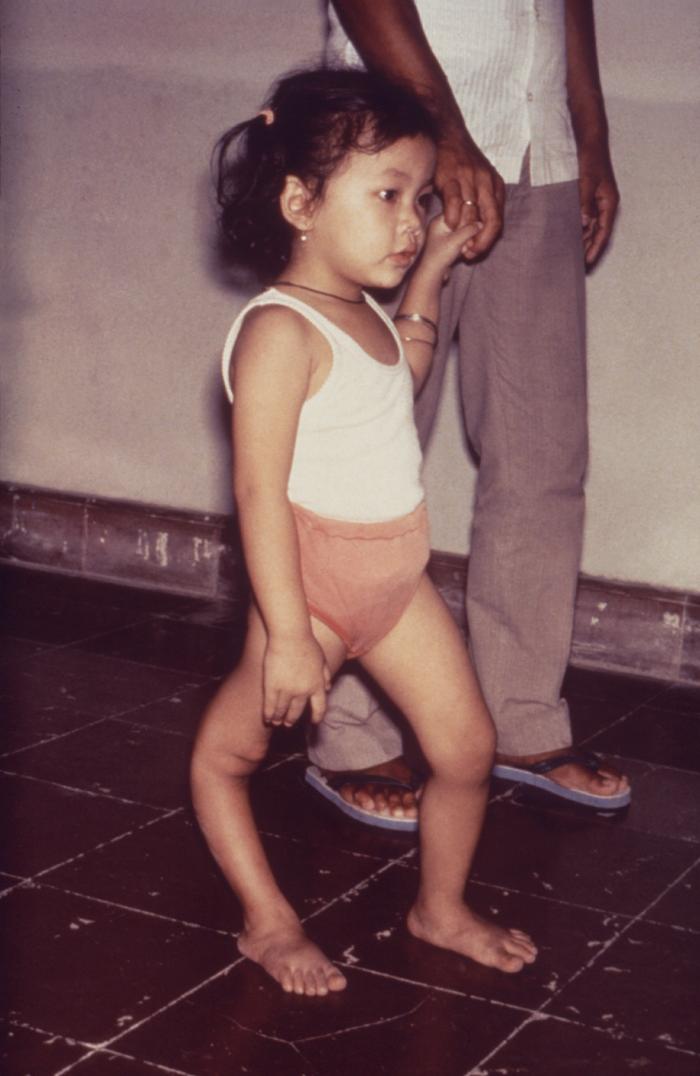
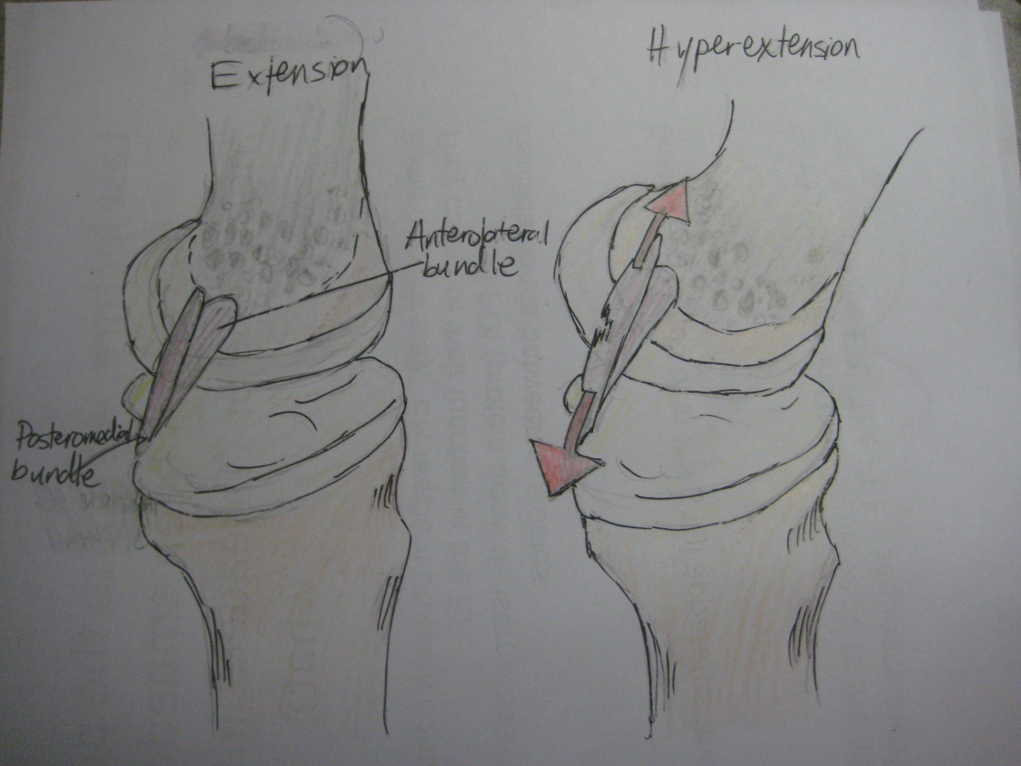
Genu recurvatum is a deformity in the knee joint, so that the knee bends backwards. In this deformity, excessive extension occurs in the tibiofemoral joint. Genu recurvatum is also called knee hyperextension and back knee. This deformity is more common in women and is correlated with men with extremely high testosterone. and people with familial ligamentous laxity. Hyperextension of the knee may be mild, moderate or severe. The normal range of motion (ROM) of the knee joint is from 0 to 135 degrees in an adult. Full knee extension should be no more than 10 degrees. In genu recurvatum, normal extension is increased. The development of genu recurvatum may lead to knee pain and knee osteoarthritis. Causes The following factors may be involved in causing this deformity: * Inherent laxity of the knee ligaments * Weakness of biceps femoris muscle * Instability of the knee joint due to ligaments and joint capsule injuries * Inappropriate alignment of the tibia and femur * Malunion of ... [...More Info...] [...Related Items...] OR: [Wikipedia] [Google] [Baidu] |
Ella Harper
Ella Harper (January 5, 1870 – December 19, 1921), known professionally as The Camel Girl, was born with an extremely rare orthopedic condition that caused her knees to bend backwards, called ''congenital genu recurvatum''. Her preference to walk on all fours resulted in her nickname "Camel Girl". In 1886 she was featured as the star in W. H. Harris's Nickel Plate Circus, appearing in newspapers wherever the circus visited. The back of her pitch card reads: Harper received a $200 per week salary for her appearances (). The money she earned via this role likely afforded her opportunities in life she may not otherwise have had. Harper married a schoolteacher named Robert Savely in 1905; she died in 1921 at the age of fifty-one. She is buried in Spring Hill Cemetery in Nashville, Tennessee Nashville, often known as Music City, is the capital and List of municipalities in Tennessee, most populous city in the U.S. state of Tennessee. It is the county seat, seat of Davidson ... [...More Info...] [...Related Items...] OR: [Wikipedia] [Google] [Baidu] |
Dorsiflexion
Motion, the process of movement, is described using specific anatomical terms. Motion includes movement of organs, joints, limbs, and specific sections of the body. The terminology used describes this motion according to its direction relative to the anatomical position of the body parts involved. Anatomists and others use a unified set of terms to describe most of the movements, although other, more specialized terms are necessary for describing unique movements such as those of the hands, feet, and eyes. In general, motion is classified according to the anatomical plane it occurs in. ''Flexion'' and ''extension'' are examples of ''angular'' motions, in which two axes of a joint are brought closer together or moved further apart. ''Rotational'' motion may occur at other joints, for example the shoulder, and are described as ''internal'' or ''external''. Other terms, such as ''elevation'' and ''depression'', describe movement above or below the horizontal plane. Many anatomica ... [...More Info...] [...Related Items...] OR: [Wikipedia] [Google] [Baidu] |
Joint Capsule
In anatomy, a joint capsule or articular capsule is an envelope surrounding a synovial joint. Each joint capsule has two parts: an outer fibrous layer or membrane, and an inner synovial layer or membrane. Membranes Each capsule consists of two layers or membranes: * an outer (fibrous membrane, ''fibrous stratum'') composed of avascular white fibrous tissue * an inner ('''', ''synovial stratum'') which is a secreting layer On the inside of the capsule, articular cartilage covers the end surfaces of the bones that articulate within ...[...More Info...] [...Related Items...] OR: [Wikipedia] [Google] [Baidu] |
Fibular Collateral Ligament
The lateral collateral ligament (LCL, long external lateral ligament or fibular collateral ligament) is an extrinsic ligament of the knee located on the lateral side of the knee. Its superior attachment is at the lateral epicondyle of the femur (superoposterior to the popliteal groove); its inferior attachment is at the lateral aspect of the head of fibula (anterior to the apex). The LCL is not fused with the joint capsule. Inferiorly, the LCL splits the tendon of insertion of the biceps femoris muscle. Structure The LCL measures some 5 cm in length. It is rounded, and is more narrow and less broad compared to the medial collateral ligament. It extends obliquely inferoposteriorly from its superior attachment to its inferior attachment. In contrast to the medial collateral ligament, it is not fused with either the capsular ligament nor the lateral meniscus. Because of this, the LCL is more flexible than its medial counterpart, and is therefore less susceptible to injury. ... [...More Info...] [...Related Items...] OR: [Wikipedia] [Google] [Baidu] |
Medial Collateral Ligament
The medial collateral ligament (MCL), also called the superficial medial collateral ligament (sMCL) or tibial collateral ligament (TCL), is one of the major ligaments of the knee. It is on the medial (inner) side of the knee joint and occurs in humans and other primates. Its primary function is to resist valgus (inward bending) forces on the knee. Structure It is a broad, flat, membranous band, situated slightly posterior on the medial side of the knee joint. It is attached proximally to the medial epicondyle of the femur, immediately below the adductor tubercle; below to the medial condyle of the tibia and medial surface of its body. It resists forces that would push the knee medially, which would otherwise produce valgus deformity. It provides up to 78% of the restraining force that resists valgus (inward pressing) loads on the knee. The fibers of the posterior part of the ligament are short and incline backward as they descend; they are inserted into the tibia above t ... [...More Info...] [...Related Items...] OR: [Wikipedia] [Google] [Baidu] |
Posterior Cruciate Ligament
The posterior cruciate ligament (PCL) is a ligament in each knee of humans and various other animals. It works as a counterpart to the anterior cruciate ligament (ACL). It connects the posterior intercondylar area of the tibia to the Medial condyle of femur, medial condyle of the femur. This configuration allows the PCL to resist forces pushing the tibia posteriorly relative to the femur. The PCL and ACL are intracapsular ligaments because they lie deep within the knee joint. They are both isolated from the fluid-filled synovial cavity, with the synovial membrane wrapped around them. The PCL gets its name by attaching to the posterior portion of the tibia. The PCL, Anterior cruciate ligament, ACL, medial collateral ligament, MCL, and fibular collateral ligament, LCL are the four main ligaments of the knee in primates. Structure The PCL is located within the knee joint where it stabilizes the articulating bones, particularly the femur and the tibia, during movement. It originates ... [...More Info...] [...Related Items...] OR: [Wikipedia] [Google] [Baidu] |
Anterior Cruciate Ligament
The anterior cruciate ligament (ACL) is one of a pair of cruciate ligaments (the other being the posterior cruciate ligament) in the human knee. The two ligaments are called "cruciform" ligaments, as they are arranged in a crossed formation. In the quadruped stifle joint (analogous to the knee), based on its anatomical position, it is also referred to as the cranial cruciate ligament. The term cruciate is Latin for cross. This name is fitting because the ACL crosses the posterior cruciate ligament to form an "X". It is composed of strong, fibrous material and assists in controlling excessive motion by limiting mobility of the joint. The anterior cruciate ligament is one of the four main ligaments of the knee, providing 85% of the restraining force to anterior tibial displacement at 30 and 90° of knee flexion. The ACL is the most frequently injured ligament in the knee. Structure The ACL originates from deep within the notch of the distal femur. Its proximal fibers fan out alo ... [...More Info...] [...Related Items...] OR: [Wikipedia] [Google] [Baidu] |
Ligaments
A ligament is a type of fibrous connective tissue in the body that connects bones to other bones. It also connects flight feathers to bones, in dinosaurs and birds. All 30,000 species of amniotes (land animals with internal bones) have ligaments. It is also known as ''articular ligament'', ''articular larua'', ''fibrous ligament'', or ''true ligament''. Comparative anatomy Ligaments are similar to tendons and fasciae as they are all made of connective tissue. The differences among them are in the connections that they make: ligaments connect one bone to another bone, tendons connect muscle to bone, and fasciae connect muscles to other muscles. These are all found in the skeletal system of the human body. Ligaments cannot usually be regenerated naturally; however, there are periodontal ligament stem cells located near the periodontal ligament which are involved in the adult regeneration of periodontist ligament. The study of ligaments is known as . Humans Other ligaments ... [...More Info...] [...Related Items...] OR: [Wikipedia] [Google] [Baidu] |
Osteogenesis Imperfecta
Osteogenesis imperfecta (; OI), colloquially known as brittle bone disease, is a group of genetic disorders that all result in bones that bone fracture, break easily. The range of symptoms—on the skeleton as well as on the body's other Organ (biology), organs—may be mild to severe. Symptoms found in various types of OI include sclera, whites of the eye (sclerae) that are blue instead, short stature, joint hypermobility, loose joints, hearing loss, breathing problems and problems with the teeth (dentinogenesis imperfecta). Potentially life-threatening Complication (medicine), complications, all of which become more common in more severe OI, include: tearing (Dissection (medical), dissection) of the major arteries, such as Aortic dissection, the aorta; pulmonary insufficiency, pulmonary valve insufficiency secondary to distortion of the ribcage; and basilar invagination. The underlying mechanism is usually a problem with connective tissue due to a lack of, or poorly forme ... [...More Info...] [...Related Items...] OR: [Wikipedia] [Google] [Baidu] |
Hypermobility (joints)
Hypermobility, also known as double-jointedness, describes joints that stretch farther than normal. For example, some hypermobile people can bend their thumbs backwards to their Wrist, wrists, bend their knee joints backwards, put their leg behind the head, or perform other contortionist "tricks". It can affect one or more joints throughout the body. Hypermobile joints are common and occur in about 10 to 25% of the population. In a minority of people, pain and other symptoms are present. This may be a sign of hypermobility spectrum disorder (HSD). Hypermobile joints are a feature of genetic Connective tissue disease#Heritable connective tissue disorders, connective tissue disorders such as hypermobility spectrum disorder or Ehlers–Danlos syndrome (EDS). Until new diagnostic criteria were introduced, hypermobility syndrome was sometimes considered identical to hypermobile Ehlers–Danlos syndrome (hEDS), formerly called EDS Type 3. As no genetic test can distinguish the two condi ... [...More Info...] [...Related Items...] OR: [Wikipedia] [Google] [Baidu] |
Ehlers–Danlos Syndrome
Ehlers–Danlos syndromes (EDS) is a group of 14 genetic connective-tissue disorders. Symptoms often include loose joints, joint pain, stretchy velvety skin, and abnormal scar formation. These may be noticed at birth or in early childhood. Complications may include aortic dissection, joint dislocations, scoliosis, chronic pain, or early osteoarthritis. The existing classification was last updated in 2017, when a number of rarer forms of EDS were added. EDS occurs due to mutations in one or more particular genes—there are 19 genes that can contribute to the condition. The specific gene affected determines the type of EDS, though the genetic causes of hypermobile Ehlers–Danlos syndrome are still unknown. Some cases result from a new variation occurring during early development, while others are inherited in an autosomal dominant or recessive manner. Typically, these variations result in defects in the structure or processing of the protein collagen or tenascin. Diagnos ... [...More Info...] [...Related Items...] OR: [Wikipedia] [Google] [Baidu] |
Loeys–Dietz Syndrome
Loeys–Dietz syndrome (LDS) is an autosomal dominant genetic connective tissue disorder. It has features similar to Marfan syndrome and Ehlers–Danlos syndrome. The disorder is marked by aneurysms in the aorta, often in children, and the aorta may also undergo sudden dissection in the weakened layers of the wall of the aorta. Aneurysms and dissections also can occur in arteries other than the aorta. Because aneurysms in children tend to rupture early, children are at greater risk for dying if the syndrome is not identified. Surgery to repair aortic aneurysms is essential for treatment. It was previously believed that the life expectancy of an individual with this condition was around 30-40 years of age, however with progressive treatments such as possibilities for surgery and medications like losartan it is proven now that life expectancy can be full age with the correct medical attention and scans. There are five types of the syndrome, designated types I through V, caused by mu ... [...More Info...] [...Related Items...] OR: [Wikipedia] [Google] [Baidu] |


