|
Dual-axis Optical Coherence Tomography
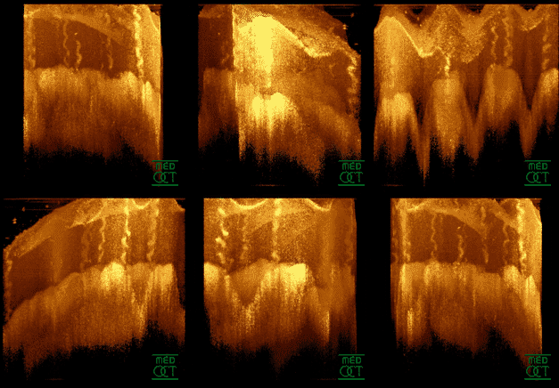
Dual-axis optical coherence tomography (DA-OCT) is an imaging modality that is based on the principles of optical coherence tomography (OCT). These techniques are largely used for medical imaging. OCT is non-invasive and non-contact. It allows for real-time, in situ imaging and provides high image resolution. OCT is analogous to ultrasound but relies on light waves (typically near-infrared), which makes it faster than ultrasound. In general, OCT has proven to be compact and portable. It is compatible with arterial catheters and endoscopes, which helps diagnose diseases within long internal cavities, including the esophagus ( Barrett's disease) and coronary arteries (cardiovascular disease). The biggest limitation with traditional OCT is that it relies on detecting ballistic (non-scattered) photons, which can have a mean free path of only 100 microns, or singly backscattered photons. This strongly restricts depth penetration in highly-scattering biological tissue. It causes unsatis ... [...More Info...] [...Related Items...] OR: [Wikipedia] [Google] [Baidu] |
Optical Coherence Tomography
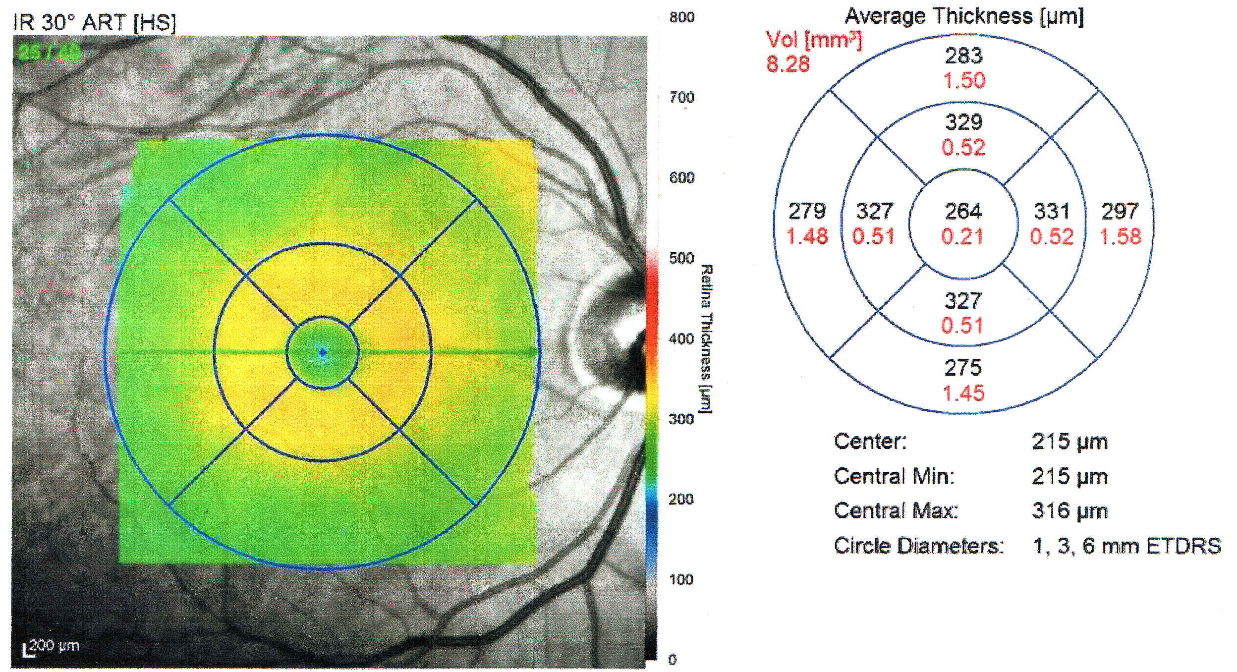
Optical coherence tomography (OCT) is an imaging technique that uses low-coherence light to capture micrometer-resolution, two- and three-dimensional images from within optical scattering media (e.g., biological tissue). It is used for medical imaging and industrial nondestructive testing (NDT). Optical coherence tomography is based on low-coherence interferometry, typically employing near-infrared light. The use of relatively long wavelength light allows it to penetrate into the scattering medium. Confocal microscopy, another optical technique, typically penetrates less deeply into the sample but with higher resolution. Depending on the properties of the light source (superluminescent diodes, ultrashort pulsed lasers, and supercontinuum lasers have been employed), optical coherence tomography has achieved sub-micrometer resolution (with very wide-spectrum sources emitting over a ~100 nm wavelength range). Optical coherence tomography is one of a class of optical tom ... [...More Info...] [...Related Items...] OR: [Wikipedia] [Google] [Baidu] |
Necrosis
Necrosis () is a form of cell injury which results in the premature death of cells in living tissue by autolysis. Necrosis is caused by factors external to the cell or tissue, such as infection, or trauma which result in the unregulated digestion of cell components. In contrast, apoptosis is a naturally occurring programmed and targeted cause of cellular death. While apoptosis often provides beneficial effects to the organism, necrosis is almost always detrimental and can be fatal. Cellular death due to necrosis does not follow the apoptotic signal transduction pathway, but rather various receptors are activated and result in the loss of cell membrane integrity and an uncontrolled release of products of cell death into the extracellular space. This initiates in the surrounding tissue an inflammatory response, which attracts leukocytes and nearby phagocytes which eliminate the dead cells by phagocytosis. However, microbial damaging substances released by leukocytes woul ... [...More Info...] [...Related Items...] OR: [Wikipedia] [Google] [Baidu] |
Multiple Scattering Low Coherence Interferometry
Multiple scattering low coherence interferometry (ms/LCI) is an imaging technique that relies on analyzing multiply scattered light in order to capture depth-resolved images from optical scattering media. With current applications primarily in medical imaging, has the advantage of a higher range since forward scattered light attenuates less with depth when compared to the specular reflection, specularly reflected light that is assessed in more conventional imaging methods such as optical coherence tomography. This allows ms/LCI to image through up to 90 mean free scattering paths, compared to roughly 27 scattering MFPs in OCT and 1–2 scattering MFPs in confocal microscopy. Design Time-domain implementation Early implementations of ms/LCI were in the time domain using lock-in amplifier, lock-in detection in order to take advantage of long scanning depths as well narrow detection bandwidths. As in traditional OCT, the beam wave interference, interference coherence gates the li ... [...More Info...] [...Related Items...] OR: [Wikipedia] [Google] [Baidu] |
Optical Coherence Tomography
Optical coherence tomography (OCT) is an imaging technique that uses low-coherence light to capture micrometer-resolution, two- and three-dimensional images from within optical scattering media (e.g., biological tissue). It is used for medical imaging and industrial nondestructive testing (NDT). Optical coherence tomography is based on low-coherence interferometry, typically employing near-infrared light. The use of relatively long wavelength light allows it to penetrate into the scattering medium. Confocal microscopy, another optical technique, typically penetrates less deeply into the sample but with higher resolution. Depending on the properties of the light source (superluminescent diodes, ultrashort pulsed lasers, and supercontinuum lasers have been employed), optical coherence tomography has achieved sub-micrometer resolution (with very wide-spectrum sources emitting over a ~100 nm wavelength range). Optical coherence tomography is one of a class of optical tom ... [...More Info...] [...Related Items...] OR: [Wikipedia] [Google] [Baidu] |
Medical Imaging
Medical imaging is the technique and process of imaging the interior of a body for clinical analysis and medical intervention, as well as visual representation of the function of some organs or tissues ( physiology). Medical imaging seeks to reveal internal structures hidden by the skin and bones, as well as to diagnose and treat disease. Medical imaging also establishes a database of normal anatomy and physiology to make it possible to identify abnormalities. Although imaging of removed organs and tissues can be performed for medical reasons, such procedures are usually considered part of pathology instead of medical imaging. Measurement and recording techniques that are not primarily designed to produce images, such as electroencephalography (EEG), magnetoencephalography (MEG), electrocardiography (ECG), and others, represent other technologies that produce data susceptible to representation as a parameter graph versus time or maps that contain data about the measurement ... [...More Info...] [...Related Items...] OR: [Wikipedia] [Google] [Baidu] |
Interferometry
Interferometry is a technique which uses the '' interference'' of superimposed waves to extract information. Interferometry typically uses electromagnetic waves and is an important investigative technique in the fields of astronomy, fiber optics, engineering metrology, optical metrology, oceanography, seismology, spectroscopy (and its applications to chemistry), quantum mechanics, nuclear and particle physics, plasma physics, remote sensing, biomolecular interactions, surface profiling, microfluidics, mechanical stress/strain measurement, velocimetry, optometry, and making holograms. Interferometers are devices that extract information from interference. They are widely used in science and industry for the measurement of microscopic displacements, refractive index changes and surface irregularities. In the case with most interferometers, light from a single source is split into two beams that travel in different optical paths, which are then combined again to prod ... [...More Info...] [...Related Items...] OR: [Wikipedia] [Google] [Baidu] |
Ballistic Photon
Ballistic light, also known as ballistic photons, is photons of light that have traveled through a scattering (turbid) medium in a straight line. When pulses of laser light pass through a turbid medium such as fog or body tissue, most of the photons are either scattered or absorbed. However, across short distances, a few photons pass through the scattering medium in straight lines. These coherent photons are referred to as ballistic photons. Photons that are slightly scattered, retaining some degree of coherence, are referred to as snake photons. The aim of ballistic imaging modalities is to efficiently detect ballistic photons that carry useful information, while rejecting non-ballistic photons. To perform this task, specific characteristics of ballistic photons vs. non-ballistic photons are used, such as time of flight through coherence-gated imaging, collimation, wavefront propagation, and polarization. Slightly scattered "quasi-ballistic" photons are often measured as well, t ... [...More Info...] [...Related Items...] OR: [Wikipedia] [Google] [Baidu] |
Angle-resolved Low-coherence Interferometry
Angle-resolved low-coherence interferometry (a/LCI) is an emerging biomedical imaging technology which uses the properties of scattered light to measure the average size of cell structures, including cell nuclei. The technology shows promise as a clinical tool for ''in situ'' detection of dysplastic, or precancerous tissue. Introduction A/LCI combines low-coherence interferometry with angle-resolved scattering to solve the inverse problem of determining scatterer geometry based on far field diffraction patterns. Similar to optical coherence domain reflectometry (OCDR) and optical coherence tomography (OCT), a/LCI uses a broadband light source in an interferometry scheme in order to achieve optical sectioning with a depth resolution set by the coherence length of the source. Angle-resolved scattering measurements capture light as a function of the scattering angle, and invert the angles to deduce the average size of the scattering objects via a computational light scattering m ... [...More Info...] [...Related Items...] OR: [Wikipedia] [Google] [Baidu] |
Hydrogel
A hydrogel is a crosslinked hydrophilic polymer that does not dissolve in water. They are highly absorbent yet maintain well defined structures. These properties underpin several applications, especially in the biomedical area. Many hydrogels are synthetic, but some are derived from nature. The term 'hydrogel' was coined in 1894. Chemistry Classification The crosslinks which bond the polymers of a hydrogel fall under two general categories: physical and chemical. Chemical hydrogels have covalent cross-linking bonds, whereas physical hydrogels have non-covalent bonds. Chemical hydrogels result in strong irreversible gels due to the covalent bonding, and they may also possess harmful properties which makes them unfavourable for medical applications. Physical hydrogels on the other hand have high biocompatibility, aren’t toxic, and are also easily reversible, by simply changing an external stimulus such as pH or temperature; thus they are favourable for use in medical applicat ... [...More Info...] [...Related Items...] OR: [Wikipedia] [Google] [Baidu] |
Medical Imaging
Medical imaging is the technique and process of imaging the interior of a body for clinical analysis and medical intervention, as well as visual representation of the function of some organs or tissues ( physiology). Medical imaging seeks to reveal internal structures hidden by the skin and bones, as well as to diagnose and treat disease. Medical imaging also establishes a database of normal anatomy and physiology to make it possible to identify abnormalities. Although imaging of removed organs and tissues can be performed for medical reasons, such procedures are usually considered part of pathology instead of medical imaging. Measurement and recording techniques that are not primarily designed to produce images, such as electroencephalography (EEG), magnetoencephalography (MEG), electrocardiography (ECG), and others, represent other technologies that produce data susceptible to representation as a parameter graph versus time or maps that contain data about the measurement ... [...More Info...] [...Related Items...] OR: [Wikipedia] [Google] [Baidu] |
MEMS
Microelectromechanical systems (MEMS), also written as micro-electro-mechanical systems (or microelectronic and microelectromechanical systems) and the related micromechatronics and microsystems constitute the technology of microscopic devices, particularly those with moving parts. They merge at the nanoscale into nanoelectromechanical systems (NEMS) and nanotechnology. MEMS are also referred to as micromachines in Japan and microsystem technology (MST) in Europe. MEMS are made up of components between 1 and 100 micrometers in size (i.e., 0.001 to 0.1 mm), and MEMS devices generally range in size from 20 micrometres to a millimetre (i.e., 0.02 to 1.0 mm), although components arranged in arrays (e.g., digital micromirror devices) can be more than 1000 mm2. They usually consist of a central unit that processes data (an integrated circuit chip such as microprocessor) and several components that interact with the surroundings (such as microsensors). Because of th ... [...More Info...] [...Related Items...] OR: [Wikipedia] [Google] [Baidu] |
Michelson Interferometer
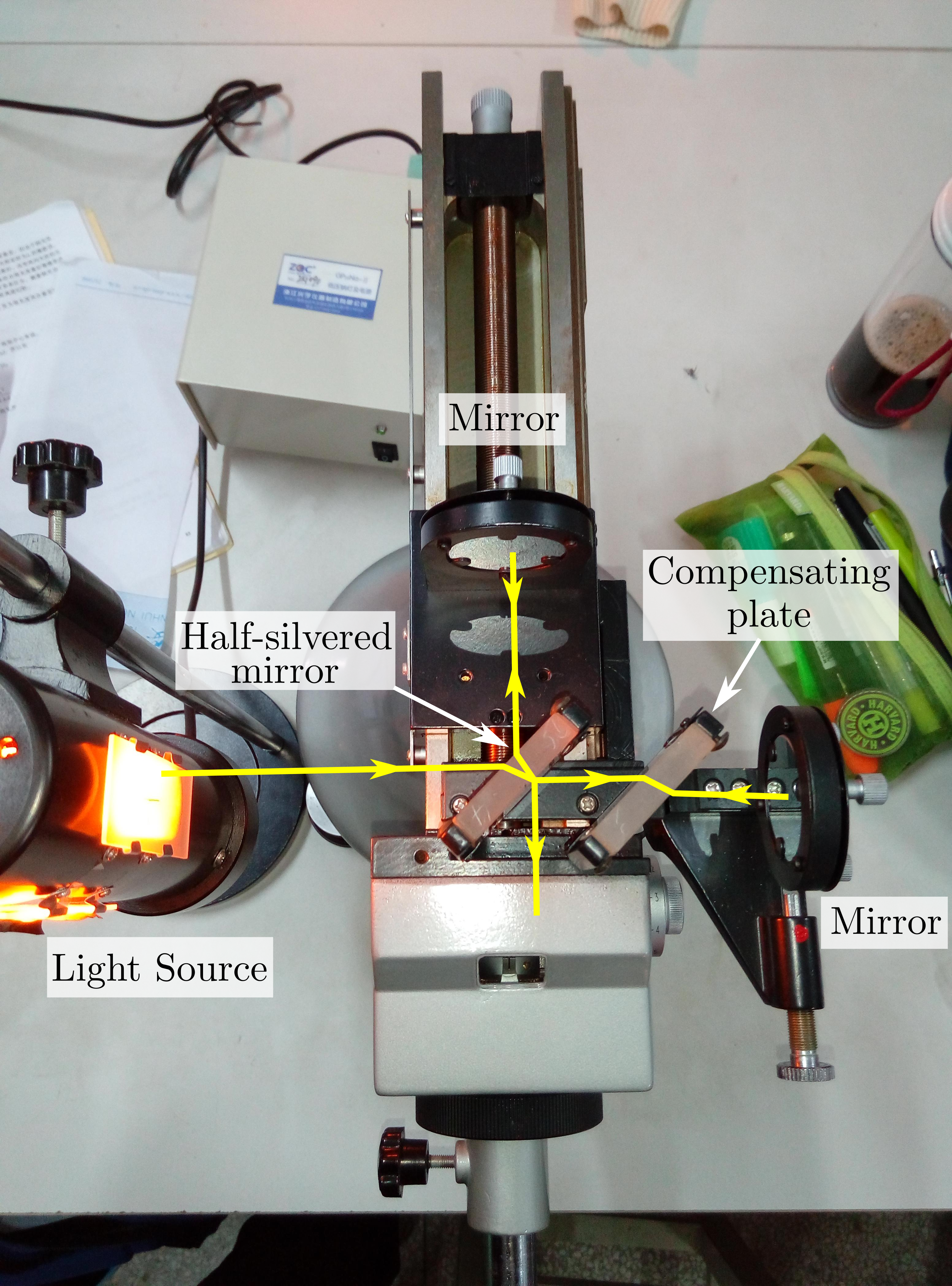
The Michelson interferometer is a common configuration for optical interferometry and was invented by the 19/20th-century American physicist Albert Abraham Michelson. Using a beam splitter, a light source is split into two arms. Each of those light beams is reflected back toward the beamsplitter which then combines their amplitudes using the superposition principle. The resulting interference pattern that is not directed back toward the source is typically directed to some type of photoelectric detector or camera. For different applications of the interferometer, the two light paths can be with different lengths or incorporate optical elements or even materials under test. The Michelson interferometer (among other interferometer configurations) is employed in many scientific experiments and became well known for its use by Michelson and Edward Morley in the famous Michelson–Morley experiment (1887) in a configuration which would have detected the Earth's motion through the ... [...More Info...] [...Related Items...] OR: [Wikipedia] [Google] [Baidu] |


