|
Acute Idiopathic Blind Spot Enlargement Syndrome
Acute idiopathic blind spot enlargement syndrome (AIBSE) is a rare eye disease affecting the retina of the eye. It is basically a type of retinopathy which affects females more than males. Currently there is no treatment for this condition, but, it is usually self limiting. Cause Though exact etiology of AIBSE syndrome is unknown, studies shows that viral illness like influenza and vaccinations like MMR may trigger the condition. Demographics AIBSE syndrome affects females more than males. Higher incidence is seen in Caucasian people. Signs and Symptoms Enlargement of blind spot area in the visual field of the eye is the main sign and acute onset photopsia is the main symptom of AIBSE syndrome. Other symptoms include monocular scotoma and reduced light perception. Diagnosis Diagnostic techniques like ophthalmoscopy, visual field test, optical coherence tomography, fluorescein angiography, multifocal electroretinography and electrophysiology may be used in diagnosing AIBSE syn ... [...More Info...] [...Related Items...] OR: [Wikipedia] [Google] [Baidu] |
Ophthalmology
Ophthalmology (, ) is the branch of medicine that deals with the diagnosis, treatment, and surgery of eye diseases and disorders. An ophthalmologist is a physician who undergoes subspecialty training in medical and surgical eye care. Following a medical degree, a doctor specialising in ophthalmology must pursue additional postgraduate residency training specific to that field. In the United States, following graduation from medical school, one must complete a four-year residency in ophthalmology to become an ophthalmologist. Following residency, additional specialty training (or fellowship) may be sought in a particular aspect of eye pathology. Ophthalmologists prescribe medications to treat ailments, such as eye diseases, implement laser therapy, and perform surgery when needed. Ophthalmologists provide both primary and specialty eye care—medical and surgical. Most ophthalmologists participate in academic research on eye diseases at some point in their training and many inc ... [...More Info...] [...Related Items...] OR: [Wikipedia] [Google] [Baidu] |
Visual Field Test
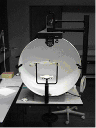
A visual field test is an eye examination that can detect dysfunction in central and peripheral vision which may be caused by various medical conditions such as glaucoma, stroke, pituitary disease, brain tumours or other neurological deficits. Visual field testing can be performed clinically by keeping the subject's gaze fixed while presenting objects at various places within their visual field. Simple manual equipment can be used such as in the tangent screen test or the Amsler grid. When dedicated machinery is used it is called a perimeter. The exam may be performed by a technician in one of several ways. The test may be performed by a technician directly, with the assistance of a machine, or completely by an automated machine. Machine-based tests aid diagnostics by allowing a detailed printout of the patient's visual field. Other names for this test may include perimetry, Tangent screen exam, Automated perimetry exam, Goldmann visual field exam, or brand names such as the H ... [...More Info...] [...Related Items...] OR: [Wikipedia] [Google] [Baidu] |
Disorders Of Choroid And Retina
Disorder may refer to randomness, a lack of intelligible pattern, or: Healthcare * Disorder (medicine), a functional abnormality or disturbance * Mental disorder or psychological disorder, a psychological pattern associated with distress or disability that occurs in a person and is not a part of normal development or culture: :* Anxiety disorder, different forms of abnormal and pathological fear and anxiety :* Attention-deficit hyperactivity disorder :* Autism spectrum disorder :* Conversion disorder, neurological symptoms such as numbness, blindness, paralysis, or fits, where no neurological explanation is possible :* Obsessive–compulsive disorder, an anxiety disorder characterized by repetitive behaviors aimed at reducing anxiety :* Obsessive–compulsive personality disorder, obsession with perfection, rules, and organization :* Personality disorder, an enduring pattern of inner experience and behavior that deviates markedly from the expectations of the culture of the person ... [...More Info...] [...Related Items...] OR: [Wikipedia] [Google] [Baidu] |
Fundus Fluorescein Angiography
Fluorescein angiography (FA), fluorescent angiography (FAG), or fundus fluorescein angiography (FFA) is a technique for examining the circulation of the retina and choroid (parts of the fundus) using a fluorescent dye and a specialized camera. Sodium fluorescein is added into the systemic circulation, the retina is illuminated with blue-green light at a wavelength of 490 nanometers, and an angiogram is obtained by photographing the fluorescent green light that is emitted by the dye. The fluorescein is administered intravenously in intravenous fluorescein angiography (IVFA) and orally in oral fluorescein angiography (OFA). The test is a dye tracing method. The fluorescein dye also reappears in the patient urine, causing the urine to appear darker, and sometimes orange. It can also cause discolouration of the saliva. Fluorescein angiography is one of several health care applications of this dye, all of which have a risk of severe adverse effects. See fluorescein safety in health c ... [...More Info...] [...Related Items...] OR: [Wikipedia] [Google] [Baidu] |
Blind Spot (vision)
A blind spot, scotoma, is an obscuration of the visual field. A particular blind spot known as the ''physiological blind spot'', "blind point", or ''punctum caecum'' in medical literature, is the place in the visual field that corresponds to the lack of light-detecting photoreceptor cells on the optic disc of the retina where the optic nerve passes through the optic disc.Gregory, R., & Cavanagh, P. (2011)"The Blind Spot" Scholarpedia. Retrieved on 2011-05-21. Because there are no cells to detect light on the optic disc, the corresponding part of the field of vision is invisible. Via processes in the brain, the blind spot is interpolated based on surrounding detail and information from the other eye, so it is not normally perceived. Although all vertebrates have this blind spot, cephalopod eyes, which are only superficially similar because they evolved independently, do not. In them, the optic nerve approaches the receptors from behind, so it does not create a break in t ... [...More Info...] [...Related Items...] OR: [Wikipedia] [Google] [Baidu] |
Electrophysiology
Electrophysiology (from [see the Electron#Etymology, etymology of "electron"]; ; and ) is the branch of physiology that studies the electrical properties of biological cell (biology), cells and tissues. It involves measurements of voltage changes or electric current or manipulations on a wide variety of scales from single ion channel proteins to whole organs like the heart. In neuroscience, it includes measurements of the electrical activity of neurons, and, in particular, action potential activity. Recordings of large-scale electric signals from the nervous system, such as electroencephalography, may also be referred to as electrophysiological recordings. They are useful for electrodiagnostic medicine, electrodiagnosis and monitoring (medicine), monitoring. Definition and scope Classical electrophysiological techniques Principle and mechanisms Electrophysiology is the branch of physiology that pertains broadly to the flow of ions (ion current) in biological tissues and, in p ... [...More Info...] [...Related Items...] OR: [Wikipedia] [Google] [Baidu] |
Electroretinography
Electroretinography measures the electrical responses of various cell types in the retina, including the Photoreceptor cell, photoreceptors (rod cell, rods and cone cell, cones), inner retinal cells (Retinal bipolar cell, bipolar and amacrine cell, amacrine cells), and the Retinal ganglion cell, ganglion cells. Electrodes are placed on the surface of the cornea (DTL silver/nylon fiber string or ERG jet) or on the skin beneath the eye (sensor strips) to measure retinal responses. Retinal pigment epithelium (RPE) responses are measured with an Electrooculography, EOG test with skin-contact electrodes placed near the Canthus, canthi. During a recording, the patient's eyes are exposed to standardized stimulus (physiology), stimuli and the resulting signal is displayed showing the time course of the signal's amplitude (voltage). Signals are very small, and typically are measured in microvolts or nanovolts. The ERG is composed of electrical potentials contributed by different cell type ... [...More Info...] [...Related Items...] OR: [Wikipedia] [Google] [Baidu] |
Fluorescein Angiography
Fluorescein angiography (FA), fluorescent angiography (FAG), or fundus fluorescein angiography (FFA) is a technique for examining the circulation of the retina and choroid (parts of the fundus) using a fluorescent dye and a specialized camera. Sodium fluorescein is added into the systemic circulation, the retina is illuminated with blue-green light at a wavelength of 490 nanometers, and an angiogram is obtained by photographing the fluorescent green light that is emitted by the dye. The fluorescein is administered intravenously in intravenous fluorescein angiography (IVFA) and orally in oral fluorescein angiography (OFA). The test is a dye tracing method. The fluorescein dye also reappears in the patient urine, causing the urine to appear darker, and sometimes orange. It can also cause discolouration of the saliva. Fluorescein angiography is one of several health care applications of this dye, all of which have a risk of severe adverse effects. See fluorescein safety in h ... [...More Info...] [...Related Items...] OR: [Wikipedia] [Google] [Baidu] |
Optical Coherence Tomography
Optical coherence tomography (OCT) is a high-resolution imaging technique with most of its applications in medicine and biology. OCT uses coherent near-infrared light to obtain micrometer-level depth resolved images of biological tissue or other scattering media. It uses interferometry techniques to detect the amplitude and time-of-flight of reflected light. OCT uses transverse sample scanning of the light beam to obtain two- and three-dimensional images. Short-coherence-length light can be obtained using a superluminescent diode (SLD) with a broad spectral bandwidth or a broadly tunable laser with narrow linewidth. The first demonstration of OCT imaging (in vitro) was published by a team from MIT and Harvard Medical School in a 1991 article in the journal ''Science (journal), Science''. The article introduced the term "OCT" to credit its derivation from optical coherence-domain reflectometry, in which the axial resolution is based on temporal coherence. The first demonstrat ... [...More Info...] [...Related Items...] OR: [Wikipedia] [Google] [Baidu] |
Ophthalmoscopy
Ophthalmoscopy, also called funduscopy, is a test that allows a health professional to see inside the fundus of the eye and other structures using an ophthalmoscope (or funduscope). It is done as part of an eye examination and may be done as part of a routine physical examination. It is crucial in determining the health of the retina, optic disc, and vitreous humor. The pupil is a hole through which the eye's interior can be viewed. For better viewing, the pupil can be opened wider (dilated; mydriasis) before ophthalmoscopy using medicated eye drops ( dilated fundus examination). However, undilated examination is more convenient (albeit not as comprehensive), and is the most common type in primary care. An alternative or complement to ophthalmoscopy is to perform a fundus photography, where the image can be analysed later by a professional. Types There are two major types of ophthalmoscopy: * direct ophthalmoscopy, which produces an upright (unreversed) image of approxima ... [...More Info...] [...Related Items...] OR: [Wikipedia] [Google] [Baidu] |
Eye Examination
An eye examination, commonly known as an eye test, is a series of tests performed to assess Visual acuity, vision and ability to Focus (optics), focus on and discern objects. It also includes other tests and examinations of the human eye, eyes. Eye examinations are primarily performed by an optometrist, ophthalmologist, or an orthoptist. Health care professionals often recommend that all people should have periodic and thorough eye examinations as part of routine primary care, especially since many eye diseases are asymptomatic. Typically, a healthy individual who otherwise has no concerns with their eyes receives an eye exam once in their 20s and twice in their 30s. Eye examinations may detect potentially treatable blindness, blinding eye diseases, ocular manifestation of systemic disease, ocular manifestations of systemic disease, or signs of tumour, tumors or other anomalies of the Human brain, brain. A full eye examination consists of a comprehensive evaluation of medical h ... [...More Info...] [...Related Items...] OR: [Wikipedia] [Google] [Baidu] |
Scotoma
A scotoma is an area of partial alteration in the field of vision consisting of a partially diminished or entirely degenerated visual acuity that is surrounded by a field of normal – or relatively well-preserved – vision. Every normal mammalian eye has a scotoma in its field of vision, usually termed its blind spot. This is a location with no photoreceptor cells, where the retinal ganglion cell axons that compose the optic nerve exit the retina. This location is called the optic disc. There is no direct conscious awareness of visual scotomas. They are simply regions of reduced information within the visual field. Rather than recognizing an incomplete image, patients with scotomas report that things "disappear" on them. The presence of the blind spot scotoma can be demonstrated subjectively by covering one eye, carefully holding fixation with the open eye, and placing an object (such as one's thumb) in the lateral and horizontal visual field, about 15 degrees from fixati ... [...More Info...] [...Related Items...] OR: [Wikipedia] [Google] [Baidu] |


