|
╬▒-actinin
Actinin is a microfilament protein. The functional protein is an anti-parallel dimer, which cross-links the thin filaments in adjacent sarcomeres, and therefore coordinates contractions between sarcomeres in the horizontal axis. Alpha-actinin is a part of the spectrin superfamily. This superfamily is made of spectrin, dystrophin, and their homologous and isoforms. In non-muscle cells, it is found by the actin filaments and at the adhesion sites. The lattice like arrangement provides stability to the muscle contractile apparatus. Specifically, it helps bind actin filaments to the cell membrane. There is a binding site at each end of the rod and with bundles of actin filaments. The non-sarcomeric alpha-actinins, encoded by '' ACTN1'' and '' ACTN4'', are widely expressed. '' ACTN2'' expression is found in both cardiac and skeletal muscle, whereas ''ACTN3'' is limited to the latter. Both ends of the rod-shaped alpha-actinin dimer contain actin-binding domains. Six different protein ... [...More Info...] [...Related Items...] OR: [Wikipedia] [Google] [Baidu] |
Actin
Actin is a family of globular multi-functional proteins that form microfilaments in the cytoskeleton, and the thin filaments in muscle fibrils. It is found in essentially all eukaryotic cells, where it may be present at a concentration of over 100 ╬╝M; its mass is roughly 42 kDa, with a diameter of 4 to 7 nm. An actin protein is the monomeric subunit of two types of filaments in cells: microfilaments, one of the three major components of the cytoskeleton, and thin filaments, part of the contractile apparatus in muscle cells. It can be present as either a free monomer called G-actin (globular) or as part of a linear polymer microfilament called F-actin (filamentous), both of which are essential for such important cellular functions as the mobility and contraction of cells during cell division. Actin participates in many important cellular processes, including muscle contraction, cell motility, cell division and cytokinesis, vesicle and organelle mov ... [...More Info...] [...Related Items...] OR: [Wikipedia] [Google] [Baidu] |
Smooth Muscle Cells
Smooth muscle is one of the three major types of vertebrate muscle tissue, the others being skeletal muscle, skeletal and cardiac muscle. It can also be found in invertebrates and is controlled by the autonomic nervous system. It is non-striated muscle tissue, striated, so-called because it has no sarcomeres and therefore no striations (''bands'' or ''stripes''). It can be divided into two subgroups, ''single-unit'' and ''multi-unit'' smooth muscle. Within single-unit muscle, the whole bundle or sheet of #Smooth muscle cells, smooth muscle cells muscle contraction, contracts as a syncytium. Smooth muscle is found in the walls of hollow organs, including the stomach, intestines, urinary bladder, bladder and uterus. In the walls of blood vessels, and lymph vessels, (excluding blood and lymph capillaries) it is known as vascular smooth muscle. There is smooth muscle in the tracts of the respiratory, urinary, and reproductive Organ system, systems. In the eyes, the ciliary muscles, ir ... [...More Info...] [...Related Items...] OR: [Wikipedia] [Google] [Baidu] |
ACTN3
Alpha-actinin-3, also known as alpha-actinin skeletal muscle isoform 3 or F-actin cross-linking protein, is a protein that in humans is encoded by the ''ACTN3'' gene (named sprinter gene, speed gene or athlete gene) located on chromosome 11. All people have two copies (alleles) of this gene. Alpha-actinin is an actin-binding protein with multiple roles in different cell types. This gene expression is limited to skeletal muscle. It is localized to the Z-disc and analogous dense bodies, where it helps to anchor the myofibrillar actin filaments. Fast versus slow twitch muscle fibers Skeletal muscle is composed of long cylindrical cells called muscle fibers. There are two types of muscle fibers, slow twitch or muscle contraction (type I) and fast twitch (type II). Slow twitch fibers are more efficient in using oxygen to generate energy, while fast twitch fibers are less efficient. However, fast twitch fibers fire more rapidly, allowing them to generate more power than slow twitc ... [...More Info...] [...Related Items...] OR: [Wikipedia] [Google] [Baidu] |
Microfilament
Microfilaments, also called actin filaments, are protein filaments in the cytoplasm of eukaryotic cells that form part of the cytoskeleton. They are primarily composed of polymers of actin, but are modified by and interact with numerous other proteins in the cell. Microfilaments are usually about 7 nm in diameter and made up of two strands of actin. Microfilament functions include cytokinesis, amoeboid movement, cell motility, changes in cell shape, endocytosis and exocytosis, cell contractility, and mechanical stability. Microfilaments are flexible and relatively strong, resisting buckling by multi-piconewton compressive forces and filament fracture by nanonewton tensile forces. In inducing cell motility, one end of the actin filament elongates while the other end contracts, presumably by myosin II molecular motors. Additionally, they function as part of actomyosin-driven contractile molecular motors, wherein the thin filaments serve as tensile platforms for myosin's ... [...More Info...] [...Related Items...] OR: [Wikipedia] [Google] [Baidu] |
ACTN2
Alpha-actinin-2 is a protein which in humans is encoded by the ''ACTN2'' gene. This gene encodes an alpha-actinin isoform that is expressed in both skeletal and cardiac muscles and functions to anchor myofibrillar actin thin filaments and titin to Z-discs. Structure Alpha-actinin-2 is a 103.8 kDa protein composed of 894 amino acids. Each molecule is rod-shaped (35 nm in length) and it homodimerizes in an anti-parallel fashion. Each monomer has an N-terminal actin-binding region composed of two calponin homology domains, two C-terminal EF hand domains, and four tandem spectrin-like repeats form the rod domain in the central region of the molecule. The high-resolution crystal structure of human alpha-actinin 2 at 3.5 ├ģ was recently resolved. Alpha actinins belong to the spectrin gene superfamily which represents a diverse group of actin-binding cytoskeletal proteins, including spectrin, dystrophin, utrophin and fimbrin. Skeletal, cardiac, and smooth muscle isoforms are local ... [...More Info...] [...Related Items...] OR: [Wikipedia] [Google] [Baidu] |
Cardiac Muscle
Cardiac muscle (also called heart muscle or myocardium) is one of three types of vertebrate muscle tissues, the others being skeletal muscle and smooth muscle. It is an involuntary, striated muscle that constitutes the main tissue of the wall of the heart. The cardiac muscle (myocardium) forms a thick middle layer between the outer layer of the heart wall (the pericardium) and the inner layer (the endocardium), with blood supplied via the coronary circulation. It is composed of individual cardiac muscle cells joined by intercalated discs, and encased by collagen fibers and other substances that form the extracellular matrix. Cardiac muscle contracts in a similar manner to skeletal muscle, although with some important differences. Electrical stimulation in the form of a cardiac action potential triggers the release of calcium from the cell's internal calcium store, the sarcoplasmic reticulum. The rise in calcium causes the cell's myofilaments to slide past each other i ... [...More Info...] [...Related Items...] OR: [Wikipedia] [Google] [Baidu] |
ACTN4
Alpha-actinin-4 is a protein that in humans is encoded by the ''ACTN4'' gene. Alpha actinins belong to the spectrin gene superfamily which represents a diverse group of cytoskeletal proteins, including the alpha and beta spectrins and dystrophins. Alpha actinin is an actin-binding protein with multiple roles in different cell types. In nonmuscle cells, the cytoskeletal isoform is found along microfilament bundles and adherens-type junctions, where it is involved in binding actin to the membrane. In contrast, skeletal, cardiac, and smooth muscle isoforms are localized to the Z-disc and analogous dense bodies, where they help anchor the myofibrillar actin filaments. This gene encodes a nonmuscle, alpha actinin isoform which is concentrated in the cytoplasm, and thought to be involved in metastatic processes. Mutations in this gene have been associated with focal and segmental glomerulosclerosis. Interactions Alpha-actinin-4 has been shown to interact with PDLIM1, Sodium-hydro ... [...More Info...] [...Related Items...] OR: [Wikipedia] [Google] [Baidu] |
Alpha-actinin-1
Alpha-actinin-1 is a protein that in humans is encoded by the ''ACTN1'' gene. Function Alpha actinins belong to the spectrin gene superfamily which represents a diverse group of cytoskeletal proteins, including the alpha and beta spectrins and dystrophins. Alpha-actinin-1 is an F-actin cross-linking protein ŌĆō a bundling protein that is thought to anchor actin to a number of intracellular structures. Alpha-actinin-1 is a non-muscle cytoskeletal isoform found along microfilament bundles and adherens-type junctions, where it is involved in binding actin to the membrane. In contrast, skeletal, cardiac, and smooth muscle isoforms are localized to the Z-disc and analogous dense bodies, where they help anchor the myofibrillar actin filaments. Interactions Alpha-actinin-1 has been shown to interact with: * CDK5R1, * CDK5R2, * Collagen, type XVII, alpha 1, * GIPC1, * PDLIM1, * Protein kinase N1, * SSX2IP, and * Zyxin. *PTPRT (PTPrho) See also * Actinin Actinin is a microfilam ... [...More Info...] [...Related Items...] OR: [Wikipedia] [Google] [Baidu] |
Protein Dimer
In biochemistry, a protein dimer is a macromolecular complex or protein multimer, multimer formed by two protein monomers, or single proteins, which are usually Non-covalent interaction, non-covalently bound. Many macromolecules, such as proteins or nucleic acids, form dimers. The word ''dimer'' has roots meaning "two parts", ''wikt:di-#Prefix, di-'' + ''wikt:-mer#Suffix, -mer''. A protein dimer is a type of protein quaternary structure. A protein homodimer is formed by two identical proteins while a protein heterodimer is formed by two different proteins. Most protein dimers in biochemistry are not connected by covalent bonds. An example of a non-covalent heterodimer is the enzyme reverse transcriptase, which is composed of two different amino acid chains. An exception is dimers that are linked by disulfide bridges such as the homodimeric protein IKBKG, NEMO. Some proteins contain specialized domains to ensure dimerization (dimerization domains) and specificity. The G protein- ... [...More Info...] [...Related Items...] OR: [Wikipedia] [Google] [Baidu] |
Myofilament
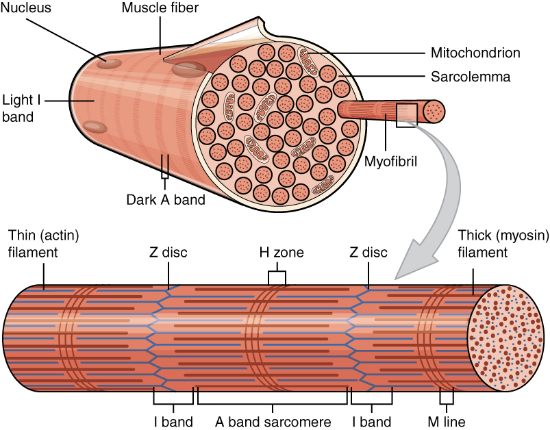
Myofilaments are the three protein filaments of myofibrils in muscle cells. The main proteins involved are myosin, actin, and titin. Myosin and actin are the ''contractile proteins'' and titin is an elastic protein. The myofilaments act together in muscle contraction, and in order of size are a thick one of mostly myosin, a thin one of mostly actin, and a very thin one of mostly titin. Types of muscle tissue are striated skeletal muscle and cardiac muscle, obliquely striated muscle (found in some invertebrates), and non-striated smooth muscle. Various arrangements of myofilaments create different muscles. Striated muscle has transverse bands of filaments. In obliquely striated muscle, the filaments are staggered. Smooth muscle has irregular arrangements of filaments. Structure There are three different types of myofilaments: thick, thin, and elastic filaments. *Thick filaments consist primarily of a type of myosin, a motor protein ŌĆō myosin II. Each thick filament is appr ... [...More Info...] [...Related Items...] OR: [Wikipedia] [Google] [Baidu] |


