|
Tonofilament
Tonofibrils are cytoplasmic protein structures in epithelial tissues that converge at desmosomes and hemidesmosomes. They consist of fine fibrils in epithelial cells that are anchored to the cytoskeleton. They were discovered by Rudolf Heidenhain, and first described in detail by Louis-Antoine Ranvier in 1897. Composition Tonofilaments are keratin intermediate filaments that make up tonofibrils in the epithelial tissue. In epithelial cells, tonofilaments loop through desmosomes. Electron microscopy has advanced now to illustrate the tonofilaments more clearly. The protein filaggrin is believed to be synthesized as a giant precursor protein, profilaggrin (>400 kDA in humans). When filaggrin binds to keratin intermediate filaments, the keratin aggregates into macrofibrils. References External links Diagram at ultrakohl.com Keratins Cytoskeleton {{cell-biology-stub ... [...More Info...] [...Related Items...] OR: [Wikipedia] [Google] [Baidu] |
Epithelial
Epithelium or epithelial tissue is a thin, continuous, protective layer of cells with little extracellular matrix. An example is the epidermis, the outermost layer of the skin. Epithelial ( mesothelial) tissues line the outer surfaces of many internal organs, the corresponding inner surfaces of body cavities, and the inner surfaces of blood vessels. Epithelial tissue is one of the four basic types of animal tissue, along with connective tissue, muscle tissue and nervous tissue. These tissues also lack blood or lymph supply. The tissue is supplied by nerves. There are three principal shapes of epithelial cell: squamous (scaly), columnar, and cuboidal. These can be arranged in a singular layer of cells as simple epithelium, either simple squamous, simple columnar, or simple cuboidal, or in layers of two or more cells deep as stratified (layered), or ''compound'', either squamous, columnar or cuboidal. In some tissues, a layer of columnar cells may appear to be stratified due ... [...More Info...] [...Related Items...] OR: [Wikipedia] [Google] [Baidu] |
Desmosomes
A desmosome (; "binding body"), also known as a macula adherens (plural: maculae adherentes) (Latin for ''adhering spot''), is a cell structure specialized for cell-to-cell adhesion. A type of junctional complex, they are localized spot-like adhesions randomly arranged on the lateral sides of plasma membranes. Desmosomes are one of the stronger cell-to-cell adhesion types and are found in tissue that experience intense mechanical stress, such as cardiac muscle tissue, bladder tissue, gastrointestinal mucosa, and epithelia. Structure Desmosomes are composed of desmosome-intermediate filament complexes (DIFCs), a network of cadherin proteins, linker proteins and intermediate filaments. The DIFCs can be broken into three regions: the extracellular core region ("desmoglea"), the outer dense plaque (ODP), and the inner dense plaque (IDP). The extracellular core region, approximately 34 nm in length, contains desmoglein and desmocollin, which are in the cadherin family of ... [...More Info...] [...Related Items...] OR: [Wikipedia] [Google] [Baidu] |
Hemidesmosomes
Hemidesmosomes are very small stud-like structures found in keratinocytes of the epidermis of skin that attach to the extracellular matrix. They are similar in form to desmosomes when visualized by electron microscopy; however, desmosomes attach to adjacent cells. Hemidesmosomes are also comparable to focal adhesions, as they both attach cells to the extracellular matrix. Instead of desmogleins and desmocollins in the extracellular space, hemidesmosomes utilize integrins. Hemidesmosomes are found in epithelial cells connecting the basal epithelial cells to the lamina lucida, which is part of the basal lamina. Hemidesmosomes are also involved in signaling pathways, such as keratinocyte migration or carcinoma cell intrusion. Structure Hemidesmosomes can be categorized into two types based on their protein constituents. Type 1 hemidesmosomes are found in stratified and pseudo-stratified epithelium. Type 1 hemidesmosomes have five main elements: integrin α6 β4, plectin in its ... [...More Info...] [...Related Items...] OR: [Wikipedia] [Google] [Baidu] |
Fibril
Fibrils () are structural biological materials found in nearly all living organisms. Not to be confused with fibers or protein filament, filaments, fibrils tend to have diameters ranging from 10 to 100 nanometers (whereas fibers are micro to milli-scale structures and filaments have diameters approximately 10–50 nanometers in size). Fibrils are not usually found alone but rather are parts of greater hierarchical structures commonly found in biological systems. Due to the prevalence of fibrils in biological systems, their study is of great importance in the fields of microbiology, biomechanics, and materials science. Structure and mechanics Fibrils are composed of linear biopolymers, and are characterized by rod-like structures with high length-to-diameter ratios. They often spontaneously arrange into helical structures. In biomechanics problems, fibrils can be characterized as classical beams with a roughly circular cross-sectional area on the nanometer scale. As such, s ... [...More Info...] [...Related Items...] OR: [Wikipedia] [Google] [Baidu] |
Cytoskeleton
The cytoskeleton is a complex, dynamic network of interlinking protein filaments present in the cytoplasm of all cells, including those of bacteria and archaea. In eukaryotes, it extends from the cell nucleus to the cell membrane and is composed of similar proteins in the various organisms. It is composed of three main components: microfilaments, intermediate filaments, and microtubules, and these are all capable of rapid growth and or disassembly depending on the cell's requirements. Cytoskeleton can perform many functions. Its primary function is to give the cell its shape and mechanical resistance to deformation, and through association with extracellular connective tissue and other cells it stabilizes entire tissues. The cytoskeleton can also contract, thereby deforming the cell and the cell's environment and allowing cells to migrate. Moreover, it is involved in many cell signaling pathways and in the uptake of extracellular material ( endocytosis), the segregation of ... [...More Info...] [...Related Items...] OR: [Wikipedia] [Google] [Baidu] |
Rudolf Heidenhain
Rudolf Peter Heinrich Heidenhain (; 29 January 1834 – 13 October 1897) was a German physiologist born in Marienwerder, Province of Prussia (now Kwidzyn, Poland). His son, Martin Heidenhain, was a highly regarded anatomist. Academic career He studied medicine at the Universities of Halle and Berlin. After receiving his doctorate, he remained in Berlin as an assistant to Emil du Bois-Reymond (1818-1896). In 1856 he returned to Halle, and worked in the laboratory of Alfred Wilhelm Volkmann (1801-1877). While serving an assistant at Halle, he made improvements on Hermann Welcker’s procedure for measurement of blood volume. In 1859 he attained the chair of physiology at the University of Breslau, where he remained for the rest of his career. Three of his famous students at Breslau were Karl Weigert (1845-1904), Ivan Petrovich Pavlov (1849-1936), and Albert Wojciech Adamkiewicz (1850-1921). His laboratory was the source of voluminous contributions by himself, his pupils, ... [...More Info...] [...Related Items...] OR: [Wikipedia] [Google] [Baidu] |
Louis-Antoine Ranvier
Louis-Antoine Ranvier (2 October 1835 – 22 March 1922) was a French physician, pathologist, anatomist and histologist, who discovered the nodes of Ranvier, regularly spaced discontinuities of the myelin sheath, occurring at varying intervals along the length of a nerve fiber. Career Ranvier was born and studied medicine at Lyon, graduating in 1865 from the Ecole Préparatoire de Médecine et de Pharmacie. He moved to Paris after receiving the internship of Parisian hospitals. Here he founded a small private research laboratory on Rue Christine along with fellow intern Victor André Cornil, and together they later offered a course in histology to medical students which involved the careful examination of tissues under a microscope. Their course was unique in the time as microscopy had not been viewed favourably in medicine especially by Henri Ducrotay de Blainville (1777-1850) and Auguste Comte (1798-1857). Their histology course material became an influential textbook o ... [...More Info...] [...Related Items...] OR: [Wikipedia] [Google] [Baidu] |
Keratin
Keratin () is one of a family of structural fibrous proteins also known as ''scleroproteins''. It is the key structural material making up Scale (anatomy), scales, hair, Nail (anatomy), nails, feathers, horn (anatomy), horns, claws, Hoof, hooves, and the outer layer of skin in vertebrates. Keratin also protects epithelial cells from damage or stress. Keratin is extremely insoluble in water and organic solvents. Keratin monomers assemble into bundles to form intermediate filaments, which are tough and form strong mineralization (biology), unmineralized epidermal appendages found in reptiles, birds, amphibians, and mammals. Excessive keratinization participate in fortification of certain tissues such as in horns of cattle and rhinos, and armadillos' osteoderm. The only other biology, biological matter known to approximate the toughness of keratinized tissue is chitin. Keratin comes in two types: the primitive, softer forms found in all vertebrates and the harder, derived forms fou ... [...More Info...] [...Related Items...] OR: [Wikipedia] [Google] [Baidu] |
Intermediate Filaments
Intermediate filaments (IFs) are cytoskeletal structural components found in the cells of vertebrates, and many invertebrates. Homologues of the IF protein have been noted in an invertebrate, the cephalochordate ''Branchiostoma''. Intermediate filaments are composed of a family of related proteins sharing common structural and sequence features. Initially designated 'intermediate' because their average diameter (10 nm) is between those of narrower microfilaments (actin) and wider myosin filaments found in muscle cells, the diameter of intermediate filaments is now commonly compared to actin microfilaments (7 nm) and microtubules (25 nm). Animal intermediate filaments are subcategorized into six types based on similarities in amino acid sequence and protein structure. Most types are cytoplasmic, but one type, Type V is a nuclear lamin. Unlike microtubules, IF distribution in cells shows no good correlation with the distribution of either mitochondria or endop ... [...More Info...] [...Related Items...] OR: [Wikipedia] [Google] [Baidu] |
Chiton Epidermis TEM
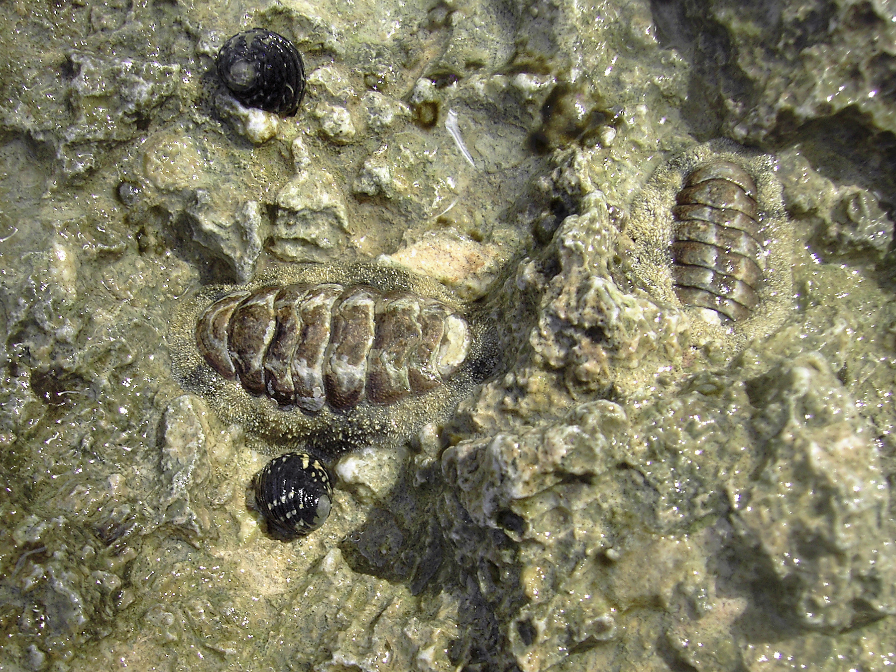
Chitons () are marine molluscs of varying size in the class Polyplacophora ( ), formerly known as Amphineura. About 940 extant and 430 fossil species are recognized. They are also sometimes known as sea cradles or coat-of-mail shells or suck-rocks, or more formally as loricates, polyplacophorans, and occasionally as polyplacophores. Chitons have a shell composed of eight separate shell plates or valves. These plates overlap slightly at the front and back edges, and yet articulate well with one another. Because of this, the shell provides protection at the same time as permitting the chiton to flex upward when needed for locomotion over uneven surfaces, and even allows the animal to curl up into a ball when dislodged from rocks. The shell plates are encircled by a skirt known as a girdle. Habitat Chitons live worldwide, from cold waters through to the tropics. They live on hard surfaces, such as on or under rocks, or in rock crevices. Some species live quite high in the int ... [...More Info...] [...Related Items...] OR: [Wikipedia] [Google] [Baidu] |
Filaggrin
Filaggrin (filament aggregating protein) is a filament-associated protein that binds to keratin fibers in epithelial cells. Ten to twelve filaggrin units are post-translationally hydrolyzed from a large profilaggrin precursor protein during terminal differentiation of epidermal cells. In humans, profilaggrin is encoded by the ''FLG'' gene, which is part of the S100 fused-type protein (SFTP) family within the epidermal differentiation complex on chromosome 1q21. In cetaceans and sirenians, the ''FLG'' family has lost its function, with the curious exception of manatees in the latter clade: manatees still retain some functional ''FLG'' genes. Profilaggrin Filaggrin monomers are tandemly clustered into a large, 350kDa protein precursor known as profilaggrin. In the epidermis, these structures are present in the keratohyalin granules in cells of the stratum granulosum. Profilaggrin undergoes proteolytic processing to yield individual filaggrin monomers at the transition between the ... [...More Info...] [...Related Items...] OR: [Wikipedia] [Google] [Baidu] |


