|
Dilator Naris Muscle
The dilator naris muscle (or alae nasi muscle) is a part of the nasalis muscle. It has an anterior and a posterior part. It has origins from the nasal notch of the maxilla and the major alar cartilage, and a single insertion near the margin of the nostril. It controls nostril width, including changes during breathing. Its function can be tested as an analogue for the function of the facial nerve (VII), which supplies it. Structure The dilator naris muscle is divided into posterior and anterior parts. * The ''dilator naris posterior'' is placed partly beneath the levator labii superioris muscle. It arises from the margin of the nasal notch of the maxilla, and from the minor alar cartilages. It is inserted into the skin near the margin of the nostril. * The ''dilator naris anterior'' is a delicate fasciculus. It originates from the lateral crus of the major alar cartilage, more laterally. It inserts into the margin of the nostril, the alar groove. It is situated in front of the ... [...More Info...] [...Related Items...] OR: [Wikipedia] [Google] [Baidu] |
Maxilla
In vertebrates, the maxilla (: maxillae ) is the upper fixed (not fixed in Neopterygii) bone of the jaw formed from the fusion of two maxillary bones. In humans, the upper jaw includes the hard palate in the front of the mouth. The two maxillary bones are fused at the intermaxillary suture, forming the anterior nasal spine. This is similar to the mandible (lower jaw), which is also a fusion of two mandibular bones at the mandibular symphysis. The mandible is the movable part of the jaw. Anatomy Structure The maxilla is a paired bone - the two maxillae unite with each other at the intermaxillary suture. The maxilla consists of: * The body of the maxilla: pyramid-shaped; has an orbital, a nasal, an infratemporal, and a facial surface; contains the maxillary sinus. * Four processes: ** the zygomatic process ** the frontal process ** the alveolar process ** the palatine process It has three surfaces: * the anterior, posterior, medial Features of the maxilla include: * t ... [...More Info...] [...Related Items...] OR: [Wikipedia] [Google] [Baidu] |
Human Skin
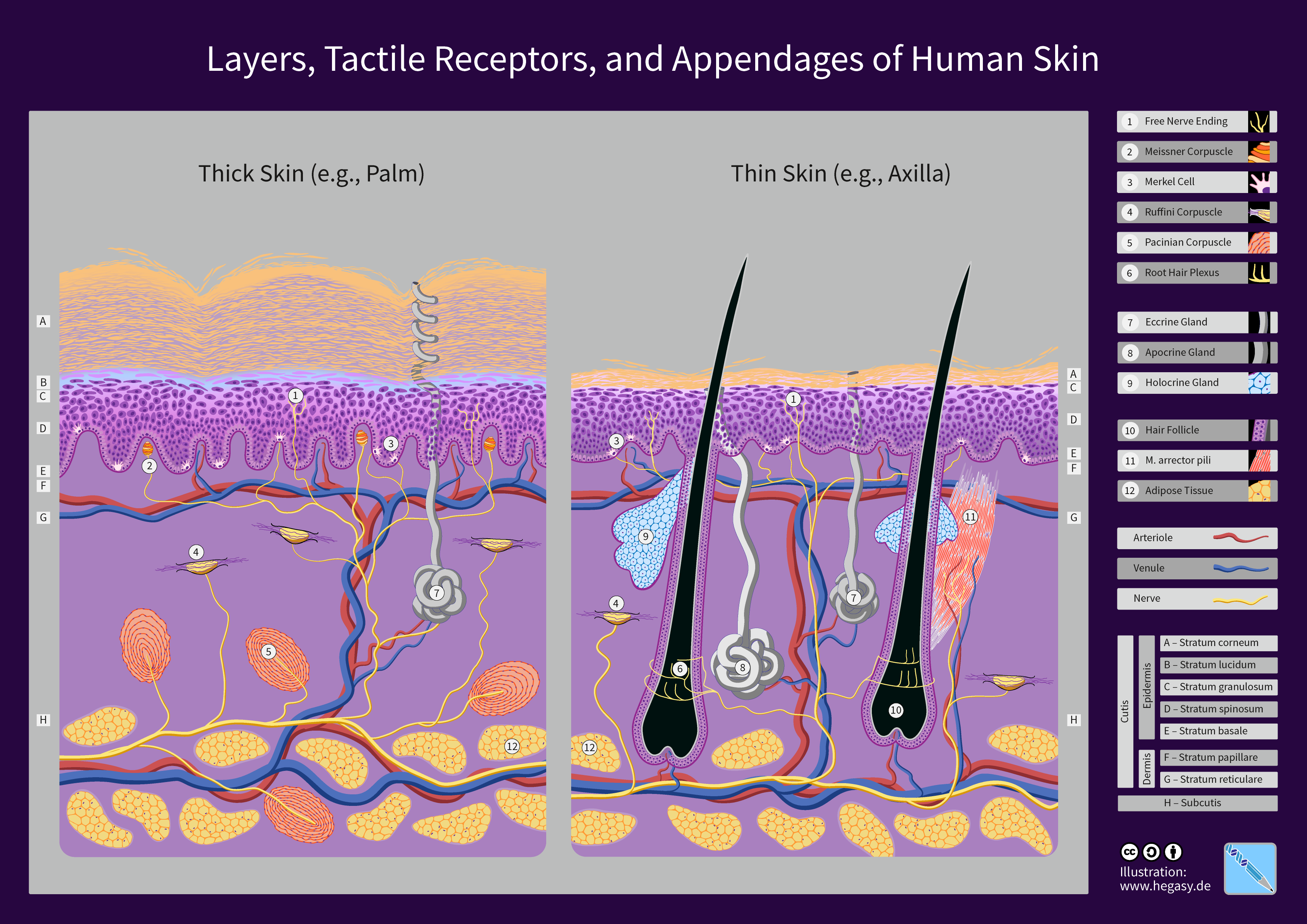
The human skin is the outer covering of the body and is the largest organ of the integumentary system. The skin has up to seven layers of ectodermal tissue (biology), tissue guarding Skeletal muscle, muscles, bones, ligaments and organ (anatomy), internal organs. Human skin is similar to most of the other mammals' skin, and it is very similar to pig skin. Though nearly all human skin is covered with hair follicles, it can appear Nudity#Evolution of hairlessness, hairless. There are two general types of skin: hairy and glabrous skin (hairless). The adjective List of medical roots, suffixes and prefixes#C, ''cutaneous'' literally means "of the skin" (from Latin ''cutis'', skin). Skin plays an important immunity (medical), immunity role in protecting the body against pathogens and excessive transepidermal water loss, water loss. Its other functions are Thermal insulation, insulation, thermoregulation, temperature regulation, sensation, synthesis of vitamin D, and the protection ... [...More Info...] [...Related Items...] OR: [Wikipedia] [Google] [Baidu] |
Muscles Of The Head And Neck
Muscle is a soft tissue, one of the four basic types of animal tissue. There are three types of muscle tissue in vertebrates: skeletal muscle, cardiac muscle, and smooth muscle. Muscle tissue gives skeletal muscles the ability to contract. Muscle tissue contains special contractile proteins called actin and myosin which interact to cause movement. Among many other muscle proteins, present are two regulatory proteins, troponin and tropomyosin. Muscle is formed during embryonic development, in a process known as myogenesis. Skeletal muscle tissue is striated consisting of elongated, multinucleate muscle cells called muscle fibers, and is responsible for movements of the body. Other tissues in skeletal muscle include tendons and perimysium. Smooth and cardiac muscle contract involuntarily, without conscious intervention. These muscle types may be activated both through the interaction of the central nervous system as well as by innervation from peripheral plexus or endocri ... [...More Info...] [...Related Items...] OR: [Wikipedia] [Google] [Baidu] |
Human Nose
The human nose is the first organ of the respiratory system. It is also the principal organ in the olfactory system. The shape of the nose is determined by the nasal bones and the nasal cartilages, including the nasal septum, which separates the nostrils and divides the nasal cavity into two. The nose has an important function in breathing. The nasal mucosa lining the nasal cavity and the paranasal sinuses carries out the necessary conditioning of inhaled air by warming and moistening it. Nasal conchae, shell-like bones in the walls of the cavities, play a major part in this process. Filtering of the air by nasal hair in the nostrils prevents large particles from entering the lungs. Sneezing is a reflex to expel unwanted particles from the nose that irritate the mucosal lining. Sneezing can Transmission (medicine), transmit infections, because aerosols are created in which the Respiratory droplets, droplets can harbour pathogens. Another major function of the nose is olfactio ... [...More Info...] [...Related Items...] OR: [Wikipedia] [Google] [Baidu] |
Breathing
Breathing (spiration or ventilation) is the rhythmical process of moving air into ( inhalation) and out of ( exhalation) the lungs to facilitate gas exchange with the internal environment, mostly to flush out carbon dioxide and bring in oxygen. All aerobic creatures need oxygen for cellular respiration, which extracts energy from the reaction of oxygen with molecules derived from food and produces carbon dioxide as a waste product. Breathing, or external respiration, brings air into the lungs where gas exchange takes place in the alveoli through diffusion. The body's circulatory system transports these gases to and from the cells, where cellular respiration takes place. The breathing of all vertebrates with lungs consists of repetitive cycles of inhalation and exhalation through a highly branched system of tubes or airways which lead from the nose to the alveoli. The number of respiratory cycles per minute is the breathing or respiratory rate, and is one of the fou ... [...More Info...] [...Related Items...] OR: [Wikipedia] [Google] [Baidu] |
Brainstem
The brainstem (or brain stem) is the posterior stalk-like part of the brain that connects the cerebrum with the spinal cord. In the human brain the brainstem is composed of the midbrain, the pons, and the medulla oblongata. The midbrain is continuous with the thalamus of the diencephalon through the tentorial notch, and sometimes the diencephalon is included in the brainstem. The brainstem is very small, making up around only 2.6 percent of the brain's total weight. It has the critical roles of regulating heart and respiratory system, respiratory function, helping to control heart rate and breathing rate. It also provides the main motor and sensory nerve supply to the face and neck via the cranial nerves. Ten pairs of cranial nerves come from the brainstem. Other roles include the regulation of the central nervous system and the body's sleep cycle. It is also of prime importance in the conveyance of motor and sensory pathways from the rest of the brain to the body, and from the b ... [...More Info...] [...Related Items...] OR: [Wikipedia] [Google] [Baidu] |
Respiratory Center
The respiratory center is located in the medulla oblongata and pons, in the brainstem. The respiratory center is made up of three major respiratory groups of neurons, two in the medulla and one in the pons. In the medulla they are the dorsal respiratory group, and the ventral respiratory group. In the pons, the pontine respiratory group includes two areas known as the pneumotaxic center and the apneustic center. The respiratory center is responsible for generating and maintaining the rhythm of respiration, and also of adjusting this in homeostatic response to physiological changes. The respiratory center receives input from chemoreceptors, mechanoreceptors, the cerebral cortex, and the hypothalamus in order to regulate the rate and depth of breathing. Input is stimulated by altered levels of oxygen, carbon dioxide, and blood pH, by hormonal changes relating to stress and anxiety from the hypothalamus, and also by signals from the cerebral cortex to give a conscious con ... [...More Info...] [...Related Items...] OR: [Wikipedia] [Google] [Baidu] |
Inhalation
Inhalation (or inspiration) happens when air or other gases enter the lungs. Inhalation of air Inhalation of air, as part of the cycle of breathing, is a vital process for all human life. The process is autonomic (though there are exceptions in some disease states) and does not need conscious control or effort. However, breathing can be consciously controlled or interrupted (within limits). Breathing allows oxygen (which humans and a lot of other species need for survival) to enter the lungs, from where it can be absorbed into the bloodstream. Other substances – accidental Examples of accidental inhalation includes inhalation of water (e.g. in drowning), smoke, food, vomitus and less common foreign substances (e.g. tooth fragments, coins, batteries, small toy parts, needles). Other substances – deliberate Recreational use Nitrous oxide ("laughing gas") has been used recreationally since 1899 for its ability to induce euphoria, hallucinogenic states and relaxa ... [...More Info...] [...Related Items...] OR: [Wikipedia] [Google] [Baidu] |
Muscle Fascicle
A muscle fascicle is a bundle of skeletal muscle fibers surrounded by perimysium, a type of connective tissue. Structure Muscle cells are grouped into muscle fascicles by enveloping perimysium connective tissue. Fascicles are bundled together by epimysium connective tissue. Muscle fascicles typically only contain one type of muscle cell (either type I fibres or type II fibres), but can contain a mixture of both types. Function In the heart, specialized cardiac muscle cells transmit electrical impulses from the atrioventricular node (AV node) to the Purkinje fibers – fascicles, also referred to as bundle branches. These start as a single fascicle of fibers at the AV node called the bundle of His that then splits into three bundle branches: the right fascicular branch, left anterior fascicular branch, and left posterior fascicular branch. Clinical significance Myositis may cause thickening of the muscle fascicles. This may be detected with ultrasound scans. ... [...More Info...] [...Related Items...] OR: [Wikipedia] [Google] [Baidu] |
Minor Alar Cartilage
In human anatomy, the part of the nose which forms the lateral wall is curved to correspond with the ala of the nose; it is oval and flattened, narrow behind, where it is connected with the frontal process of the maxilla by a tough fibrous membrane, in which are found three or four small nasal cartilages The nasal cartilages are structures within the nose that provide form and support to the nasal cavity. The nasal cartilages are made up of a flexible material called hyaline cartilage (packed collagen) in the distal portion of the nose. There are f ... the minor alar cartilages, also referred to as lesser alar or sesamoid cartilages or accessory cartilages. ''An atlas of human anatomy for students and physicians'' (Rebman, 1919; by Carl Toldt, Alois Dalla Rosa, Eden Paul, p. 942-944)- Retr ... [...More Info...] [...Related Items...] OR: [Wikipedia] [Google] [Baidu] |
Greater Alar Cartilage
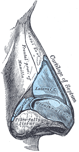
The major alar cartilage (greater alar cartilage) (lower lateral cartilage) is a thin, flexible plate, situated immediately below the lateral nasal cartilage, and bent upon itself in such a manner as to form the Nasal septum, medial wall and lateral wall of the nostril of its own side. The portion which forms the Nasal septum, medial wall (crus mediale) is loosely connected with the corresponding portion of the opposite cartilage, the two forming, together with the thickened integument and subjacent tissue, the nasal septum. The part which forms the lateral wall (crus laterale) is curved to correspond with the Human nose#External nose, ala of the nose; it is oval and flattened, narrow behind, where it is connected with the frontal process of the maxilla by a tough fibrous membrane, in which are found three or four small cartilaginous plates, the lesser alar cartilages (cartilagines alares minores; sesamoid cartilages). Above, it is connected by fibrous tissue to the lateral carti ... [...More Info...] [...Related Items...] OR: [Wikipedia] [Google] [Baidu] |
Levator Labii Superioris
The levator labii superioris (: ''levatores labii superioris'', also called quadratus labii superioris, : ''quadrati labii superioris'') is a muscle of the human body used in facial expression. It is a broad sheet, the origin of which extends from the side of the nose to the zygomatic bone. Structure Its medial fibers form the ''angular head'' (also known as the levator labii superioris alaeque nasi muscle) which arises by a pointed extremity from the upper part of the frontal process of the maxilla and passing obliquely downward and lateralward divides into two slips. One of these is inserted into the greater alar cartilage and skin of the nose; the other is prolonged into the lateral part of the upper lip, blending with the infraorbital head and with the orbicularis oris. The intermediate portion or ''infraorbital head'' arises from the lower margin of the orbit immediately above the infraorbital foramen, some of its fibers being attached to the maxilla, others to the zygo ... [...More Info...] [...Related Items...] OR: [Wikipedia] [Google] [Baidu] |


