Syndesmoses on:
[Wikipedia]
[Google]
[Amazon]
In

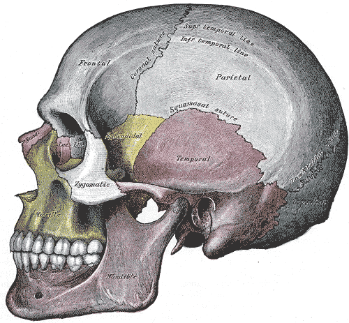
 A suture is a type of fibrous joint that is only found in the skull (cranial suture). The bones are bound together by Sharpey's fibres. A tiny amount of movement is permitted at sutures, which contributes to the compliance and elasticity of the skull.
These joints are synarthroses. It is normal for many of the bones of the skull to remain unfused at birth. The fusion of the skull's bones before birth is known as craniosynostosis. The term " fontanelle" is used to describe the resulting "soft spots". The relative positions of the bones continue to change during the life of the adult (though less rapidly), which can provide useful information in
File:Lambdoid suture.png, Lambdoid suture
File:SchaedelSeitlichSutur1.png, Coronal suture
File:SchaedelSeitlichSutur3.png, Squamosal suture
File:SchaedelSeitlichSutur4.png, Zygomaticotemporal suture
File:Sagittal suture.jpg, Sagittal suture.
File:Sagittal suture 2.jpg, Sagittal suture.
File:Sagittal suture 3.jpg, Sagittal suture.
File:Kort-lang-skalle.gif, Top view of cranial suture.
Age at Death Estimation from Cranial Suture Closures
Cranial suture closure and its implications for age estimation
from Douglas College {{Authority control Joints Skull it:Articolazione#Sindesmosi
anatomy
Anatomy () is the branch of morphology concerned with the study of the internal structure of organisms and their parts. Anatomy is a branch of natural science that deals with the structural organization of living things. It is an old scien ...
, fibrous joints are joint
A joint or articulation (or articular surface) is the connection made between bones, ossicles, or other hard structures in the body which link an animal's skeletal system into a functional whole.Saladin, Ken. Anatomy & Physiology. 7th ed. McGraw- ...
s connected by fibrous tissue
Connective tissue is one of the four primary types of animal tissue, a group of cells that are similar in structure, along with epithelial tissue, muscle tissue, and nervous tissue. It develops mostly from the mesenchyme, derived from the mesode ...
, consisting mainly of collagen
Collagen () is the main structural protein in the extracellular matrix of the connective tissues of many animals. It is the most abundant protein in mammals, making up 25% to 35% of protein content. Amino acids are bound together to form a trip ...
. These are fixed joints where bone
A bone is a rigid organ that constitutes part of the skeleton in most vertebrate animals. Bones protect the various other organs of the body, produce red and white blood cells, store minerals, provide structure and support for the body, ...
s are united by a layer of white fibrous tissue of varying thickness. In the skull
The skull, or cranium, is typically a bony enclosure around the brain of a vertebrate. In some fish, and amphibians, the skull is of cartilage. The skull is at the head end of the vertebrate.
In the human, the skull comprises two prominent ...
, the joints between the bones are called sutures. Such immovable joints are also referred to as synarthroses.
Types
Most fibrous joints are also called "fixed" or "immovable". These joints have no joint cavity and are connected via fibrous connective tissue. * Sutures: The skull bones are connected by fibrous joints called '' sutures''. In fetal skulls, the sutures are wide to allow slight movement during birth. They later become rigid ( synarthrodial). *Syndesmosis
A syndesmosis (“fastened with a band”) is a type of fibrous joint in which two bones are united to each other by fibrous connective tissue. The gap between the bones may be narrow, with the bones joined by ligaments, or the gap may be wide a ...
: Some of the long bone
The long bones are those that are longer than they are wide. They are one of five types of bones: long, short, flat, irregular and sesamoid. Long bones, especially the femur and tibia, are subjected to most of the load during daily activities ...
s in the body such as the radius
In classical geometry, a radius (: radii or radiuses) of a circle or sphere is any of the line segments from its Centre (geometry), center to its perimeter, and in more modern usage, it is also their length. The radius of a regular polygon is th ...
and ulna
The ulna or ulnar bone (: ulnae or ulnas) is a long bone in the forearm stretching from the elbow to the wrist. It is on the same side of the forearm as the little finger, running parallel to the Radius (bone), radius, the forearm's other long ...
in the forearm are joined by a ''syndesmosis
A syndesmosis (“fastened with a band”) is a type of fibrous joint in which two bones are united to each other by fibrous connective tissue. The gap between the bones may be narrow, with the bones joined by ligaments, or the gap may be wide a ...
'' (along the interosseous membrane). Syndemoses are slightly moveable ( amphiarthrodial). The distal tibiofibular joint is another example.
* A ''gomphosis
In anatomy, fibrous joints are joints connected by fibrous tissue, consisting mainly of collagen. These are fixed joints where bones are united by a layer of white fibrous tissue of varying thickness. In the skull, the joints between the bones ...
'' is a joint between the root of a tooth
A tooth (: teeth) is a hard, calcified structure found in the jaws (or mouths) of many vertebrates and used to break down food. Some animals, particularly carnivores and omnivores, also use teeth to help with capturing or wounding prey, tea ...
and the socket in the maxilla
In vertebrates, the maxilla (: maxillae ) is the upper fixed (not fixed in Neopterygii) bone of the jaw formed from the fusion of two maxillary bones. In humans, the upper jaw includes the hard palate in the front of the mouth. The two maxil ...
or mandible
In jawed vertebrates, the mandible (from the Latin ''mandibula'', 'for chewing'), lower jaw, or jawbone is a bone that makes up the lowerand typically more mobilecomponent of the mouth (the upper jaw being known as the maxilla).
The jawbone i ...
(jawbones).
Sutures

forensics
Forensic science combines principles of law and science to investigate criminal activity. Through crime scene investigations and laboratory analysis, forensic scientists are able to link suspects to evidence. An example is determining the time and ...
and archaeology
Archaeology or archeology is the study of human activity through the recovery and analysis of material culture. The archaeological record consists of Artifact (archaeology), artifacts, architecture, biofact (archaeology), biofacts or ecofacts, ...
. In old age, cranial sutures may ossify (turn to bone) completely.
The joints between the teeth and jaws (gomphoses) and the joint between the mandible and the cranium, the temporomandibular joint
In anatomy, the temporomandibular joints (TMJ) are the two joints connecting the jawbone to the skull. It is a bilateral Synovial joint, synovial articulation between the temporal bone of the skull above and the condylar process of mandible be ...
, form the only non-sutured joints in the skull.
Types of sutures
*Serrate sutures – similar to a denticulate suture but the interlocking regions are serrated rather than square. Eg: Coronal suture, sagittal Sutures. *Plane sutures – edges of the bones are flush with each other as in a normalbutt joint
A butt joint is a joinery, wood joint in which the end of a piece of material is simply placed (or “butted”) against another piece. The butt joint is the simplest joint. An unreinforced butt joint is also the weakest joint, as it provides a ...
. Eg: Internasal suture.
*Limbous sutures – edges are bevelled so the plane of the suture is sloping as in a mitre joint. Eg: Temporo-parietal suture.
*Schindylesis – formed by two bones fitting into each other similar to a bridle joint. Eg: Palatomaxillary suture.
*Denticulate sutures – the edges slot into each other as in a finger joint
A finger joint, also known as a comb joint, is a woodworking joint made by cutting a set of complementary, interlocking profiles in two pieces of wood, which are then Adhesive, glued. The cross-section of the joint resembles the interlocking of ...
. Eg: Lambdoid suture.
*
List of sutures
Most sutures are named for the bones they articulate, but some have special names of their own.Visible from the side
*Coronal suture
The coronal suture is a dense, fibrous connective tissue joint that separates the two parietal bones from the frontal bone of the skull.
Structure
The coronal suture lies between the paired parietal bones and the frontal bone of the skull ...
– between the frontal and parietal bone
The parietal bones ( ) are two bones in the skull which, when joined at a fibrous joint known as a cranial suture, form the sides and roof of the neurocranium. In humans, each bone is roughly quadrilateral in form, and has two surfaces, four bord ...
s
* Lambdoid suture – between the parietal and occipital bones and continuous with the occipitomastoid suture
*Occipitomastoid suture
The occipitomastoid suture, or occipitotemporal suture, is the cranial suture between the occipital bone and the mastoid portion of the temporal bone.
It is continuous with the lambdoidal suture.
See also
* Jugular foramen
Additional im ...
– between the occipital and temporal bones and continuous with the lambdoid suture
* Sphenofrontal suture
* Sphenoparietal suture
* Sphenosquamosal suture
* Sphenozygomatic suture
* Squamosal suture – between the parietal and the temporal bone
The temporal bone is a paired bone situated at the sides and base of the skull, lateral to the temporal lobe of the cerebral cortex.
The temporal bones are overlaid by the sides of the head known as the temples where four of the cranial bone ...
* Zygomaticotemporal suture
* Zygomaticofrontal suture
Visible from the front or above
* Frontal suture /Metopic suture
The frontal suture is a fibrous joint that divides the two halves of the frontal bone of the human skull, skull in infants and children. Typically, it completely fuses between three and nine months of age, with the two halves of the frontal bone ...
– between the two frontal bones, prior to the fusion of the two into a single bone
* Sagittal suture – along the midline, between parietal bone
The parietal bones ( ) are two bones in the skull which, when joined at a fibrous joint known as a cranial suture, form the sides and roof of the neurocranium. In humans, each bone is roughly quadrilateral in form, and has two surfaces, four bord ...
s
Visible from below or inside
* Frontoethmoidal suture * Petrosquamous suture * Sphenoethmoidal suture * Sphenopetrosal sutureGallery
Syndesmosis
A syndesmosis is a slightly mobile fibrous joint in which bones such as the tibia and fibula are joined together by connective tissue. An example is the distal tibiofibular joint. Injuries to the ankle syndesmosis are commonly known as a "high ankle sprain". Although the syndesmosis is a joint, in the literature the term syndesmotic injury is used to describe injury of the syndesmotic ligaments. It comes from the Greek σύν, ''syn'' (meaning "with") and δεσμός, ''desmos'' (meaning "a band"). Syndesmosis sprains have received increasing recognition during recent years because of a heightened awareness of the mechanism, symptoms, and signs of injury.Diagnosis of a syndesmotic injury
Diagnosis of syndesmosis injuries by physical examination is often straightforward. Physical examination findings that are often positive include the squeeze test and the external rotation test. Patients with high-grade syndesmosis injuries often cannot perform a single-leg heel raise. Patients report pain in varying degrees over the anterior and often posterior distal fibular joint.Syndesmotic tear
The severity of acute syndesmosis injury is rated from grade I to III by several authors. A grade I injury is a partial anteroinferior tibiofibular ligament tear, meaning the exorotation and squeeze tests are negative for this grade. Grade II injury is a complete anteroinferior tibiofibular ligament and inferior interosseous ligament tear, meaning that squeeze test and exorotation are positive. This results in the injury being stabilized with immobilization but not operatively stabilized. A grade III injury is a complete anteroinferior tibiofibular ligament tear including a (partial) interosseous ligament tear and deltoid ligament avulsion, meaning the joint is unstable and positive on the exorotation and squeeze tests. This grade requires operative stabilization. If the syndesmosis is torn apart as result of bone fracture, surgeons will sometimes fix the relevant bones together with asyndesmotic screw
A syndesmotic screw is a metal screw designed to replace the syndesmosis of the human body, usually temporarily. If the syndosmosis is torn apart as result of bone fracture, surgeons will sometimes fix the relevant bones together with a syndesmot ...
, temporarily replacing the syndesmosis, or with a tightrope fixation, which is called syndesmosis procedure. The screw inhibits normal movement of the bones and, thereby, the corresponding joint(s). When the natural articulation is healed, the screw may be removed. The tightrope fixation with elastic fiberwire suture on the other hand allows physiologic motion of the ankle and may be permanent.
Gomphosis
A gomphosis, also known as a dentoalveolarsyndesmosis
A syndesmosis (“fastened with a band”) is a type of fibrous joint in which two bones are united to each other by fibrous connective tissue. The gap between the bones may be narrow, with the bones joined by ligaments, or the gap may be wide a ...
, or 'peg and socket joint' is a joint that binds the teeth
A tooth (: teeth) is a hard, calcified structure found in the jaws (or mouths) of many vertebrates and used to break down food. Some animals, particularly carnivores and omnivores, also use teeth to help with capturing or wounding prey, tear ...
to bony teeth sockets in the maxillary bone and mandible
In jawed vertebrates, the mandible (from the Latin ''mandibula'', 'for chewing'), lower jaw, or jawbone is a bone that makes up the lowerand typically more mobilecomponent of the mouth (the upper jaw being known as the maxilla).
The jawbone i ...
. Gomphos is the Greek word for "bolt". The fibrous connection between a tooth and its socket is a periodontal ligament. Specifically, the connection is made between the maxilla or mandible to the cementum of the tooth.
The motion of a gomphosis is minimal, though considerable movement can be achieved over time—the basis of using braces to realign teeth. The joint can be considered a synarthrosis
A synarthrosis is a type of joint which allows no movement under normal conditions. Sutures and gomphoses are both synarthroses. Joints which allow more movement are called amphiarthroses or diarthroses. Syndesmoses are considered to be amph ...
.
The gomphosis is the only joint-type in which a bone does not join another bone, as teeth are not technically bone. In modern, more anatomical, joint classification, the gomphosis is simply considered a fibrous joint because the tissue linking the structures is ligamentous. It has been suggested that this permanent soft-tissue attachment was a critical requisite in the evolution of the mammalian (synapsid
Synapsida is a diverse group of tetrapod vertebrates that includes all mammals and their extinct relatives. It is one of the two major clades of the group Amniota, the other being the more diverse group Sauropsida (which includes all extant rept ...
) tusk
Tusks are elongated, continuously growing front teeth that protrude well beyond the mouth of certain mammal species. They are most commonly canine tooth, canine teeth, as with Narwhal, narwhals, chevrotains, musk deer, water deer, muntjac, pigs, ...
.
References
External links
*Age at Death Estimation from Cranial Suture Closures
Cranial suture closure and its implications for age estimation
from Douglas College {{Authority control Joints Skull it:Articolazione#Sindesmosi