|
Craniosynostosis
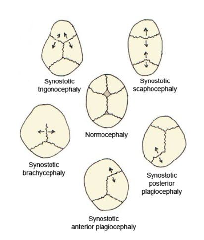
Craniosynostosis is a condition in which one or more of the fibrous Suture (joint), sutures in a young infant's skull prematurely fuses by turning into bone (ossification), thereby changing the growth pattern of the skull. Because the skull cannot expand perpendicular to the fused suture, it compensates by growing more in the direction parallel to the closed sutures. Sometimes the resulting growth pattern provides the necessary space for the growing brain, but results in an abnormal head shape and abnormal facial features. In cases in which the compensation does not effectively provide enough space for the growing brain, craniosynostosis results in increased intracranial pressure leading possibly to visual impairment, sleeping impairment, eating difficulties, or an impairment of mental development combined with a significant reduction in IQ. Craniosynostosis occurs in one in 2000 births. Craniosynostosis is part of a syndrome in 15% to 40% of affected patients, but it usually occ ... [...More Info...] [...Related Items...] OR: [Wikipedia] [Google] [Baidu] |
Trigonocephaly
Trigonocephaly is a congenital condition of premature fusion of the metopic suture (from the Greek , "forehead"), leading to a triangular forehead. The merging of the two frontal bones leads to transverse growth restriction and parallel growth expansion. It may occur syndromic, involving other abnormalities, or isolated. The term is from the Greek , "triangle", and , "head". Cause Trigonocephaly can either occur syndromatic or isolated. Trigonocephaly is associated with the following syndromes: Opitz syndrome, Muenke syndrome, Jacobsen syndrome, Baller–Gerold syndrome and Say–Meyer syndrome. The etiology of trigonocephaly is mostly unknown although there are three main theories. Trigonocephaly is probably a multifactorial congenital condition, but due to limited proof of these theories this cannot safely be concluded. Intrinsic bone malformation The first theory assumes that the origin of pathological synostosis lies within disturbed bone formation early on in the preg ... [...More Info...] [...Related Items...] OR: [Wikipedia] [Google] [Baidu] |
Cranium
The skull is a bone protective cavity for the brain. The skull is composed of four types of bone i.e., cranial bones, facial bones, ear ossicles and hyoid bone. However two parts are more prominent: the cranium and the mandible. In humans, these two parts are the neurocranium and the viscerocranium (facial skeleton) that includes the mandible as its largest bone. The skull forms the anterior-most portion of the skeleton and is a product of cephalisation—housing the brain, and several sensory structures such as the eyes, ears, nose, and mouth. In humans these sensory structures are part of the facial skeleton. Functions of the skull include protection of the brain, fixing the distance between the eyes to allow stereoscopic vision, and fixing the position of the ears to enable sound localisation of the direction and distance of sounds. In some animals, such as horned ungulates (mammals with hooves), the skull also has a defensive function by providing the mount (on the ... [...More Info...] [...Related Items...] OR: [Wikipedia] [Google] [Baidu] |
Turricephaly
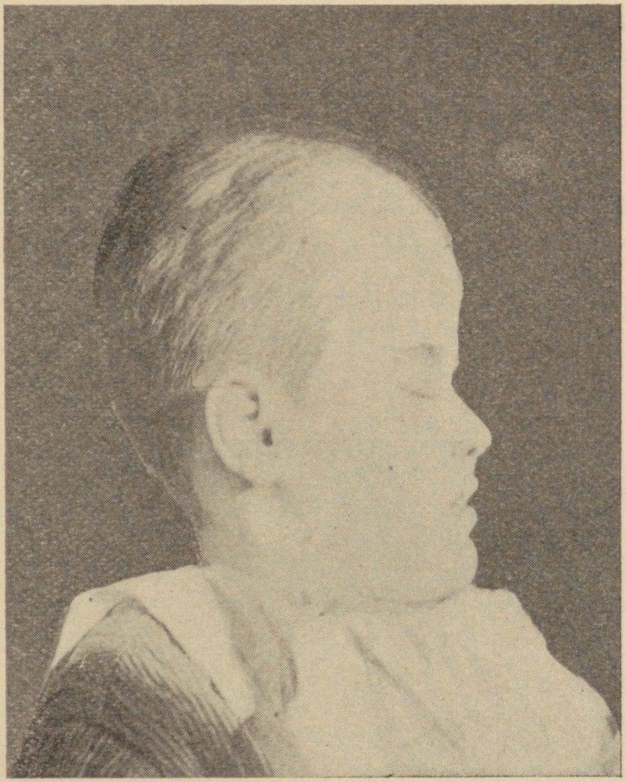
Turricephaly is a type of cephalic disorder where the head appears tall with a small length and width. It is due to premature closure of the coronal suture plus any other suture, like the lambdoid, or it may be used to describe the premature fusion of all sutures. It should be differentiated from Crouzon syndrome. Oxycephaly (or acrocephaly) is a form of turricephaly where the head is cone-shaped, and is the most severe of the craniosynostoses. Presentation Common associations It may be associated with: * 8th cranial nerve lesion * Optic nerve compression * Intellectual disability * Syndactyly Diagnosis Treatment See also * Acrocephalosyndactylia Acrocephalosyndactyly is a group of autosomal dominant congenital disorders characterized by craniofacial (craniosynostosis) and hand and foot (syndactyly) abnormalities. When polydactyly is present, the classification is acrocephalopolysyndactyl ... References Further reading NINDS Overview* External links Congenit ... [...More Info...] [...Related Items...] OR: [Wikipedia] [Google] [Baidu] |
Intracranial Pressure
Intracranial pressure (ICP) is the pressure exerted by fluids such as cerebrospinal fluid (CSF) inside the skull and on the brain tissue. ICP is measured in millimeters of mercury ( mmHg) and at rest, is normally 7–15 mmHg for a supine adult. The body has various mechanisms by which it keeps the ICP stable, with CSF pressures varying by about 1 mmHg in normal adults through shifts in production and absorption of CSF. Changes in ICP are attributed to volume changes in one or more of the constituents contained in the cranium. CSF pressure has been shown to be influenced by abrupt changes in intrathoracic pressure during coughing (which is induced by contraction of the diaphragm and abdominal wall muscles, the latter of which also increases intra-abdominal pressure), the valsalva maneuver, and communication with the vasculature (venous and arterial systems). Intracranial hypertension (IH), also called increased ICP (IICP) or raised intracranial pressure (RICP), is elevation of ... [...More Info...] [...Related Items...] OR: [Wikipedia] [Google] [Baidu] |
High-head Syndrome
Turricephaly is a type of cephalic disorder where the head appears tall with a small length and width. It is due to premature closure of the coronal suture plus any other suture, like the lambdoid, or it may be used to describe the premature fusion of all sutures. It should be differentiated from Crouzon syndrome. Oxycephaly (or acrocephaly) is a form of turricephaly where the head is cone-shaped, and is the most severe of the craniosynostoses. Presentation Common associations It may be associated with: * 8th cranial nerve lesion * Optic nerve compression * Intellectual disability * Syndactyly Diagnosis Treatment See also * Acrocephalosyndactylia Acrocephalosyndactyly is a group of autosomal dominant congenital disorders characterized by craniofacial (craniosynostosis) and hand and foot (syndactyly) abnormalities. When polydactyly is present, the classification is acrocephalopolysyndactyl ... References Further reading NINDS Overview* External links Congenit ... [...More Info...] [...Related Items...] OR: [Wikipedia] [Google] [Baidu] |
Brachycephaly
Brachycephaly (derived from the Ancient Greek '' βραχύς'', 'short' and '' κεφαλή'', 'head') is the shape of a skull shorter than typical for its species. It is perceived as a desirable trait in some domesticated dog and cat breeds, notably the pug and Persian, and can be normal or abnormal in other animal species. In humans, the cephalic disorder is known as flat head syndrome, and results from premature fusion of the coronal sutures, or from external deformation. The coronal suture is the fibrous joint that unites the frontal bone with the two parietal bones of the skull. The parietal bones form the top and sides of the skull. This feature can be seen in Down syndrome. In anthropology, human populations have been characterized as either dolichocephalic (long-headed), mesaticephalic (moderate-headed), or brachycephalic (short-headed). The usefulness of the cephalic index was questioned by Giuseppe Sergi, who argued that cranial morphology provided a better ... [...More Info...] [...Related Items...] OR: [Wikipedia] [Google] [Baidu] |
Orbit (anatomy)
In anatomy, the orbit is the cavity or socket of the skull in which the eye and its appendages are situated. "Orbit" can refer to the bony socket, or it can also be used to imply the contents. In the adult human, the volume of the orbit is , of which the eye occupies . The orbital contents comprise the eye, the orbital and retrobulbar fascia, extraocular muscles, cranial nerves II, III, IV, V, and VI, blood vessels, fat, the lacrimal gland with its sac and duct, the eyelids, medial and lateral palpebral ligaments, cheek ligaments, the suspensory ligament, septum, ciliary ganglion and short ciliary nerves. Structure The orbits are conical or four-sided pyramidal cavities, which open into the midline of the face and point back into the head. Each consists of a base, an apex and four walls."eye, human."Encyclopædia Britannica from Encyclopædia Britannica 2006 Ultimate Reference Suite DVD 2009 Openings There are two important foramina, or windows, two important f ... [...More Info...] [...Related Items...] OR: [Wikipedia] [Google] [Baidu] |
Lambdoid
The lambdoid suture (or lambdoidal suture) is a dense, fibrous connective tissue joint on the posterior aspect of the skull that connects the parietal bones with the occipital bone. It is continuous with the occipitomastoid suture. Structure The lambdoid suture is between the paired parietal bones and the occipital bone of the skull. It runs from the asterion on each side. Nerve supply The lambdoid suture may be supplied by a branch of the supraorbital nerve, a branch of the frontal branch of the trigeminal nerve. Clinical significance At birth, the bones of the skull do not meet. If certain bones of the skull grow too fast, then craniosynostosis (premature closure of the sutures) may occur. This can result in skull deformities. If the lambdoid suture closes too soon on one side, the skull will appear twisted and asymmetrical, a condition called "plagiocephaly". Plagiocephaly refers to the shape and not the condition. The condition is craniosynostosis. The lambdoid sut ... [...More Info...] [...Related Items...] OR: [Wikipedia] [Google] [Baidu] |
Coronal Plane
The coronal plane (also known as the frontal plane) is an anatomical plane that divides the body into dorsal and ventral sections. It is perpendicular to the sagittal and transverse planes. Details The coronal plane is an example of a longitudinal plane. For a human, the mid-coronal plane would transect a standing body into two halves (front and back, or anterior and posterior) in an imaginary line that cuts through both shoulders. The description of the coronal plane applies to most animals as well as humans even though humans walk upright and the various planes are usually shown in the vertical orientation. The sternal plane (''planum sternale'') is a coronal plane which transects the front of the sternum. Etymology The term is derived from Latin ''corona'' ('garland, crown'), from Ancient Greek κορώνη (''korōnē'', 'garland, wreath'). The coronal plane is so-called because it lies in the direction of Coronal suture. Additional images File:Coronal plane CT scan ... [...More Info...] [...Related Items...] OR: [Wikipedia] [Google] [Baidu] |
Occiput
The occipital bone () is a cranial dermal bone and the main bone of the occiput (back and lower part of the skull). It is trapezoidal in shape and curved on itself like a shallow dish. The occipital bone overlies the occipital lobes of the cerebrum. At the base of skull in the occipital bone, there is a large oval opening called the foramen magnum, which allows the passage of the spinal cord. Like the other cranial bones, it is classed as a flat bone. Due to its many attachments and features, the occipital bone is described in terms of separate parts. From its front to the back is the basilar part, also called the basioccipital, at the sides of the foramen magnum are the lateral parts, also called the exoccipitals, and the back is named as the squamous part. The basilar part is a thick, somewhat quadrilateral piece in front of the foramen magnum and directed towards the pharynx. The squamous part is the curved, expanded plate behind the foramen magnum and is the largest par ... [...More Info...] [...Related Items...] OR: [Wikipedia] [Google] [Baidu] |
Lambdoid Suture
The lambdoid suture (or lambdoidal suture) is a dense, fibrous connective tissue joint on the posterior aspect of the skull that connects the parietal bones with the occipital bone. It is continuous with the occipitomastoid suture. Structure The lambdoid suture is between the paired parietal bones and the occipital bone of the skull. It runs from the asterion on each side. Nerve supply The lambdoid suture may be supplied by a branch of the supraorbital nerve, a branch of the frontal branch of the trigeminal nerve. Clinical significance At birth, the bones of the skull do not meet. If certain bones of the skull grow too fast, then craniosynostosis (premature closure of the sutures) may occur. This can result in skull deformities. If the lambdoid suture closes too soon on one side, the skull will appear twisted and asymmetrical, a condition called "plagiocephaly". Plagiocephaly refers to the shape and not the condition. The condition is craniosynostosis. The lambdoid s ... [...More Info...] [...Related Items...] OR: [Wikipedia] [Google] [Baidu] |


.jpg)

