|
Sphenopetrosal Suture
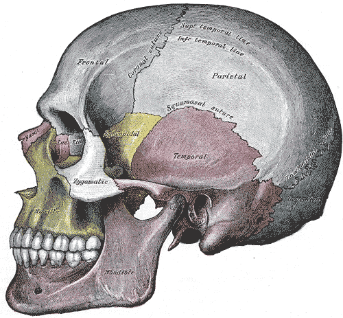
The sphenopetrosal fissure (or sphenopetrosal suture) is the cranial suture between the sphenoid bone and the petrous portion of the temporal bone. It is in the middle cranial fossa The middle cranial fossa is formed by the sphenoid bones, and the temporal bones. It lodges the temporal lobes, and the pituitary gland. It is deeper than the anterior cranial fossa, is narrow medially and widens laterally to the sides of the skull .... External links Skull {{musculoskeletal-stub ... [...More Info...] [...Related Items...] OR: [Wikipedia] [Google] [Baidu] |
Sphenoid Bone
The sphenoid bone is an unpaired bone of the neurocranium. It is situated in the middle of the skull towards the front, in front of the basilar part of occipital bone, basilar part of the occipital bone. The sphenoid bone is one of the seven bones that articulate to form the orbit (anatomy), orbit. Its shape somewhat resembles that of a butterfly, bat or wasp with its wings extended. The name presumably originates from this shape, since () means in Ancient Greek. Structure It is divided into the following parts: * a median portion, known as the body of sphenoid bone, containing the sella turcica, which houses the pituitary gland as well as the paired paranasal sinuses, the sphenoidal sinuses * two Greater wing of sphenoid bone, greater wings on the lateral side of the body and two Lesser wing of sphenoid bone, lesser wings from the anterior side. * Pterygoid processes of the sphenoides, directed downwards from the junction of the body and the greater wings. Two sphenoidal co ... [...More Info...] [...Related Items...] OR: [Wikipedia] [Google] [Baidu] |
Temporal Bone
The temporal bone is a paired bone situated at the sides and base of the skull, lateral to the temporal lobe of the cerebral cortex. The temporal bones are overlaid by the sides of the head known as the temples where four of the cranial bones fuse. Each temple is covered by a temporal muscle. The temporal bones house the structures of the ears. The lower seven cranial nerves and the major vessels to and from the brain traverse the temporal bone. Structure The temporal bone consists of four parts—the squamous, mastoid, petrous and tympanic parts. The squamous part is the largest and most superiorly positioned relative to the rest of the bone. The zygomatic process is a long, arched process projecting from the lower region of the squamous part and it articulates with the zygomatic bone. Posteroinferior to the squamous is the mastoid part. Fused with the squamous and mastoid parts and between the sphenoid and occipital bones lies the petrous part, which is shaped li ... [...More Info...] [...Related Items...] OR: [Wikipedia] [Google] [Baidu] |
Zygomatic Bone
In the human skull, the zygomatic bone (from ), also called cheekbone or malar bone, is a paired irregular bone, situated at the upper and lateral part of the face and forming part of the lateral wall and floor of the orbit, of the temporal fossa and the infratemporal fossa. It presents a malar and a temporal surface; four processes (the frontosphenoidal, orbital, maxillary, and temporal), and four borders. Etymology The term ''zygomatic'' derives from the Ancient Greek , ''zygoma'', meaning "yoke". The zygomatic bone is occasionally referred to as the zygoma, but this term may also refer to the zygomatic arch. Structure Surfaces The ''malar surface'' is convex and perforated near its center by a small aperture, the zygomaticofacial foramen, for the passage of the zygomaticofacial nerve and vessels; below this foramen is a slight elevation, which gives origin to the zygomaticus muscle. The ''temporal surface'', directed posteriorly and medially, is concave, prese ... [...More Info...] [...Related Items...] OR: [Wikipedia] [Google] [Baidu] |
Cranial Suture
In anatomy, fibrous joints are joints connected by fibrous tissue, consisting mainly of collagen. These are fixed joints where bones are united by a layer of white fibrous tissue of varying thickness. In the skull, the joints between the bones are called sutures. Such immovable joints are also referred to as synarthroses. Types Most fibrous joints are also called "fixed" or "immovable". These joints have no joint cavity and are connected via fibrous connective tissue. * Sutures: The skull bones are connected by fibrous joints called '' sutures''. In fetal skulls, the sutures are wide to allow slight movement during birth. They later become rigid ( synarthrodial). * Syndesmosis: Some of the long bones in the body such as the radius and ulna in the forearm are joined by a ''syndesmosis'' (along the interosseous membrane). Syndemoses are slightly moveable ( amphiarthrodial). The distal tibiofibular joint is another example. * A ''gomphosis'' is a joint between the root of a ... [...More Info...] [...Related Items...] OR: [Wikipedia] [Google] [Baidu] |
Petrous Part Of The Temporal Bone
The petrous part of the temporal bone is pyramid-shaped and is wedged in at the base of the skull between the sphenoid and occipital bones. Directed medially, forward, and a little upward, it presents a base, an apex, three surfaces, and three angles, and houses in its interior the components of the inner ear. The petrous portion is among the most basal elements of the skull and forms part of the endocranium. Petrous comes from the Latin word ''petrosus'', meaning "stone-like, hard". It is one of the densest bones in the body. In other mammals, it is a separate bone, the petrosal bone. The petrous bone is important for studies of ancient DNA from skeletal remains, as it tends to contain extremely well-preserved DNA. Base The base is fused with the internal surfaces of the squamous, tympanic, and mastoid parts. Apex The apex, which is rough and uneven, is received into the angular interval between the posterior border of the great wing of the sphenoid bone and the basi ... [...More Info...] [...Related Items...] OR: [Wikipedia] [Google] [Baidu] |
Middle Cranial Fossa
The middle cranial fossa is formed by the sphenoid bones, and the temporal bones. It lodges the temporal lobes, and the pituitary gland. It is deeper than the anterior cranial fossa, is narrow medially and widens laterally to the sides of the skull. It is separated from the posterior cranial fossa by the clivus and the petrous crest. It is bounded in front by the posterior margins of the lesser wings of the sphenoid bone, the anterior clinoid processes, and the ridge forming the anterior margin of the chiasmatic groove; behind, by the superior angles of the petrous portions of the temporal bones and the dorsum sellae; laterally by the temporal squamae, sphenoidal angles of the parietals, and greater wings of the sphenoid. It is traversed by the squamosal, sphenoparietal, sphenosquamosal, and sphenopetrosal sutures. Anatomy Features Middle part The middle part of the fossa presents, in front, the chiasmatic groove and tuberculum sellae; the chiasmatic groove ends o ... [...More Info...] [...Related Items...] OR: [Wikipedia] [Google] [Baidu] |


