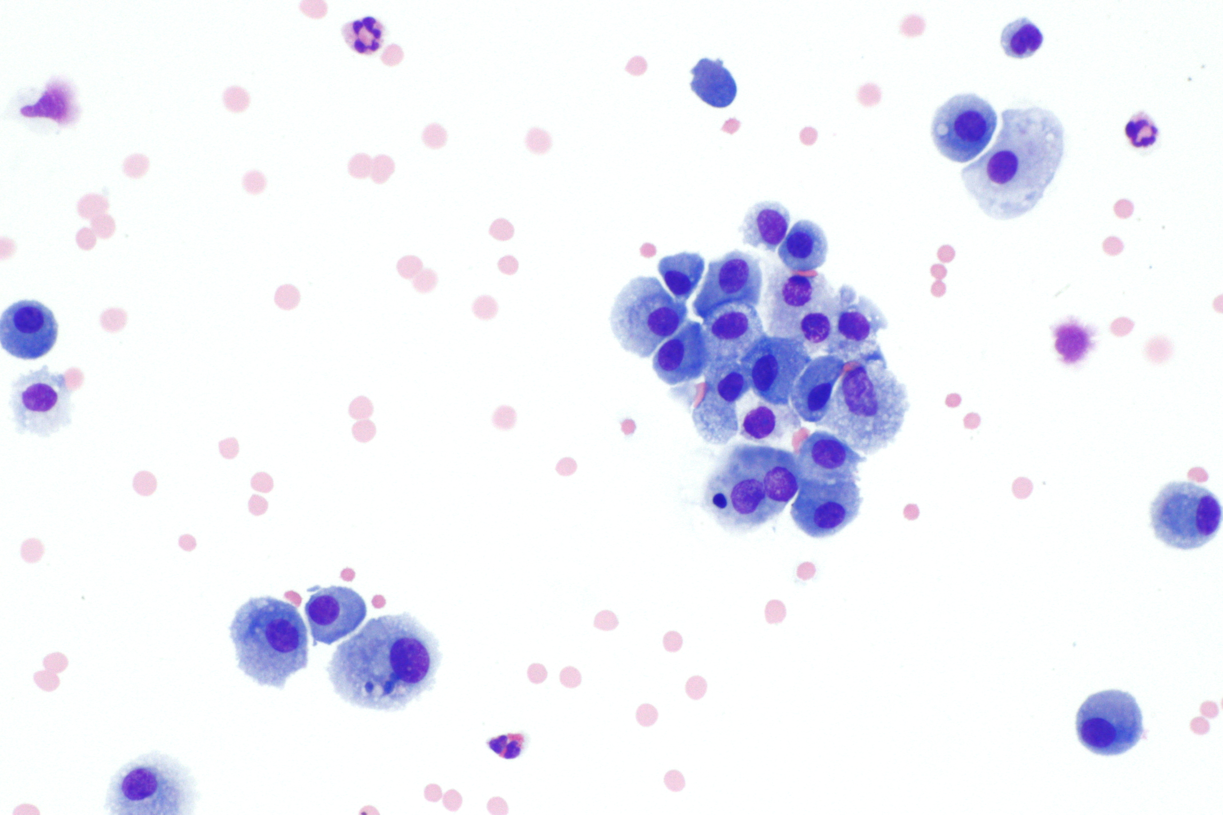
Diff-Quik on:
[Wikipedia]
[Google]
[Amazon]
 Diff-Quik is a commercial
 Diff-Quik is a commercial
 Romanowsky stain
Romanowsky staining is a prototypical staining technique that was the forerunner of several distinct but similar stains widely used in hematology (the study of blood) and cytopathology (the study of diseased cells). Romanowsky-type stains are use ...
variant used to rapidly stain and differentiate a variety of pathology
Pathology is the study of disease. The word ''pathology'' also refers to the study of disease in general, incorporating a wide range of biology research fields and medical practices. However, when used in the context of modern medical treatme ...
specimens. It is most frequently used for blood films and cytopathological smears, including fine needle aspirates. The Diff-Quik procedure is based on a modification of the Wright-Giemsa stain pioneered by Harleco in the 1970s, and has advantages over the routine Wright-Giemsa staining technique in that it reduces the 4-minute process into a much shorter operation and allows for selective increased eosinophilic or basophilic staining depending upon the time the smear is left in the staining solutions.
There are generic brands of such stain, and the trade name is sometimes used loosely to refer to any such stain (much as "Coke" or "Band-Aid" are sometimes used imprecisely).
Usage
Diff-Quik may be utilized on material which is ''air-dried'' prior to alcohol fixation rather than immersed immediately (i.e. "wet-fixed"), although immediate alcohol fixation results in improved microscopic detail. The primary use of Romanowsky-type stains in cytopathology is for cytoplasmic detail, while Papanicolaou stain is used for nuclear detail. Diff-Quik stain highlights cytoplasmic elements such asmucins
Mucins () are a family of high molecular weight, heavily glycosylation, glycosylated proteins (glycoconjugates) produced by epithelial tissues in most animals. Mucins' key characteristic is their ability to form gels; therefore they are a key com ...
, fat droplets and neurosecretory granules. Extracellular substances, such as free mucin, colloid, and ground substance
Ground substance is an amorphous gel-like substance in the extracellular space of animals that contains all components of the extracellular matrix (ECM) except for fibrous materials such as collagen and elastin. Ground substance is active in the d ...
, are also easily stained, and appear metachromatic. Major applications include blood smear
A blood smear, peripheral blood smear or blood film is a thin layer of blood smeared on a glass microscope slide and then stained in such a way as to allow the various blood cells to be examined microscopically. Blood smears are examined in the i ...
s, bone marrow
Bone marrow is a semi-solid biological tissue, tissue found within the Spongy bone, spongy (also known as cancellous) portions of bones. In birds and mammals, bone marrow is the primary site of new blood cell production (or haematopoiesis). It i ...
aspirates, semen analysis
A semen analysis (plural: semen analyses), also called seminogram or spermiogram, evaluates certain characteristics of a male's semen and the spermatozoon, sperm contained therein. It is done to help evaluate male fertility, whether for those see ...
and cytology of various body fluid
Body fluids, bodily fluids, or biofluids, sometimes body liquids, are liquids within the Body (biology), body of an organism. In lean healthy adult men, the total body water is about 60% (60–67%) of the total Human body weight, body weight; it ...
s including urine
Urine is a liquid by-product of metabolism in humans and many other animals. In placental mammals, urine flows from the Kidney (vertebrates), kidneys through the ureters to the urinary bladder and exits the urethra through the penile meatus (mal ...
and cerebrospinal fluid
Cerebrospinal fluid (CSF) is a clear, colorless Extracellular fluid#Transcellular fluid, transcellular body fluid found within the meninges, meningeal tissue that surrounds the vertebrate brain and spinal cord, and in the ventricular system, ven ...
. Microbiologic agents, such as bacteria and fungi, also appear more easily in Diff-Quik. This is useful for the detection of for example ''Helicobacter pylori
''Helicobacter pylori'', previously known as ''Campylobacter pylori'', is a gram-negative, Flagellum#bacterial, flagellated, Bacterial cellular morphologies#Helical, helical bacterium. Mutants can have a rod or curved rod shape that exhibits l ...
'' from gastric and pyloric specimens.
Due to its short staining time, Diff-Quik stain is often used for initial screening of cytopathology specimens. This staining technique allows the cytotechnologist or pathologist
Pathology is the study of disease. The word ''pathology'' also refers to the study of disease in general, incorporating a wide range of biology research fields and medical practices. However, when used in the context of modern medical treatme ...
to quickly assess the adequacy of the specimen, identify possible neoplastic
A neoplasm () is a type of abnormal and excessive growth of tissue. The process that occurs to form or produce a neoplasm is called neoplasia. The growth of a neoplasm is uncoordinated with that of the normal surrounding tissue, and persists ...
or inflammatory changes, and decide whether or not additional staining is required.
Components
The Diff-Quik stain consists of 3 solutions: *Diff-Quik fixative reagent ** Triarylmethane dye **Methanol
Methanol (also called methyl alcohol and wood spirit, amongst other names) is an organic chemical compound and the simplest aliphatic Alcohol (chemistry), alcohol, with the chemical formula (a methyl group linked to a hydroxyl group, often ab ...
*Diff-Quik solution I (eosinophilic
Eosinophilic (Greek suffix '' -phil'', meaning ''eosin-loving'') describes the staining of tissues, cells, or organelles after they have been washed with eosin, a dye commonly used in histological staining.
Eosin is an acidic dye for stainin ...
)
**Xanthene
Xanthene (9''H''-xanthene, 10''H''-9-oxaanthracene) is the organic compound with the formula CH2 6H4sub>2O. It is a yellow solid that is soluble in common organic solvents. Xanthene itself is an obscure compound, but many of its derivatives are u ...
dye (Eosin Y
Eosin Y, also called C.I. 45380 or C.I. Acid Red 87, is a member of the triarylmethane dyes. It is produced from fluorescein by bromination.
Use
Eosin Y is commonly used as the red dye in red inks.
It is commonly used in histology, most nota ...
)
**pH buffer
A buffer solution is a solution where the pH does not change significantly on dilution or if an acid or base is added at constant temperature. Its pH changes very little when a small amount of strong acid or base is added to it. Buffer solutions ...
*Diff-Quik solution II (basophilic
Basophilic is a technical term used by pathologists. It describes the appearance of cells, tissues and cellular structures as seen through the microscope after a histological section has been stained with a basic dye. The most common such dye ...
thiazine dyes)
**Methylene blue
Methylthioninium chloride, commonly called methylene blue, is a salt used as a dye and as a medication. As a medication, it is mainly used to treat methemoglobinemia. It has previously been used for treating cyanide poisoning and urinary trac ...
**Azure A
Azure A is an organic compound with the chemical formula
A chemical formula is a way of presenting information about the chemical proportions of atoms that constitute a particular chemical compound or molecule, using chemical element symbo ...
**pH buffer
Results
Alternatives
* Wright Giemsa stain * Papanicolaou stainReferences
{{Romanowsky stains Staining Histopathology Hematopathology Cytopathology Romanowsky stains