|
Transthyretin Amyloid Cardiomyopathy
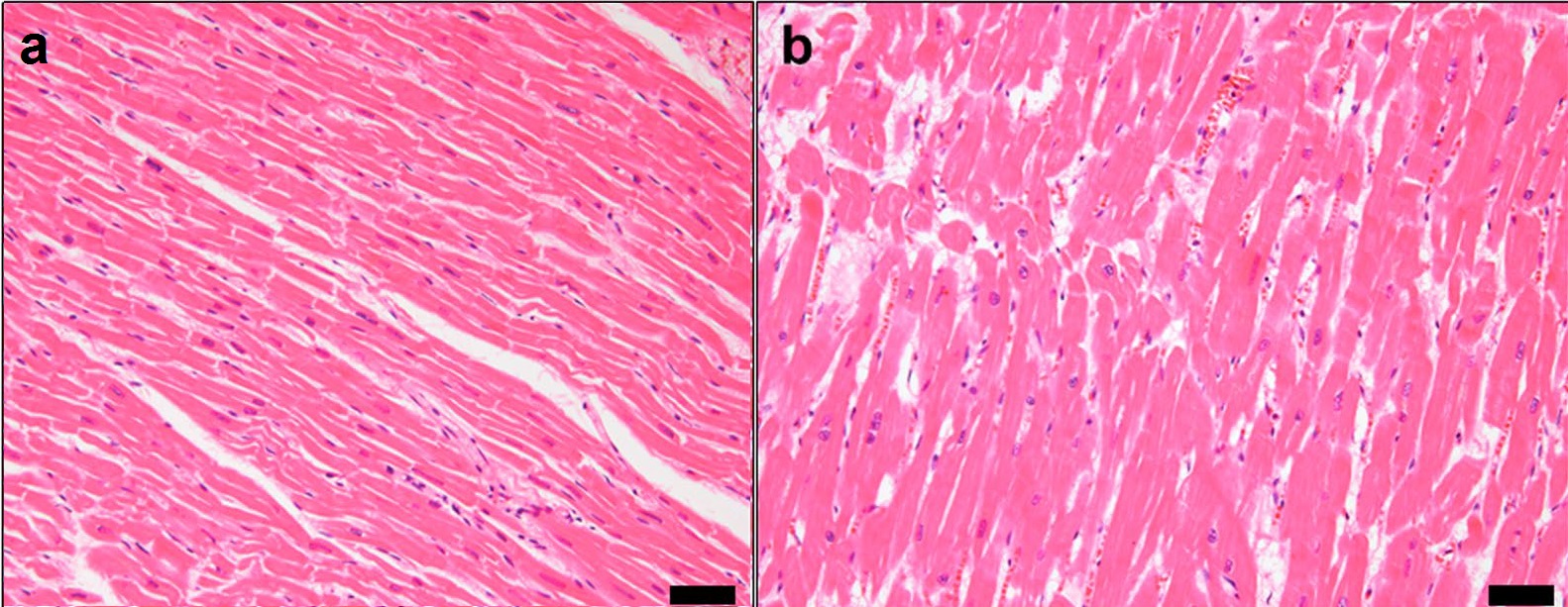
Amyloid cardiomyopathy ( stiff heart syndrome) is a condition resulting in the death of part of the myocardium (heart muscle). It is associated with the systemic production and release of many amyloidogenic proteins, especially immunoglobulin light chain or transthyretin (TTR). It can be characterized by the extracellular deposition of amyloids, foldable proteins that stick together to build fibrils in the heart. Symptoms Amyloid cardiomyopathy is associated with a number of symptoms: * diastolic dysfunction * congestive heart failure * arrhythmia * cardiac nervous conduction block * fatigue * dyspnea Pathophysiology Amyloid proteins are deposited in the myocardium. This limits ventricular filling during diastole, which increases end-diastolic volume. This can lead to a variety of cardiac issues, such as congestive heart failure, atrial arrhythmia, ventricular arrhythmia, and blocks to cardiac nervous conduction. Diagnosis Diagnosis is often delayed, because its symptoms a ... [...More Info...] [...Related Items...] OR: [Wikipedia] [Google] [Baidu] |
Ventricular Hypertrophy
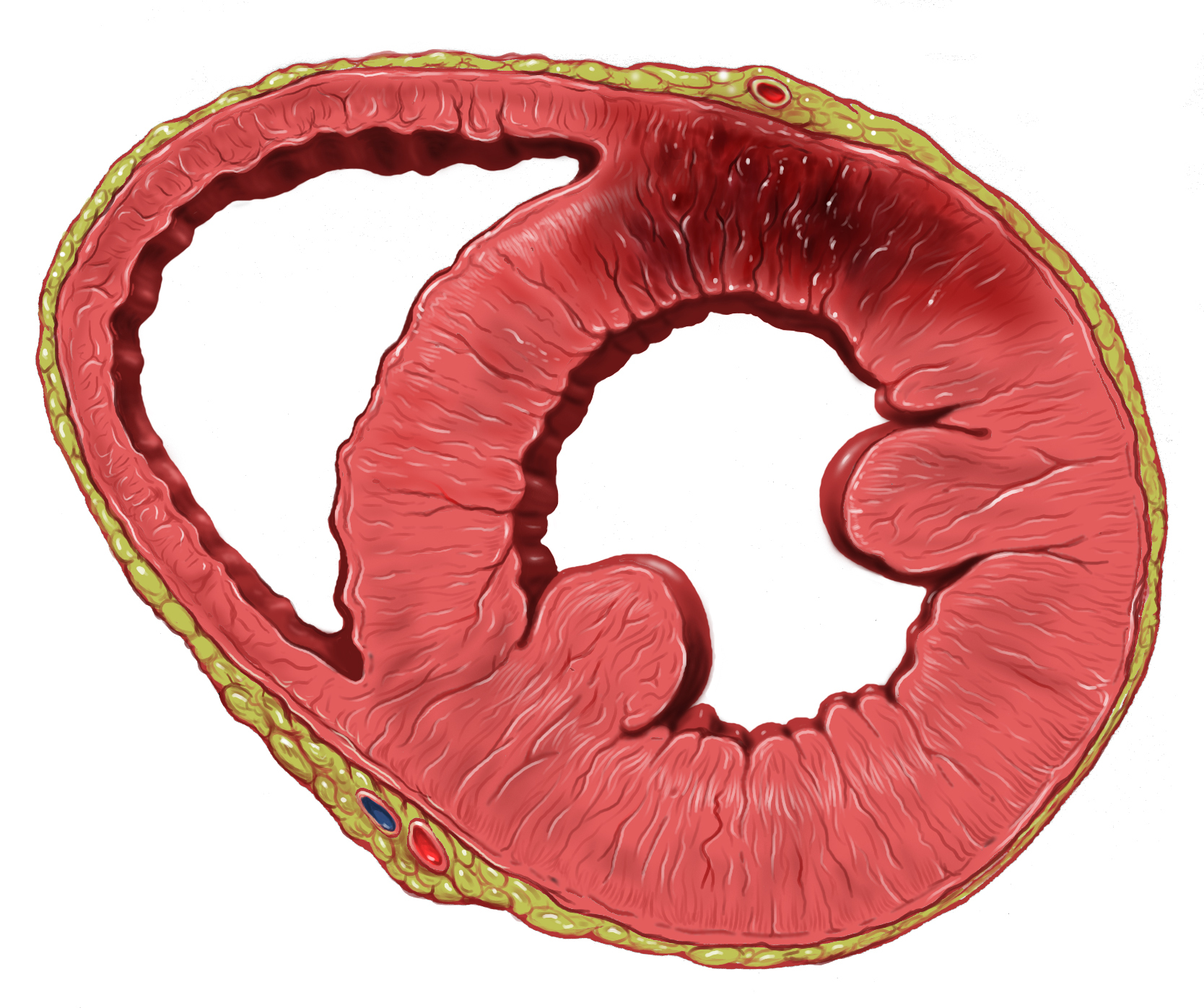
Ventricular hypertrophy (VH) is thickening of the walls of a ventricle (lower chamber) of the heart. Although left ventricular hypertrophy (LVH) is more common, right ventricular hypertrophy (RVH), as well as concurrent hypertrophy of both ventricles can also occur. Ventricular hypertrophy can result from a variety of conditions, both adaptive and maladaptive. For example, it occurs in what is regarded as a physiologic, adaptive process in pregnancy in response to increased blood volume; but can also occur as a consequence of ventricular remodeling following a heart attack. Importantly, pathologic and physiologic remodeling engage different cellular pathways in the heart and result in different gross cardiac phenotypes. Presentation In individuals with eccentric hypertrophy there may be little or no indication that hypertrophy has occurred as it is generally a healthy response to increased demands on the heart. Conversely, concentric hypertrophy can make itself known in a vari ... [...More Info...] [...Related Items...] OR: [Wikipedia] [Google] [Baidu] |
Protein
Proteins are large biomolecules and macromolecules that comprise one or more long chains of amino acid residue (biochemistry), residues. Proteins perform a vast array of functions within organisms, including Enzyme catalysis, catalysing metabolic reactions, DNA replication, Cell signaling, responding to stimuli, providing Cytoskeleton, structure to cells and Fibrous protein, organisms, and Intracellular transport, transporting molecules from one location to another. Proteins differ from one another primarily in their sequence of amino acids, which is dictated by the Nucleic acid sequence, nucleotide sequence of their genes, and which usually results in protein folding into a specific Protein structure, 3D structure that determines its activity. A linear chain of amino acid residues is called a polypeptide. A protein contains at least one long polypeptide. Short polypeptides, containing less than 20–30 residues, are rarely considered to be proteins and are commonly called pep ... [...More Info...] [...Related Items...] OR: [Wikipedia] [Google] [Baidu] |
Cardiac Magnetic Resonance Imaging
Cardiac magnetic resonance imaging (cardiac MRI, CMR), also known as cardiovascular MRI, is a magnetic resonance imaging (MRI) technology used for non-invasive assessment of the function and structure of the cardiovascular system. Conditions in which it is performed include congenital heart disease, cardiomyopathies and valvular heart disease, diseases of the aorta such as dissection, aneurysm and coarctation, coronary heart disease. It can also be used to look at pulmonary veins. It is contraindicated if there are some implanted metal or electronic devices such as some intracerebral clips or claustrophobia. Conventional MRI sequences are adapted for cardiac imaging by using ECG gating and high temporal resolution protocols. The development of cardiac MRI is an active field of research and continues to see a rapid expansion of new and emerging techniques. Uses Cardiovascular MRI is complementary to other imaging techniques, such as echocardiography, cardiac CT, and nuclear med ... [...More Info...] [...Related Items...] OR: [Wikipedia] [Google] [Baidu] |
Myocardial Infarction
A myocardial infarction (MI), commonly known as a heart attack, occurs when Ischemia, blood flow decreases or stops in one of the coronary arteries of the heart, causing infarction (tissue death) to the heart muscle. The most common symptom is retrosternal Angina, chest pain or discomfort that classically radiates to the left shoulder, arm, or jaw. The pain may occasionally feel like heartburn. This is the dangerous type of acute coronary syndrome. Other symptoms may include shortness of breath, nausea, presyncope, feeling faint, a diaphoresis, cold sweat, Fatigue, feeling tired, and decreased level of consciousness. About 30% of people have atypical symptoms. Women more often present without chest pain and instead have neck pain, arm pain or feel tired. Among those over 75 years old, about 5% have had an MI with little or no history of symptoms. An MI may cause heart failure, an Cardiac arrhythmia, irregular heartbeat, cardiogenic shock or cardiac arrest. Most MIs occur d ... [...More Info...] [...Related Items...] OR: [Wikipedia] [Google] [Baidu] |
Ventricle (heart)
A ventricle is one of two large chambers located toward the bottom of the heart that collect and expel blood towards the peripheral beds within the body and lungs. The blood pumped by a ventricle is supplied by an atrium, an adjacent chamber in the upper heart that is smaller than a ventricle. Interventricular means between the ventricles (for example the interventricular septum), while intraventricular means within one ventricle (for example an intraventricular block). In a four-chambered heart, such as that in humans, there are two ventricles that operate in a double circulatory system: the right ventricle pumps blood into the pulmonary circulation to the lungs, and the left ventricle pumps blood into the systemic circulation through the aorta. Structure Ventricles have thicker walls than atria and generate higher blood pressures. The physiological load on the ventricles requiring pumping of blood throughout the body and lungs is much greater than the pressure generated by ... [...More Info...] [...Related Items...] OR: [Wikipedia] [Google] [Baidu] |
Atrium (heart)
The atrium (; : atria) is one of the two Heart#Chambers, upper chambers in the heart that receives blood from the circulatory system. The blood in the atria is pumped into the Ventricle (heart), heart ventricles through the atrioventricular valve, atrioventricular mitral valve, mitral and tricuspid valve, tricuspid heart valves. There are two atria in the human heart – the left atrium receives blood from the pulmonary circulation, and the right atrium receives blood from the venae cavae of the systemic circulation. During the cardiac cycle, the atria receive blood while relaxed in diastole, then contract in systole to move blood to the ventricles. Each atrium is roughly cube-shaped except for an ear-shaped projection called an atrial appendage, previously known as an auricle. All animals with a closed circulatory system have at least one atrium. The atrium was formerly called the 'auricle'. That term is still used to describe this chamber in some other animals, such as the ''Mo ... [...More Info...] [...Related Items...] OR: [Wikipedia] [Google] [Baidu] |
Congestive Heart Failure
Heart failure (HF), also known as congestive heart failure (CHF), is a syndrome caused by an impairment in the heart's ability to fill with and pump blood. Although symptoms vary based on which side of the heart is affected, HF typically presents with shortness of breath, excessive fatigue, and bilateral leg swelling. The severity of the heart failure is mainly decided based on ejection fraction and also measured by the severity of symptoms. Other conditions that have symptoms similar to heart failure include obesity, kidney failure, liver disease, anemia, and thyroid disease. Common causes of heart failure include coronary artery disease, heart attack, high blood pressure, atrial fibrillation, valvular heart disease, excessive alcohol consumption, infection, and cardiomyopathy. These cause heart failure by altering the structure or the function of the heart or in some cases both. There are different types of heart failure: right-sided heart failure, which affect ... [...More Info...] [...Related Items...] OR: [Wikipedia] [Google] [Baidu] |
End-diastolic Volume
In cardiovascular physiology, end-diastolic volume (EDV) is the volume of blood in the right or left ventricle at end of filling in diastole which is amount of blood present in ventricle at the end of diastole. Because greater EDVs cause greater distention of the ventricle, ''EDV'' is often used synonymously with '' preload'', which refers to the length of the sarcomeres in cardiac muscle prior to contraction (systole). An increase in EDV increases the preload on the heart and, through the Frank-Starling mechanism of the heart, increases the amount of blood ejected from the ventricle during systole (stroke volume). __TOC__ Sample values The right ventricular end-diastolic volume (RVEDV) ranges between 100 and 160 mL. The right ventricular end-diastolic volume index (RVEDVI) is calculated by RVEDV/ BSA and ranges between 60 and 100 mL/m2. See also * End-systolic volume * Stroke volume In cardiovascular physiology, stroke volume (SV) is the volume of blood pumped from the ventr ... [...More Info...] [...Related Items...] OR: [Wikipedia] [Google] [Baidu] |
Diastole
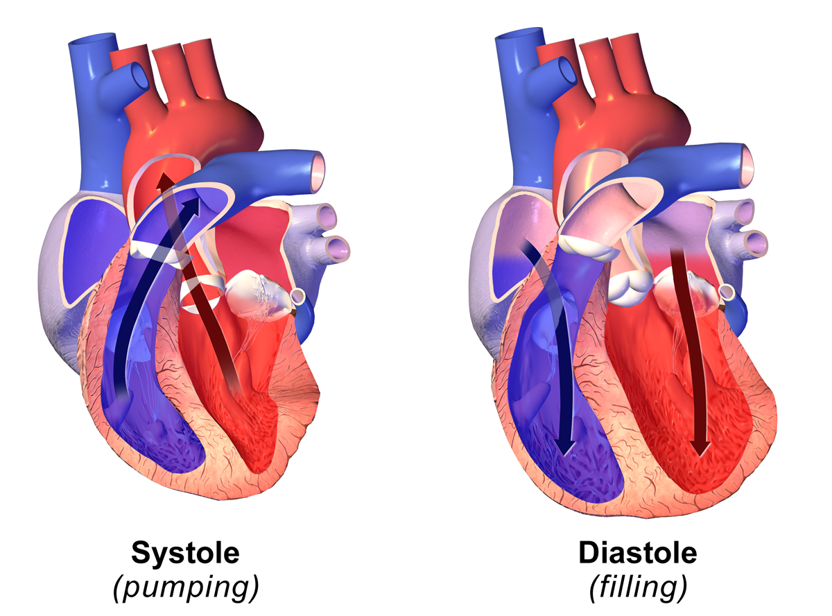
Diastole ( ) is the relaxed phase of the cardiac cycle when the chambers of the heart are refilling with blood. The contrasting phase is systole when the heart chambers are contracting. Atrial diastole is the relaxing of the atria, and ventricular diastole the relaxing of the ventricles. The term originates from the Greek word (''diastolē''), meaning "dilation", from (''diá'', "apart") + (''stéllein'', "to send"). Role in cardiac cycle A typical heart rate is 75 beats per minute (bpm), which means that the cardiac cycle that produces one heartbeat, lasts for less than one second. The cycle requires 0.3 sec in ventricular systole (contraction)—pumping blood to all body systems from the two ventricles; and 0.5 sec in diastole (dilation), re-filling the four chambers of the heart, for a total of 0.8 sec to complete the cycle. Early ventricular diastole During early ventricular diastole, pressure in the two ventricles begins to drop from the peak reached during systole ... [...More Info...] [...Related Items...] OR: [Wikipedia] [Google] [Baidu] |
Amyloid
Amyloids are aggregates of proteins characterised by a fibrillar morphology of typically 7–13 nm in diameter, a β-sheet secondary structure (known as cross-β) and ability to be stained by particular dyes, such as Congo red. In the human body, amyloids have been linked to the development of various diseases. Pathogenic amyloids form when previously healthy proteins lose their normal structure and physiological functions ( misfolding) and form fibrous deposits within and around cells. These protein misfolding and deposition processes disrupt the healthy function of tissues and organs. Such amyloids have been associated with (but not necessarily as the cause of) more than 50 human diseases, known as amyloidosis, and may play a role in some neurodegenerative diseases. Some of these diseases are mainly sporadic and only a few cases are familial. Others are only familial. Some result from medical treatment. Prions are an infectious form of amyloids that can act as a templa ... [...More Info...] [...Related Items...] OR: [Wikipedia] [Google] [Baidu] |
Heart (organ)
The heart is a muscular organ found in humans and other animals. This organ pumps blood through the blood vessels. The heart and blood vessels together make the circulatory system. The pumped blood carries oxygen and nutrients to the tissue, while carrying metabolic waste such as carbon dioxide to the lungs. In humans, the heart is approximately the size of a closed fist and is located between the lungs, in the middle compartment of the chest, called the mediastinum. In humans, the heart is divided into four chambers: upper left and right atria and lower left and right ventricles. Commonly, the right atrium and ventricle are referred together as the right heart and their left counterparts as the left heart. In a healthy heart, blood flows one way through the heart due to heart valves, which prevent backflow. The heart is enclosed in a protective sac, the pericardium, which also contains a small amount of fluid. The wall of the heart is made up of three layers: epicardium, ... [...More Info...] [...Related Items...] OR: [Wikipedia] [Google] [Baidu] |


