|
Optic Disk Drusen
Optic disc drusen (ODD) are globules of mucoproteins and mucopolysaccharides that progressively calcify in the optic disc.Golnik, K. (2006). Congenital anomalies and acquired abnormalities of the optic nerve, (Version 14.3). UptoDate (On-Line Serial) They are thought to be the remnants of the axonal transport system of degenerated retinal ganglion cells. ODD have also been referred to as congenitally elevated or anomalous discs, pseudopapilledema, pseudoneuritis, buried disc drusen, and disc hyaline bodies. Anatomy The optic nerve is a cable connection that transmits images from the retina to the brain. It consists of over one million retinal ganglion cell axons. The optic nerve head, or optic disc is the anterior end of the nerve that is in the eye and hence is visible with an ophthalmoscope. It is located nasally and slightly inferior to the macula of the eye. There is a blind spot at the optic disc because there are no rods or cones beneath it to detect light. The central ... [...More Info...] [...Related Items...] OR: [Wikipedia] [Google] [Baidu] |
Mucoprotein
A mucoprotein is a glycoprotein composed primarily of mucopolysaccharides. Mucoproteins can be found throughout the body, including the gastrointestinal tract, reproductive organs, airways, and the synovial fluid of the knees. They are called mucoproteins because the carbohydrate quantity is more than 4% unlike glycoproteins where the carbohydrate quantity is less than 4%. Mucoprotein is produced in the cecum of the gastrointestinal tract. During gallbladder cancer, mucoprotein is over expressed. Sustaining a brain injury will lead to decreased mucoprotein production. The result is an alteration of gut microbiota as seen in mice. Function Mucoproteins are the proteins that are the building blocks of mucus, which is a protective barrier to the epithelia of cells. It is semipermeable, so it acts as a barrier to most bacteria and pathogens, while allowing for the uptake of nutrients, water, and hormones. Protein Structure Mucoproteins are composed of o-linked carbohydrates as ... [...More Info...] [...Related Items...] OR: [Wikipedia] [Google] [Baidu] |
Choroid
The choroid, also known as the choroidea or choroid coat, is a part of the uvea, the vascular layer of the eye. It contains connective tissues, and lies between the retina and the sclera. The human choroid is thickest at the far extreme rear of the eye (at 0.2 mm), while in the outlying areas it narrows to 0.1 mm. The choroid provides oxygen and nourishment to the outer layers of the retina. Along with the ciliary body and iris, the choroid forms the uveal tract. The structure of the choroid is generally divided into four layers (classified in order of furthest away from the retina to closest): *Haller's layer – outermost layer of the choroid consisting of larger diameter blood vessels; * Sattler's layer – layer of medium diameter blood vessels; * Choriocapillaris – layer of capillaries; and * Bruch's membrane (synonyms: Lamina basalis, Complexus basalis, Lamina vitra) – innermost layer of the choroid. Blood supply There are two circulations of the eye: ... [...More Info...] [...Related Items...] OR: [Wikipedia] [Google] [Baidu] |
Optical Coherence Tomography
Optical coherence tomography (OCT) is a high-resolution imaging technique with most of its applications in medicine and biology. OCT uses coherent near-infrared light to obtain micrometer-level depth resolved images of biological tissue or other scattering media. It uses interferometry techniques to detect the amplitude and time-of-flight of reflected light. OCT uses transverse sample scanning of the light beam to obtain two- and three-dimensional images. Short-coherence-length light can be obtained using a superluminescent diode (SLD) with a broad spectral bandwidth or a broadly tunable laser with narrow linewidth. The first demonstration of OCT imaging (in vitro) was published by a team from MIT and Harvard Medical School in a 1991 article in the journal ''Science (journal), Science''. The article introduced the term "OCT" to credit its derivation from optical coherence-domain reflectometry, in which the axial resolution is based on temporal coherence. The first demonstrat ... [...More Info...] [...Related Items...] OR: [Wikipedia] [Google] [Baidu] |
Perimetry
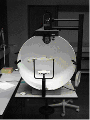
A visual field test is an eye examination that can detect dysfunction in central and peripheral vision which may be caused by various medical conditions such as glaucoma, stroke, pituitary disease, brain tumours or other neurological deficits. Visual field testing can be performed clinically by keeping the subject's gaze fixed while presenting objects at various places within their visual field. Simple manual equipment can be used such as in the tangent screen test or the Amsler grid. When dedicated machinery is used it is called a perimeter. The exam may be performed by a technician in one of several ways. The test may be performed by a technician directly, with the assistance of a machine, or completely by an automated machine. Machine-based tests aid diagnostics by allowing a detailed printout of the patient's visual field. Other names for this test may include perimetry, Tangent screen exam, Automated perimetry exam, Goldmann visual field exam, or brand names such as the H ... [...More Info...] [...Related Items...] OR: [Wikipedia] [Google] [Baidu] |
Intraocular Pressure
Intraocular pressure (IOP) is the fluid pressure inside the eye. Tonometry is the method eye care professionals use to determine this. IOP is an important aspect in the evaluation of patients at risk of glaucoma. Most tonometers are calibrated to measure pressure in millimeters of mercury (mmHg). Physiology Intraocular pressure is determined by the production and drainage of aqueous humour by the ciliary body and its drainage via the trabecular meshwork and uveoscleral outflow. The reason for this is because the vitreous humour in the posterior segment has a relatively fixed volume and thus does not affect intraocular pressure regulation. An important quantitative relationship (Goldmann's equation) is as follows: :P_o = \frac + P_v Where: * P_o is the IOP in millimeters of mercury (mmHg) * F the rate of aqueous humour formation in microliters per minute (μL/min) * U the resorption of aqueous humour through the uveoscleral route (μL/min) * C is the facility of outflow in mic ... [...More Info...] [...Related Items...] OR: [Wikipedia] [Google] [Baidu] |
Color Vision
Color vision, a feature of visual perception, is an ability to perceive differences between light composed of different frequencies independently of light intensity. Color perception is a part of the larger visual system and is mediated by a complex process between neurons that begins with differential stimulation of different types of photoreceptors by light entering the eye. Those photoreceptors then emit outputs that are propagated through many layers of neurons ultimately leading to higher cognitive functions in the brain. Color vision is found in many animals and is mediated by similar underlying mechanisms with common types of biological molecules and a complex history of the evolution of color vision within different animal taxa. In primates, color vision may have evolved under selective pressure for a variety of visual tasks including the foraging for nutritious young leaves, ripe fruit, and flowers, as well as detecting predator camouflage and emotional states in othe ... [...More Info...] [...Related Items...] OR: [Wikipedia] [Google] [Baidu] |
Contrast Sensitivity
Contrast is the difference in luminance or color that makes an object (or its representation in an image or display) visible against a background of different luminance or color. The human visual system is more sensitive to contrast than to absolute luminance; thus, we can perceive the world similarly despite significant changes in illumination throughout the day or across different locations. The maximum contrast of an image is termed the contrast ratio or dynamic range. In images where the contrast ratio approaches the maximum possible for the medium, there is a ''conservation of contrast''. In such cases, increasing contrast in certain parts of the image will necessarily result in a decrease in contrast elsewhere. Brightening an image increases contrast in darker areas but decreases it in brighter areas; conversely, darkening the image will have the opposite effect. Bleach bypass reduces contrast in the darkest and brightest parts of an image while enhancing luminance contras ... [...More Info...] [...Related Items...] OR: [Wikipedia] [Google] [Baidu] |
Visual Acuity
Visual acuity (VA) commonly refers to the clarity of visual perception, vision, but technically rates an animal's ability to recognize small details with precision. Visual acuity depends on optical and neural factors. Optical factors of the eye influence the sharpness of an image on its retina. Neural factors include the health and functioning of the retina, of the neural pathways to the brain, and of the interpretative faculty of the brain. The most commonly referred-to visual acuity is ''distance acuity'' or ''far acuity'' (e.g., "20/20 vision"), which describes someone's ability to recognize small details at a far distance. This ability is compromised in people with myopia, also known as short-sightedness or near-sightedness. Another visual acuity is ''Near visual acuity, near acuity'', which describes someone's ability to recognize small details at a near distance. This ability is compromised in people with hyperopia, also known as long-sightedness or far-sightedness. A com ... [...More Info...] [...Related Items...] OR: [Wikipedia] [Google] [Baidu] |
Ophthalmoscopy
Ophthalmoscopy, also called funduscopy, is a test that allows a health professional to see inside the fundus of the eye and other structures using an ophthalmoscope (or funduscope). It is done as part of an eye examination and may be done as part of a routine physical examination. It is crucial in determining the health of the retina, optic disc, and vitreous humor. The pupil is a hole through which the eye's interior can be viewed. For better viewing, the pupil can be opened wider (dilated; mydriasis) before ophthalmoscopy using medicated eye drops ( dilated fundus examination). However, undilated examination is more convenient (albeit not as comprehensive), and is the most common type in primary care. An alternative or complement to ophthalmoscopy is to perform a fundus photography, where the image can be analysed later by a professional. Types There are two major types of ophthalmoscopy: * direct ophthalmoscopy, which produces an upright (unreversed) image of approxima ... [...More Info...] [...Related Items...] OR: [Wikipedia] [Google] [Baidu] |
LHON
Leber's hereditary optic neuropathy (LHON) is a mitochondrially inherited (transmitted from mother to offspring) degeneration of retinal ganglion cells (RGCs) and their axons that leads to an acute or subacute loss of central vision; it predominantly affects adult males, and onset is more likely in younger adults. LHON is transmitted only through the mother, as it is primarily due to mutations in the mitochondrial (not nuclear) genome, and only the egg contributes mitochondria to the embryo. Men cannot pass on the disease to their offspring. LHON is usually due to one of three pathogenic mitochondrial DNA (mtDNA) point mutations. These mutations are at nucleotide positions 11778 G to A, 3460 G to A and 14484 T to C, respectively in the ND4, ND1 and ND6 subunit genes of complex I of the oxidative phosphorylation chain in mitochondria. Signs and symptoms Clinically, there is an acute onset of visual loss, first in one eye, and then a few weeks to months later in the other. O ... [...More Info...] [...Related Items...] OR: [Wikipedia] [Google] [Baidu] |
Alagille Syndrome
Alagille syndrome (ALGS) is a genetic disorder that affects primarily the liver and the heart. Problems associated with the disorder generally become evident in infancy or early childhood. The disorder is inherited in an autosomal dominant pattern, and the estimated prevalence of Alagille syndrome is 1 in every 30,000 to 1 in every 40,000 live births. It is named after the French pediatrician Daniel Alagille, who first described the condition in 1969. Children with Alagille syndrome live to the age of 18 in about 90% of the cases. Signs and symptoms The severity of the disorder can vary within the same family, with symptoms ranging from so mild as to go unnoticed, to severe heart and/or liver disease that requires transplantation. It is uncommon, but Alagille syndrome can be a life-threatening disease with a mortality rate of 10%. The majority of deaths from ALGS are typically due to heart complications or chronic liver failure. Liver Signs and symptoms arising from liver d ... [...More Info...] [...Related Items...] OR: [Wikipedia] [Google] [Baidu] |


