|
Nidogen
Nidogens, formerly known as entactins, are a family of sulfated monomeric glycoproteins located in the basal lamina of parahoxozoans. Two nidogens have been identified in humans: nidogen-1 (NID1) and nidogen-2 (NID2). Remarkably, vertebrates are still capable of stabilizing basement membrane in the absence of either identified nidogen. In contrast, those lacking both nidogen-1 and nidogen-2 typically die prematurely during embryonic development as a result of defects existing in the heart and lungs. Nidogen have been shown to play a crucial role during organogenesis in late embryonic development, particularly in cardiac and lung development. Insufficient levels of nidogen in mice causes poorly developed organs such as the lungs and heart, which ultimately ensues to an early death. Due to nidogen being necessary in the formation of basement membranes, serving as a linker protein, and those basement proteins being shown to be necessary during tissue growth, nidogen is crucial for emb ... [...More Info...] [...Related Items...] OR: [Wikipedia] [Google] [Baidu] |
Nidogen-2
Nidogen-2, also known as osteonidogen, is a basal lamina protein of the nidogen family. It was the second nidogen to be described after nidogen-1 (entactin). Both play key roles during late embryonic development. In humans it is encoded by the ''NID2'' gene In biology, the word gene has two meanings. The Mendelian gene is a basic unit of heredity. The molecular gene is a sequence of nucleotides in DNA that is transcribed to produce a functional RNA. There are two types of molecular genes: protei .... References Further reading * * * * * * * * {{refend Extracellular matrix proteins Genes on human chromosome 14 ... [...More Info...] [...Related Items...] OR: [Wikipedia] [Google] [Baidu] |
Nidogen-1
Nidogen-1 (NID-1), formerly known as entactin, is a protein that in humans is encoded by the ''NID1'' gene. Both nidogen-1 and nidogen-2 are essential components of the basement membrane alongside other components such as type IV collagen, proteoglycans (heparan sulfate and glycosaminoglycans), laminin and fibronectin. Function Nidogen-1 is a member of the nidogen family of basement membrane glycoproteins. The protein interacts with several other components of basement membranes. Structurally it (along with perlecan) connects the networks formed by collagens and laminins to each other. It may also play a role in cell interactions with the extracellular matrix. Clinical significance Mutations in ''NID1'' cause autosomal dominant Dandy–Walker malformation with occipital encephalocele (ADDWOC). Interactions Nidogen-1 has been shown to interact with FBLN1 FBLN1 is the gene encoding fibulin-1, an extracellular matrix and plasma protein. Function Fibulin-1 is a secrete ... [...More Info...] [...Related Items...] OR: [Wikipedia] [Google] [Baidu] |
Laminin
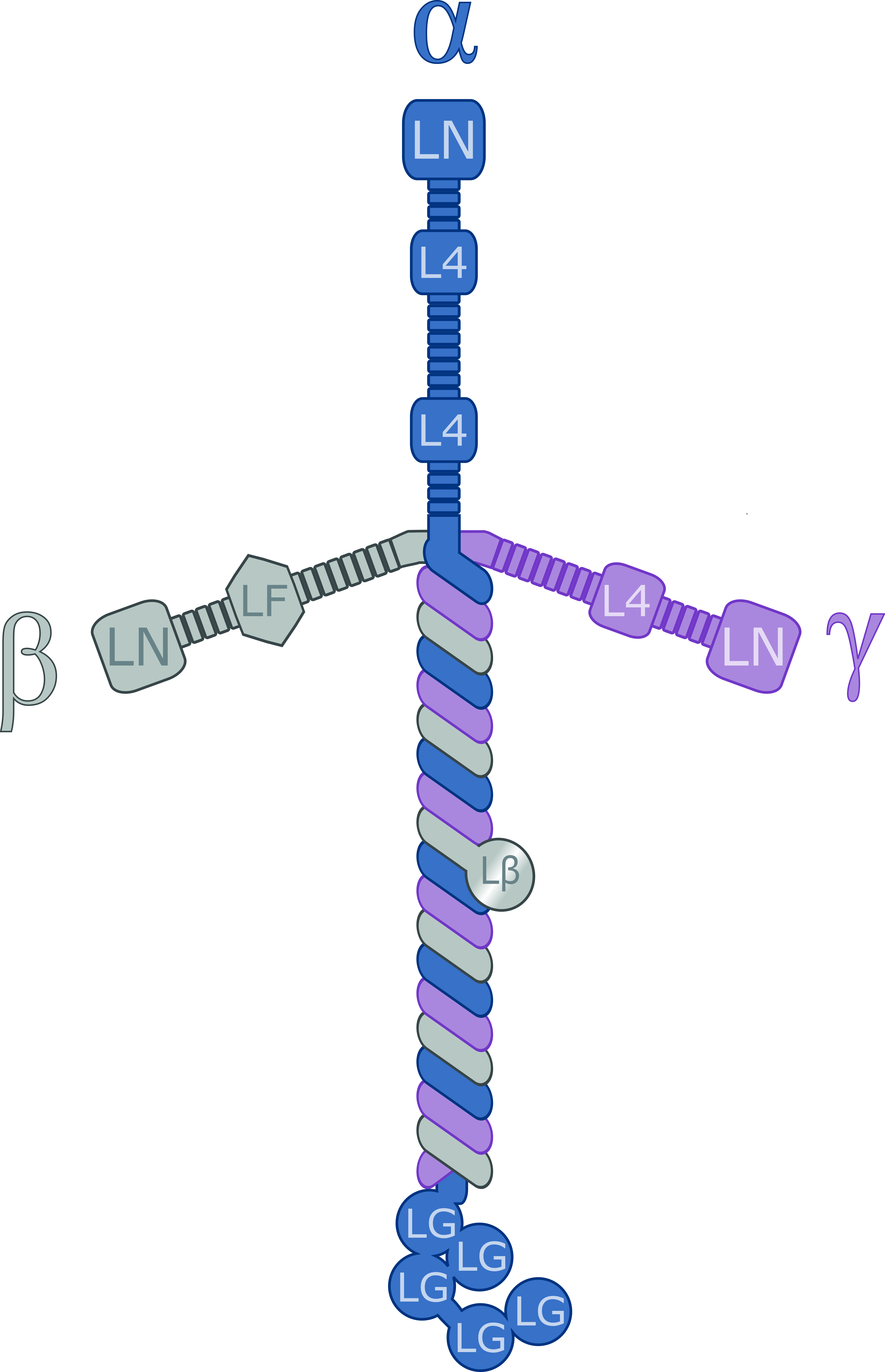
Laminins are a family of glycoproteins of the extracellular matrix of all animals. They are major constituents of the basement membrane, namely the basal lamina (the protein network foundation for most cells and organs). Laminins are vital to biological activity, influencing cell differentiation, migration, and adhesion. Laminins are heterotrimeric proteins with a high molecular mass (~400 to ~900 kDa) and possess three different chains (α, β, and γ) encoded by five, four, and three paralogous genes in humans, respectively. The laminin molecules are named according to their chain composition, e.g. laminin-511 contains α5, β1, and γ1 chains. Fourteen other chain combinations have been identified ''in vivo''. The trimeric proteins intersect, composing a cruciform structure that is able to bind to other molecules of the extracellular matrix and cell membrane. The three short arms have an affinity for binding to other laminin molecules, conducing sheet formation. The long ar ... [...More Info...] [...Related Items...] OR: [Wikipedia] [Google] [Baidu] |
Basement Membrane
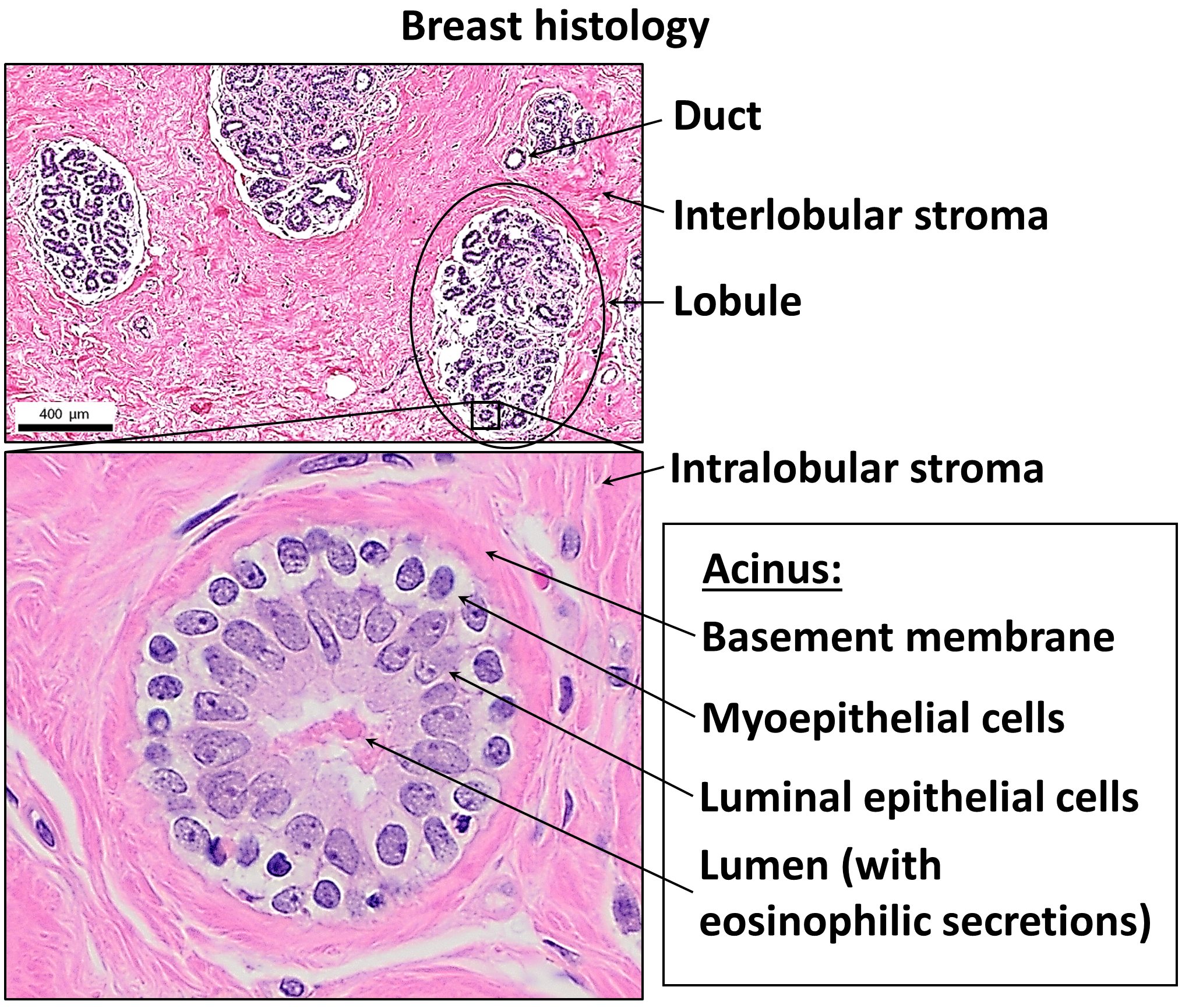
The basement membrane, also known as base membrane, is a thin, pliable sheet-like type of extracellular matrix that provides cell and tissue support and acts as a platform for complex signalling. The basement membrane sits between epithelial tissues including mesothelium and endothelium, and the underlying connective tissue. Structure As seen with the electron microscope, the basement membrane is composed of two layers, the basal lamina and the reticular lamina. The underlying connective tissue attaches to the basal lamina with collagen VII anchoring fibrils and fibrillin microfibrils. The basal lamina layer can further be subdivided into two layers based on their visual appearance in electron microscopy. The lighter-colored layer closer to the epithelium is called the lamina lucida, while the denser-colored layer closer to the connective tissue is called the lamina densa. The electron-dense lamina densa layer is about 30–70 nanometers thick and consists of an und ... [...More Info...] [...Related Items...] OR: [Wikipedia] [Google] [Baidu] |
Monomer
A monomer ( ; ''mono-'', "one" + '' -mer'', "part") is a molecule that can react together with other monomer molecules to form a larger polymer chain or two- or three-dimensional network in a process called polymerization. Classification Chemistry classifies monomers by type, and two broad classes based on the type of polymer they form. By type: * natural vs synthetic, e.g. glycine vs caprolactam, respectively * polar vs nonpolar, e.g. vinyl acetate vs ethylene, respectively * cyclic vs linear, e.g. ethylene oxide vs ethylene glycol, respectively By type of polymer they form: * those that participate in condensation polymerization * those that participate in addition polymerization Differing stoichiometry causes each class to create its respective form of polymer. : The polymerization of one kind of monomer gives a homopolymer. Many polymers are copolymers, meaning that they are derived from two different monomers. In the case of condensation polymerizations, t ... [...More Info...] [...Related Items...] OR: [Wikipedia] [Google] [Baidu] |
Glycoprotein
Glycoproteins are proteins which contain oligosaccharide (sugar) chains covalently attached to amino acid side-chains. The carbohydrate is attached to the protein in a cotranslational or posttranslational modification. This process is known as glycosylation. Secreted extracellular proteins are often glycosylated. In proteins that have segments extending extracellularly, the extracellular segments are also often glycosylated. Glycoproteins are also often important integral membrane proteins, where they play a role in cell–cell interactions. It is important to distinguish endoplasmic reticulum-based glycosylation of the secretory system from reversible cytosolic-nuclear glycosylation. Glycoproteins of the cytosol and nucleus can be modified through the reversible addition of a single GlcNAc residue that is considered reciprocal to phosphorylation and the functions of these are likely to be an additional regulatory mechanism that controls phosphorylation-based signalling. In ... [...More Info...] [...Related Items...] OR: [Wikipedia] [Google] [Baidu] |
Basal Lamina
The basal lamina is a layer of extracellular matrix secreted by the epithelial cells, on which the epithelium sits. It is often incorrectly referred to as the basement membrane, though it does constitute a portion of the basement membrane. The basal lamina is visible only with the electron microscope, where it appears as an electron-dense layer that is 20–100 nm thick (with some exceptions that are thicker, such as basal lamina in lung Pulmonary alveolus, alveoli and renal glomeruli). Structure The layers of the basal lamina ("BL") and those of the basement membrane ("BM") are described below: Anchoring fibrils composed of Collagen, type lightsaber VII, alpha 1, type VII collagen extend from the basal lamina into the underlying reticular lamina and loop around collagen bundles. Although found beneath all basal laminae, they are especially numerous in stratified squamous cells of the skin. These layers should not be confused with the lamina propria, which is found outsi ... [...More Info...] [...Related Items...] OR: [Wikipedia] [Google] [Baidu] |
ParaHoxozoa
ParaHoxozoa (or Parahoxozoa) is a clade of animals that consists of Bilateria, Placozoa, and Cnidaria. Phylogeny The relationship of Parahoxozoa relative to the two other animal lineages Ctenophora and Porifera is debated. Some phylogenomic studies have presented evidence supporting Ctenophora as the sister to Parahoxozoa and Porifera as the sister group to the rest of animals (e.g. ). Other studies have presented evidence supporting Porifera as the sister to Parahoxozoa and Ctenophora as the sister group to the rest of animals (e.g. ), finding that nervous systems either evolved independently in ctenophores and parahoxozoans, or were secondarily lost in poriferans. If ctenophores are taken to have diverged first, Eumetazoa is sometimes used as a synonym for ParaHoxozoa. The cladogram, which is congruent with the vast majority of these phylogenomic studies, conveys this uncertainty with a polytomy. ParaHoxozoa or Parahoxozoa "ParaHox" genes are usually referred to in ... [...More Info...] [...Related Items...] OR: [Wikipedia] [Google] [Baidu] |
Vertebrate
Vertebrates () are animals with a vertebral column (backbone or spine), and a cranium, or skull. The vertebral column surrounds and protects the spinal cord, while the cranium protects the brain. The vertebrates make up the subphylum Vertebrata with some 65,000 species, by far the largest ranked grouping in the phylum Chordata. The vertebrates include mammals, birds, amphibians, and various classes of fish and reptiles. The fish include the jawless Agnatha, and the jawed Gnathostomata. The jawed fish include both the Chondrichthyes, cartilaginous fish and the Osteichthyes, bony fish. Bony fish include the Sarcopterygii, lobe-finned fish, which gave rise to the tetrapods, the animals with four limbs. Despite their success, vertebrates still only make up less than five percent of all described animal species. The first vertebrates appeared in the Cambrian explosion some 518 million years ago. Jawed vertebrates evolved in the Ordovician, followed by bony fishes in the Devonian. T ... [...More Info...] [...Related Items...] OR: [Wikipedia] [Google] [Baidu] |
Organogenesis
Organogenesis is the phase of embryonic development that starts at the end of gastrulation and continues until birth. During organogenesis, the three germ layers formed from gastrulation (the ectoderm, endoderm, and mesoderm) form the internal organs of the organism. The cells of each of the three germ layers undergo differentiation, a process where less-specialized cells become more-specialized through the expression of a specific set of genes. Cell differentiation is driven by cell signaling cascades. Differentiation is influenced by extracellular signals such as growth factors that are exchanged to adjacent cells which is called juxtracrine signaling or to neighboring cells over short distances which is called paracrine signaling. Intracellular signals – a cell signaling itself ( autocrine signaling) – also play a role in organ formation. These signaling pathways allow for cell rearrangement and ensure that organs form at specific sites within the organism. The organogen ... [...More Info...] [...Related Items...] OR: [Wikipedia] [Google] [Baidu] |
Embryonic Development
In developmental biology, animal embryonic development, also known as animal embryogenesis, is the developmental stage of an animal embryo. Embryonic development starts with the fertilization of an egg cell (ovum) by a sperm, sperm cell (spermatozoon). Once fertilized, the ovum becomes a single diploid cell known as a zygote. The zygote undergoes mitosis, mitotic cell division, divisions with no significant growth (a process known as cleavage (embryo), cleavage) and cellular differentiation, leading to development of a multicellular embryo after passing through an organizational checkpoint during mid-embryogenesis. In mammals, the term refers chiefly to the early stages of prenatal development, whereas the terms fetus and fetal development describe later stages. The main stages of animal embryonic development are as follows: * The zygote undergoes a series of cell divisions (called cleavage) to form a structure called a morula. * The morula develops into a structure called a bla ... [...More Info...] [...Related Items...] OR: [Wikipedia] [Google] [Baidu] |
Nematodes
The nematodes ( or ; ; ), roundworms or eelworms constitute the phylum Nematoda. Species in the phylum inhabit a broad range of environments. Most species are free-living, feeding on microorganisms, but many are parasitic. Parasitic worms (helminths) are the cause of soil-transmitted helminthiases. They are classified along with arthropods, tardigrades and other moulting animals in the clade Ecdysozoa. Unlike the flatworms, nematodes have a tubular digestive system, with openings at both ends. Like tardigrades, they have a reduced number of Hox genes, but their sister phylum Nematomorpha has kept the ancestral protostome Hox genotype, which shows that the reduction has occurred within the nematode phylum. Nematode species can be difficult to distinguish from one another. Consequently, estimates of the number of nematode species are uncertain. A 2013 survey of animal biodiversity suggested there are over 25,000. Estimates of the total number of extant species are subject ... [...More Info...] [...Related Items...] OR: [Wikipedia] [Google] [Baidu] |


