|
WD-repeat
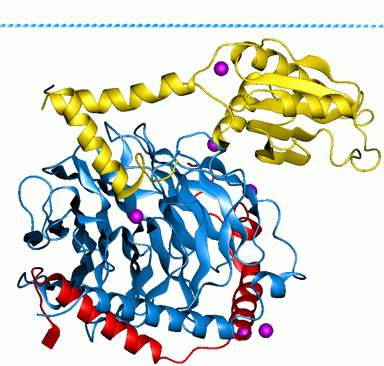
The WD40 repeat (also known as the WD or beta-transducin repeat) is a short structural motif of approximately 40 amino acids, often terminating in a tryptophan-aspartic acid (W-D) dipeptide. Tandem copies of these repeats typically fold together to form a type of circular solenoid protein domain called the WD40 domain. Structure WD40 domain-containing proteins have 4 to 16 repeating units, all of which are thought to form a circularised beta-propeller structure (see figure to the right). The WD40 domain is composed of several repeats, a variable region of around 20 residues at the beginning followed by a more common repeated set of residues. These repeats typically form a four stranded anti-parallel beta sheet or blade. These blades come together to form a propeller with the most common being a 7 bladed beta propeller. The blades interlock so that the last beta strand of one repeat forms with the first three of the next repeat to form the 3D blade structure. Function WD40-repe ... [...More Info...] [...Related Items...] OR: [Wikipedia] [Google] [Baidu] |
AAAS (gene)
Aladin, also known as adracalin, is a nuclear envelope protein that in humans is encoded by the ''AAAS'' gene. It is named after the achalasia–addisonianism–alacrima syndrome ( triple A syndrome) which occurs when the gene is mutated. Function Aladin is a component of the nuclear pore complex, to which it is attached by nucleoporin NDC1. Mutant aladin causes selective failure of nuclear protein import and hypersensitivity to oxidative stress. Mutant aladin also causes decreased nuclear import of aprataxin, a repair protein for single-strand breaks, and DNA ligase I, employed in DNA base excision repair. These decreases in DNA repair proteins may increase the susceptibility of cells to oxidative stress by allowing accumulation of oxidative DNA damages that trigger cell death. Clinical significance Mutations in the AAAS gene are responsible for Triple A syndrome (also known as Allgrove Syndrome). Triple-A syndrome is an autosomal An autosome is any chromos ... [...More Info...] [...Related Items...] OR: [Wikipedia] [Google] [Baidu] |
ATG16L1
Autophagy related 16 like 1 is a protein that in humans is encoded by the ''ATG16L1'' gene. This protein is characterized as a subunit of the autophagy-related ATG12- ATG5/ATG16 complex and is essentially important for the LC3 ( ATG8) lipidation and autophagosome formation. This complex localizes to the membrane and is released just before or after autophagosome completion. Furthermore, ATG16L1 appears to have other autophagy-independent functions, e.g., intracellular membrane trafficking regulation and inflammation. Autophagy in general plays a crucial role in pathways leading to innate and adaptive immunity activation. That is why many autophagy-related proteins, including ATG16L1, their gene expression and its role in autoimmune diseases are studied in-depth nowadays. Function Autophagy is the major intracellular degradation system delivering cytoplasmic components to lysosomes, and it accounts for degradation of most long-lived proteins and some organelles. Cytoplasmic ... [...More Info...] [...Related Items...] OR: [Wikipedia] [Google] [Baidu] |
Beta-propeller
In structural biology, a beta-propeller (β-propeller) is a type of all-β protein architecture characterized by 4 to 8 highly symmetrical blade-shaped beta sheets arranged toroidally around a central axis. Together the beta-sheets form a funnel-like active site. Structure Each beta-sheet typically has four anti-parallel β-strands arranged in the beta-zigzag motif. The strands are twisted so that the first and fourth strands are almost perpendicular to each other. There are five classes of beta-propellers, each arrangement being a highly symmetrical structure with 4–8 beta sheets, all of which generally form a central tunnel that yields pseudo-symmetric axes. While, the protein's official active site for ligand-binding is formed at one end of the central tunnel by loops between individual beta-strands, protein-protein interactions can occur at multiple areas around the domain. Depending on the packing and tilt of the beta-sheets and beta-strands, the beta-propeller may hav ... [...More Info...] [...Related Items...] OR: [Wikipedia] [Google] [Baidu] |
Autophagy
Autophagy (or autophagocytosis; from the Greek language, Greek , , meaning "self-devouring" and , , meaning "hollow") is the natural, conserved degradation of the cell that removes unnecessary or dysfunctional components through a lysosome-dependent regulated mechanism. It allows the orderly degradation and recycling of cellular components. Although initially characterized as a primordial degradation pathway induced to protect against starvation, it has become increasingly clear that autophagy also plays a major role in the homeostasis of non-starved cells. Defects in autophagy have been linked to various human diseases, including neurodegeneration and cancer, and interest in modulating autophagy as a potential treatment for these diseases has grown rapidly. Four forms of autophagy have been identified: macroautophagy, microautophagy, chaperone-mediated autophagy (CMA), and crinophagy. In macroautophagy (the most thoroughly researched form of autophagy), cytoplasmic components ( ... [...More Info...] [...Related Items...] OR: [Wikipedia] [Google] [Baidu] |
Ribbon Diagram
Ribbon diagrams, also known as Richardson diagrams, are three-dimensional space, 3D schematic representations of protein structure and are one of the most common methods of protein depiction used today. The ribbon depicts the general course and organization of the protein backbone in 3D and serves as a visual framework for hanging details of the entire atomic structure, such as the balls for the oxygen atoms attached to myoglobin's active site in the adjacent figure. Ribbon diagrams are generated by interpolating a smooth curve through the polypeptide backbone. Alpha helix, α-helices are shown as coiled ribbons or thick tubes, Beta sheet, β-sheets as arrows, and non-repetitive coils or loops as lines or thin tubes. The direction of the Peptide, polypeptide chain is shown locally by the arrows, and may be indicated overall by a colour ramp along the length of the ribbon. Ribbon diagrams are simple yet powerful, expressing the visual basics of a molecular structure (twist, fold an ... [...More Info...] [...Related Items...] OR: [Wikipedia] [Google] [Baidu] |
G Protein
G proteins, also known as guanine nucleotide-binding proteins, are a Protein family, family of proteins that act as molecular switches inside cells, and are involved in transmitting signals from a variety of stimuli outside a cell (biology), cell to its interior. Their activity is regulated by factors that control their ability to bind to and hydrolyze guanosine triphosphate (GTP) to guanosine diphosphate (GDP). When they are bound to GTP, they are 'on', and, when they are bound to GDP, they are 'off'. G proteins belong to the larger group of enzymes called GTPases. There are two classes of G proteins. The first function as monomeric small GTPases (small G-proteins), while the second function as heterotrimeric G protein protein complex, complexes. The latter class of complexes is made up of ''G alpha subunit, alpha'' (Gα), ''beta'' (Gβ) and ''gamma'' (Gγ) protein subunit, subunits. In addition, the beta and gamma subunits can form a stable Protein dimer, dimeric complex re ... [...More Info...] [...Related Items...] OR: [Wikipedia] [Google] [Baidu] |
BRWD1
Bromodomain and WD repeat-containing protein 1 (BRWD1) also known as WD repeat-containing protein 9 (WDR9) is a protein that in humans is encoded by the ''BRWD1'' gene. Function This gene encodes a member of the WD repeat protein family. WD repeats are minimally conserved regions of approximately 40 amino acids typically bracketed by Gly-His and Trp- Asp (GH-WD), which may facilitate formation of heterotrimeric or multiprotein complexes. Members of this family are involved in a variety of cellular processes, including cell cycle progression, signal transduction, apoptosis, and gene regulation. This protein contains 2 bromodomains and 8 WD repeats, and the function of this protein is not known. This gene is located within the Down syndrome region-2 on chromosome 21. Alternative splicing of this gene generates 3 transcript variants diverging at the 3' end Directionality, in molecular biology and biochemistry, is the end-to-end chemical orientation of a single strand of nuc ... [...More Info...] [...Related Items...] OR: [Wikipedia] [Google] [Baidu] |
BOP1
Ribosome biogenesis protein BOP1 is a protein that in humans is encoded by the ''BOP1'' gene. It is a WD40 repeat-containing nucleolar protein involved in rRNA processing, thereby controlling the cell cycle. It is required for the maturation of the 25S and 5.8S ribosomal RNAs. It may serve as an essential factor in ribosome formation that coordinates processing of the spacer regions in pre-rRNA. Function The Pes1-Bop1 complex has several components: BOP1, GRWD1, PES1, ORC6L, and RPL3 and is involved in ribosome biogenesis and altered chromosome segregation. The overexpression of BOP1 increases the percentage of multipolar spindles in human cells. Deregulation of the BOP1 pathway may contribute to colorectal tumourigenesis in humans. Elevated levels of Bop1 induces Bop1/WDR12 and Bop1/Pes1 subcomplexes and the assembly and integrity of the PeBoW complex is highly sensitive to changes in Bop1 protein levels. Nop7p- Erb1p-Ytm1p, found in yeast, is potentially the ... [...More Info...] [...Related Items...] OR: [Wikipedia] [Google] [Baidu] |
ARPC1B
Actin-related protein 2/3 complex subunit 1B is a protein that in humans is encoded by the ''ARPC1B'' gene. Function This gene encodes one of seven subunits of the human Arp2/3 protein complex. This subunit is a member of the SOP2 family of proteins and is most similar to the protein encoded by gene ARPC1A. The similarity between these two proteins suggests that they both may function as p41 subunit of the human Arp2/3 complex that facilitates branching of actin filaments in cells. Isoforms of the p41 subunit may adapt the functions of the complex to different cell types or developmental stages. Indeed, it has recently been shown that variants of the Arp2/3 complex differ in their ability to promote actin assembly, with complexes containing ARPC1B and ARPC5L being better at this than those containing ARPC1A and ARPC5. The differing functions of ARPC1A and ARPC1B are also evident in the recent discovery of patients with severe or total ARPC1B deficiency, who have platelet and ... [...More Info...] [...Related Items...] OR: [Wikipedia] [Google] [Baidu] |
ARPC1A
Actin-related protein 2/3 complex subunit 1A is a protein that in humans is encoded by the ''ARPC1A'' gene. This gene encodes one of seven subunits of the human Arp2/3 protein complex. This subunit is a member of the SOP2 family of proteins and is most similar to the protein encoded by gene ARPC1B Actin-related protein 2/3 complex subunit 1B is a protein that in humans is encoded by the ''ARPC1B'' gene. Function This gene encodes one of seven subunits of the human Arp2/3 protein complex. This subunit is a member of the SOP2 family of p .... The similarity between these two proteins suggests that they both may function as p41 subunit of the human Arp2/3 complex that has been implicated in the control of actin polymerization in cells. It is possible that the p41 subunit is involved in assembling and maintaining the structure of the Arp2/3 complex. Multiple versions of the p41 subunit may adapt the functions of the complex to different cell types or developmental stages. Ref ... [...More Info...] [...Related Items...] OR: [Wikipedia] [Google] [Baidu] |
APAF1
Apoptotic protease activating factor 1, also known as APAF1, is a human homolog of ''C. elegans'' CED-4 gene. Function The protein was identified in the laboratory of Xiaodong Wang as an activator of caspase-3 in the presence of cytochromeC and dATP. This gene encodes a cytoplasmic protein that forms one of the central hubs in the apoptosis regulatory network. This protein contains (from the N terminal) a caspase recruitment domain (CARD), an ATPase domain (NB-ARC), few short helical domains and then several copies of the WD40 repeat domain. Upon binding cytochrome c and dATP, this protein forms an oligomeric apoptosome. The apoptosome binds and cleaves Procaspase-9 protein, releasing its mature, activated form. The precise mechanism for this reaction is still debated though work published by Guy Salvesen suggests that the apoptosome may induce caspase-9 dimerization and subsequent autocatalysis. Activated caspase-9 stimulates the subsequent caspase cascade that commits th ... [...More Info...] [...Related Items...] OR: [Wikipedia] [Google] [Baidu] |
AMBRA1
AMBRA1 (activating molecule in Beclin1-regulated autophagy) is a protein that is able to regulate cancer cells through autophagy. AMBRA1 is described as a mechanism cells use to divide and there is new evidence demonstrating the role and impact of AMBRA1 as a candidate for the treatment of several disorders and diseases, including anticancer therapy. It is known to suppress tumors and plays a role in mitophagy and apoptosis. AMBRA1 can be found in the cytoskeleton and mitochondria and during the process of autophagy, it is localized at the endoplasmic reticulum. In normal conditions, AMBRA1 is dormant and will bind to BCL2 in the outer membrane. This relocation enables autophagosome nucleation. AMBRA1 protein is involved in several cellular processes and is involved in the regulation of the immune system and nervous system. Function AMBRA1 serves to regulate the process of autophagy and this is the cellular breakdown and recycling of unnecessary or damaged cellular components. ... [...More Info...] [...Related Items...] OR: [Wikipedia] [Google] [Baidu] |


