|
Vaginal Support Structures
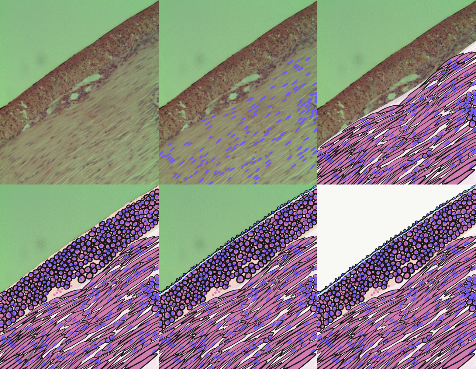
The vaginal support structures are those muscles, bones, ligaments, tendons, membranes and fascia, of the pelvic floor that maintain the position of the vagina within the pelvic cavity and allow the normal functioning of the vagina and other reproductive structures in the female. Defects or injuries to these support structures in the pelvic floor leads to pelvic organ prolapse. Anatomical and congenital variations of vaginal support structures can predispose a woman to further dysfunction and prolapse later in life. The urethra is part of the anterior wall of the vagina and damage to the support structures there can lead to incontinence and urinary retention. Pelvic bones The support for the vagina is provided by muscles, membranes, tendons and ligaments. These structures are attached to the hip bones. These bones are the pubis, ilium and ischium. The interior surface of these pelvic bones and their projections and contours are used as attachment sites for the fascia, muscles ... [...More Info...] [...Related Items...] OR: [Wikipedia] [Google] [Baidu] |
1116 Muscle Of The Female Perineum
Year 1116 ( MCXVI) was a leap year starting on Saturday (link will display the full calendar) of the Julian calendar. Events By place Byzantine Empire * Autumn – Battle of Philomelion: Emperor Alexios I (Komnenos) leads an expedition into Anatolia and meets the Seljuk army under Sultan Malik Shah (near Philomelium). The Byzantines introduce a new battle formation of Alexios' devising, the ''parataxis'' (a defensive formation, consisting of a hollow square, with the baggage in the centre). During the battle, the Seljuk Turks mount several attacks on the formations, but all are repulsed. The Byzantine cavalry makes two counterattacks; the first is unsuccessful. But a second attack, led by Nikephoros Bryennios (the Younger), breaks the Seljuk forces, who then turn to flight. The following day Malik Shah again attacks, his army completely surrounding the Byzantines from all sides. The Seljuk Turks are once more repulsed, with many losses. Alexios claims the vict ... [...More Info...] [...Related Items...] OR: [Wikipedia] [Google] [Baidu] |
Sacrospinous Ligament
The sacrospinous ligament (small or anterior sacrosciatic ligament) is a thin, triangular ligament in the human pelvis. The base of the ligament is attached to the outer edge of the sacrum and coccyx, and the tip of the ligament attaches to the spine of the ischium, a bony protuberance on the human pelvis. Its fibres are intermingled with the sacrotuberous ligament. Structure The sacrotuberous ligament passes behind the sacrospinous ligament. In its entire length, the sacrospinous ligament covers the equally triangular coccygeus muscle, to which its closely connected.Gray's Anatomy 1918 Function The presence of the ligament in the greater sciatic notch creates an opening (foramen), the greater sciatic foramen, and also converts the lesser sciatic notch into the lesser sciatic foramen.Platzer (2004), p 188 The greater sciatic foramen lies above the ligament, and the lesser sciatic foramen lies below it. The pudendal vessels and nerve pass behind the sacrospinous lig ... [...More Info...] [...Related Items...] OR: [Wikipedia] [Google] [Baidu] |
Tendinous Arch Of Pelvic Fascia
At the level of a line extending from the lower part of the pubic symphysis to the spine of the ischium The ischial spine is part of the posterior border of the body of the ischium bone of the pelvis. It is a thin and pointed triangular eminence, more or less elongated in different subjects. Structure The pudendal nerve travels close to the isch ... is a thickened whitish band in this upper layer of the diaphragmatic part of the pelvic fascia. It is termed the tendinous arch or white line of the pelvic fascia, and marks the line of attachment of the special fascia (pars endopelvina fasciae pelvis) which is associated with the pelvic viscera. It joins the fascia of the pubocervical fascia that covers the anterior wall of the vagina. If this fascia falls, the ipsilateral side of the vagina falls, carrying with it the bladder and the urethra, and thus contributing to urinary incontinence. References External links * - "The Female Pelvis: Muscles of the Pelvic Diaphragm" * ... [...More Info...] [...Related Items...] OR: [Wikipedia] [Google] [Baidu] |
Gray236
Grey (more common in British English) or gray (more common in American English) is an intermediate color between black and white. It is a neutral or achromatic color, meaning literally that it is "without color", because it can be composed of black and white. It is the color of a cloud-covered sky, of ash and of lead. The first recorded use of ''grey'' as a color name in the English language was in 700 CE.Maerz and Paul ''A Dictionary of Color'' New York:1930 McGraw-Hill Page 196 ''Grey'' is the dominant spelling in European and Commonwealth English, while ''gray'' has been the preferred spelling in American English; both spellings are valid in both varieties of English. In Europe and North America, surveys show that grey is the color most commonly associated with neutrality, conformity, boredom, uncertainty, old age, indifference, and modesty. Only one percent of respondents chose it as their favorite color. Etymology ''Grey'' comes from the Middle English or , ... [...More Info...] [...Related Items...] OR: [Wikipedia] [Google] [Baidu] |
Connective Tissue
Connective tissue is one of the four primary types of animal tissue, along with epithelial tissue, muscle tissue, and nervous tissue. It develops from the mesenchyme derived from the mesoderm the middle embryonic germ layer. Connective tissue is found in between other tissues everywhere in the body, including the nervous system. The three meninges, membranes that envelop the brain and spinal cord are composed of connective tissue. Most types of connective tissue consists of three main components: elastic and collagen fibers, ground substance, and cells. Blood, and lymph are classed as specialized fluid connective tissues that do not contain fiber. All are immersed in the body water. The cells of connective tissue include fibroblasts, adipocytes, macrophages, mast cells and leucocytes. The term "connective tissue" (in German, ''Bindegewebe'') was introduced in 1830 by Johannes Peter Müller. The tissue was already recognized as a distinct class in the 18th century. ... [...More Info...] [...Related Items...] OR: [Wikipedia] [Google] [Baidu] |
Smooth Muscle Tissue
Smooth muscle is an involuntary non- striated muscle, so-called because it has no sarcomeres and therefore no striations (''bands'' or ''stripes''). It is divided into two subgroups, single-unit and multiunit smooth muscle. Within single-unit muscle, the whole bundle or sheet of smooth muscle cells contracts as a syncytium. Smooth muscle is found in the walls of hollow organs, including the stomach, intestines, bladder and uterus; in the walls of passageways, such as blood, and lymph vessels, and in the tracts of the respiratory, urinary, and reproductive systems. In the eyes, the ciliary muscles, a type of smooth muscle, dilate and contract the iris and alter the shape of the lens. In the skin, smooth muscle cells such as those of the arrector pili cause hair to stand erect in response to cold temperature or fear. Structure Gross anatomy Smooth muscle is grouped into two types: single-unit smooth muscle, also known as visceral smooth muscle, and multiunit smooth mus ... [...More Info...] [...Related Items...] OR: [Wikipedia] [Google] [Baidu] |
Internal Anal Sphincter Muscles
The internal anal sphincter, IAS, (or sphincter ani internus) is a ring of smooth muscle that surrounds about 2.5–4.0 cm of the anal canal; its inferior border is in contact with, but quite separate from, the external anal sphincter. It is myogenic in nature and playing phasic and tonic state for relaxation and contraction. This myogenic tone is based on Ca2+/Calmodulin/MLCK/RhoA/ROCK signaling cascade pathway. Recent studies suggested, BDNF is the member of the neurotrophin family; having next target for basal IAS and NANC relaxation. It is about 5 mm thick, and is formed by an aggregation of the involuntary circular fibers of the rectum. Its lower border is about 6 mm from the orifice of the anus. Actions Its action is entirely involuntary, and it is in a state of continuous maximal contraction. It helps the Sphincter ani externus to occlude the anal aperture and aids in the expulsion of the feces. Sympathetic fibers from the superior rectal and hypogastric ple ... [...More Info...] [...Related Items...] OR: [Wikipedia] [Google] [Baidu] |
Transverse Perineal Muscles
The transverse perineal muscles (transversus perinei) are the superficial and the deep transverse perineal muscles. Superficial transverse perineal The superficial transverse perineal muscle (transversus superficialis perinei or Lloyd-Beanie muscle) is a narrow muscular slip, which passes more or less transversely across the perineal space in front of the anus. It arises by tendinous fibers from the inner and forepart of the ischial tuberosity and, running medially, is inserted into the central tendinous point of the perineum (perineal body), joining in this situation with the muscle of the opposite side, with the external anal sphincter muscle behind, and with the bulbospongiosus muscle in front. In some cases, the fibers of the deeper layer of the external anal sphincter cross over in front of the anus and are continued into this mu ... [...More Info...] [...Related Items...] OR: [Wikipedia] [Google] [Baidu] |
Bulbospongiosus Muscle
The bulbospongiosus muscle (bulbocavernosus in older texts) is one of the superficial muscles of the perineum. It has a slightly different origin, insertion and function in males and females. In males, it covers the bulb of the penis. In females, it covers the vestibular bulb. In both sexes, it is innervated by the deep or muscular branch of the perineal nerve, which is a branch of the pudendal nerve. Structure In males, the bulbospongiosus is located in the middle line of the perineum, in front of the anus. It consists of two symmetrical parts, united along the median line by a tendinous perineal raphe. It arises from the central tendinous point of the perineum and from the median perineal raphe in front. In females, there is no union, nor a tendinous perineal raphe; the parts are disjoint primarily and arise from the same central tendinous point of the perineum, which is the tendon that is formed at the point where the bulbospongiosus muscle, superficial transverse ... [...More Info...] [...Related Items...] OR: [Wikipedia] [Google] [Baidu] |
Perineal Body
The perineum in humans is the space between the anus and scrotum in the male, or between the anus and the vulva in the female. The perineum is the region of the body between the pubic symphysis (pubic arch) and the coccyx (tail bone), including the perineal body and surrounding structures. There is some variability in how the boundaries are defined. The perineal raphe is visible and pronounced to varying degrees. The perineum is an erogenous zone. The word perineum entered English from late Latin via Greek περίναιος ~ περίνεος ''perinaios, perineos'', itself from περίνεος, περίνεοι 'male genitals' and earlier περίς ''perís'' 'penis' through influence from πηρίς ''pērís'' 'scrotum'. The term was originally understood as a purely male body-part with the perineal raphe seen as a continuation of the scrotal septum since masculinization causes the development of a large anogenital distance in men, in comparison to the corresponding ... [...More Info...] [...Related Items...] OR: [Wikipedia] [Google] [Baidu] |
Perineal Membrane
The perineal membrane is an anatomical term for a fibrous membrane in the perineum. The term "inferior fascia of urogenital diaphragm", used in older texts, is considered equivalent to the perineal membrane. It is the superior border of the superficial perineal pouch, and the inferior border of the deep perineal pouch. Structure The perineal membrane is triangular in shape. It attaches to both ischiopubic rami of the pelvis. It also attaches to the perineal body. It is about 4 cm. in depth. Its apex is directed forward, and is separated from the arcuate pubic ligament by an oval opening for the transmission of the deep dorsal vein of the penis or the deep dorsal vein of the clitoris. Its lateral margins are attached on either side to the inferior rami of the pubis and ischium, above the crus penis. Its base is directed toward the rectum, and connected to the central tendinous point of the perineum. The base is fused with both the pelvic fascia and Colle's fascia. ... [...More Info...] [...Related Items...] OR: [Wikipedia] [Google] [Baidu] |

_mini.jpg)


