|
Perineal Membrane
The perineal membrane is an anatomical term for a fibrous membrane in the perineum. The term "inferior fascia of urogenital diaphragm", used in older texts, is considered equivalent to the perineal membrane. It is the superior border of the superficial perineal pouch, and the inferior border of the deep perineal pouch. Structure The perineal membrane is triangular in shape. It attaches to both ischiopubic rami of the pelvis. It also attaches to the perineal body. Female The perineal membrane has two distinct portions that span the opening of the anterior pelvic outlet. The ''ventral'' (anterior) portion is associated with the compressor urethra and urethrovaginal sphincter muscles (previously called deep transverse perineal muscles), and is continuous with the insertion of the arcus tendineus fascia pelvis. The apex is separated from the arcuate pubic ligament by an oval opening for the transmission of the deep dorsal vein of the clitoris. The ''dorsal'' (posterior) ... [...More Info...] [...Related Items...] OR: [Wikipedia] [Google] [Baidu] |
Perineum
The perineum (: perineums or perinea) in placentalia, placental mammals is the space between the anus and the genitals. The human perineum is between the anus and scrotum in the male or between the anus and vulva in the female. The perineum is the region of the body between the pubic symphysis (pubic arch) and the coccyx (tail bone), including the perineal body and surrounding structures. The perineal raphe is visible and pronounced to varying degrees. Etymology The word entered English from late Latin via Greek language, Greek περίναιος ~ περίνεος ''perinaios, perineos'', itself from περίνεος, περίνεοι 'male genitals' and earlier περίς ''perís'' 'penis' through influence from πηρίς ''pērís'' 'scrotum'. The term was originally understood as a purely male body-part with the perineal raphe seen as a continuation of the scrotal septum since Virilization, masculinization causes the development of a large anogenital distance in men, i ... [...More Info...] [...Related Items...] OR: [Wikipedia] [Google] [Baidu] |
Ischium
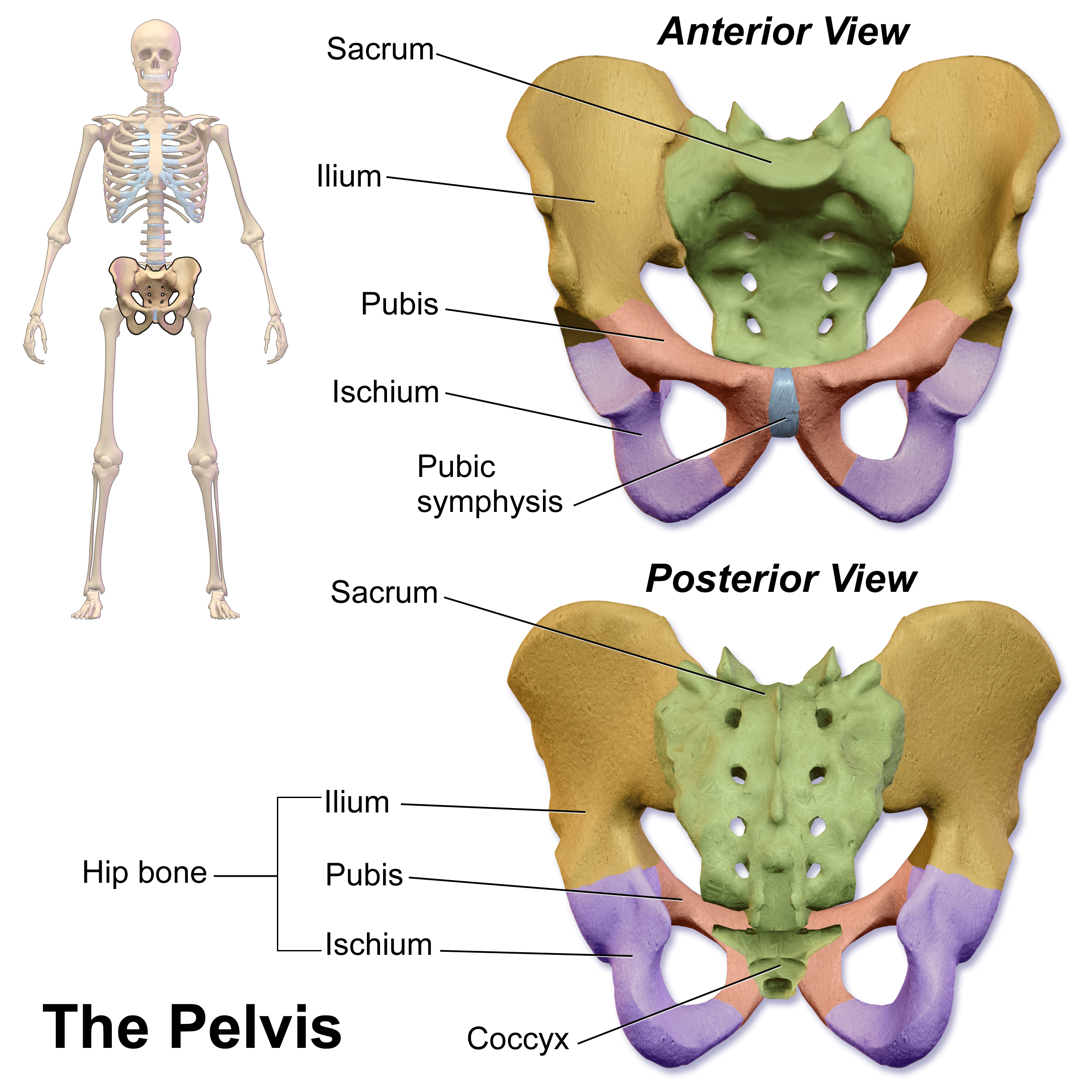
The ischium (; : ischia) is a paired bone forming the lower and back part of the hip bone. Situated below the ilium (bone), ilium and behind the pubis (bone), pubis, it is one of three regions whose fusion creates the coxal bone. The superior portion of this region forms approximately one-third of the acetabulum. Structure The ischium is made up of three parts–the body, the superior ramus and the inferior ramus. The body contains a prominent ischial spine, spine, which serves as the origin for the superior gemellus muscle. The indentation inferior to the spine is the lesser sciatic notch. Continuing down the posterior side, the ischial tuberosity is a thick, rough-surfaced prominence below the lesser sciatic notch. This is the portion ...[...More Info...] [...Related Items...] OR: [Wikipedia] [Google] [Baidu] |
Pudendal Arteries
The pudendal arteries are a group of arteries which supply many of the muscles and organs of the pelvic cavity. The arteries include the internal pudendal artery, the superficial external pudendal artery, and the deep external pudendal artery. The internal pudendal artery branches off the internal iliac artery, the main artery of the pelvis, and supplies blood to the sex organs. The internal pudendal artery gives rise to the perineal artery and the inferior rectal artery. The superficial external pudendal artery arises from the medial side of the femoral artery. It supplies the male scrotum and the female labia majora In primates, and specifically in humans, the labia majora (: labium majus), also known as the outer lips or outer labia, are two prominent Anatomical terms of location, longitudinal skin folds that extend downward and backward from the mons pubis .... References Arteries of the lower limb Arteries of the abdomen {{Portal bar, Anatomy ... [...More Info...] [...Related Items...] OR: [Wikipedia] [Google] [Baidu] |
Urethral Sphincter
The urethral sphincters are two muscles used to control the exit of urine in the urinary bladder through the urethra. The two muscles are either the external sphincter muscle of male urethra, male or external sphincter muscle of female urethra, female external urethral sphincter and the internal urethral sphincter. When either of these muscles contracts, the urethra is sealed shut. The external urethral sphincter originates at the ischiopubic ramus and inserts into the intermeshing muscle fibers from the other side. It is controlled by the deep perineal branch of the pudendal nerve. Activity in the nerve fibers constricts the urethra. * The internal sphincter muscle of urethra: located at the bladder's inferior end and the urethra's proximal end at the junction of the urethra with the urinary bladder. The internal sphincter is a continuation of the detrusor muscle and is made of smooth muscle, therefore it is under Smooth muscle tissue, involuntary or autonomic nervous system, a ... [...More Info...] [...Related Items...] OR: [Wikipedia] [Google] [Baidu] |
Deep Transverse Perineal Muscle
The transverse perineal muscles (transversus perinei) are the superficial and the deep transverse perineal muscles. Superficial transverse perineal The superficial transverse perineal muscle (transversus superficialis perinei or Lloyd-Beanie muscle) is a narrow muscular slip, which passes more or less transversely across the perineal space in front of the anus. It arises by tendinous fibers from the inner and forepart of the ischial tuberosity and, running medially, is inserted into the central tendinous point of the perineum (perineal body), joining in this situation with the muscle of the opposite side, with the external anal sphincter muscle behind, and with the bulbospongiosus muscle in front. In some cases, the fibers of the deeper layer of the external anal sphincter cross over in front of the anus and are continued into this mu ... [...More Info...] [...Related Items...] OR: [Wikipedia] [Google] [Baidu] |
Membranous Urethra
The membranous urethra or intermediate part of male urethra is the shortest, least dilatable, and, with the exception of the urinary meatus, the narrowest part of the urethra. It extends from the apex of the prostate proximally to the bulb of urethra distally. It measures some 12 mm in length. It traverses the pelvic floor. It is surrounded by the external urethral sphincter, which is in turn envelopped by the superior fascia of the urogenital diaphragm. Anatomy The mucosal internal lining of the membranous urethra features some longitudinal folds which disappear when the urethra becomes distended. Relations It extends downward and forward, with a slight anterior concavity, between the apex of the prostate and the bulb of the urethra, perforating the urogenital diaphragm about 2.5 cm below and behind the pubic symphysis. The hinder part of the urethral bulb lies in apposition with the inferior fascia of the urogenital diaphragm, but its upper portion diverges somew ... [...More Info...] [...Related Items...] OR: [Wikipedia] [Google] [Baidu] |
Bulbourethral Glands
The bulbourethral glands or Cowper's glands (named for English anatomist William Cowper (anatomist), William Cowper) are two small exocrine gland, exocrine and Male accessory gland, accessory glands in the reproductive system of many Mammal, male mammals. They are homology (biology), homologous to Bartholin's glands in females. The bulbourethral glands are responsible for producing a pre-ejaculate fluid called Pre-ejaculate, Cowper's fluid (known colloquially as ''pre-cum''), which is secreted during sexual arousal, neutralizing the acidity of the urethra in preparation for the passage of sperm cells. The paired glands are found adjacent to the urethra just below the prostate, seen best by screening (medicine) MRI as a tool in preventative healthcare in males. Screening MRI may be performed when there is a positive prostate-specific antigen on basic laboratory tests. Prostate cancer is the second-most common cause of cancer-related mortality in males in the USA. Most species of Pl ... [...More Info...] [...Related Items...] OR: [Wikipedia] [Google] [Baidu] |
Urethra
The urethra (: urethras or urethrae) is the tube that connects the urinary bladder to the urinary meatus, through which Placentalia, placental mammals Urination, urinate and Ejaculation, ejaculate. The external urethral sphincter is a striated muscle that allows voluntary control over urination. The Internal urethral sphincter, internal sphincter, formed by the involuntary smooth muscles lining the bladder neck and urethra, receives its nerve supply by the Sympathetic nervous system, sympathetic division of the autonomic nervous system. The internal sphincter is present both in males and females. Structure The urethra is a fibrous and muscular tube which connects the urinary bladder to the external urethral meatus. Its length differs between the sexes, because it passes through the penis in males. Male In the human male, the urethra is on average long and opens at the end of the external urethral meatus. The urethra is divided into four parts in men, named after the lo ... [...More Info...] [...Related Items...] OR: [Wikipedia] [Google] [Baidu] |
Pubic Symphysis
The pubic symphysis (: symphyses) is a secondary cartilaginous joint between the left and right superior rami of the pubis of the hip bones. It is in front of and below the urinary bladder. In males, the suspensory ligament of the penis attaches to the pubic symphysis. In females, the pubic symphysis is attached to the suspensory ligament of the clitoris. In most adults, it can be moved roughly 2 mm and with 1 degree rotation. This increases for women at the time of childbirth. The name comes from the Greek word ''symphysis'', meaning 'growing together'. Structure The pubic symphysis is a nonsynovial amphiarthrodial joint. The width of the pubic symphysis at the front is 3–5 mm greater than its width at the back. This joint is connected by fibrocartilage and may contain a fluid-filled cavity; the center is avascular, possibly due to the nature of the compressive forces passing through this joint, which may lead to harmful vascular disease. The ends of both pubi ... [...More Info...] [...Related Items...] OR: [Wikipedia] [Google] [Baidu] |
Inferior Fascia Of Pelvic Diaphragm
The pelvic fasciae are the fascia of the pelvis and can be divided into: * (a) the fascial sheaths of ** the obturator internus muscles ( fascia of the obturator internus) ** the piriformis muscles ( fascia of the piriformis) ** the pelvic floor * (b) fascia associated with the organs of the pelvis. Structure Fascia of pelvic organs Pelvic fascia extends to cover the organs within the pelvis. It is attached to the fascia that runs along the pelvic floor along the tendinous arch. The fascia which covers pelvic organs can be divided according to the organs that are covered: * The front is known as the "vesical layer". It forms the anterior and lateral ligaments of the bladder. * In males, its middle lamina crosses the floor of the pelvis between the rectum and vesiculæ seminales as the ''rectovesical septum''; in the female this is perforated by the cervix and is named the transverse cervical ligament. * At the back, the fascia passes to the side of the rectum; it forms a loos ... [...More Info...] [...Related Items...] OR: [Wikipedia] [Google] [Baidu] |
Superficial Transverse Perineal Muscle
The transverse perineal muscles (transversus perinei) are the superficial and the deep transverse perineal muscles. Superficial transverse perineal The superficial transverse perineal muscle (transversus superficialis perinei or Lloyd-Beanie muscle) is a narrow muscular slip, which passes more or less transversely across the perineal space in front of the anus. It arises by tendinous fibers from the inner and forepart of the ischial tuberosity and, running medially, is inserted into the central tendinous point of the perineum (perineal body), joining in this situation with the muscle of the opposite side, with the external anal sphincter muscle behind, and with the bulbospongiosus muscle in front. In some cases, the fibers of the deeper layer of the external anal sphincter cross over in front of the anus and are continued into thi ... [...More Info...] [...Related Items...] OR: [Wikipedia] [Google] [Baidu] |
Superficial Fascia
A fascia (; : fasciae or fascias; adjective fascial; ) is a generic term for macroscopic membranous bodily structures. Fasciae are classified as superficial, visceral or deep, and further designated according to their anatomical location. The knowledge of fascial structures is essential in surgery, as they create borders for infectious processes (for example Psoas abscess) and haematoma. An increase in pressure may result in a compartment syndrome, where a prompt fasciotomy may be necessary. For this reason, profound descriptions of fascial structures are available in anatomical literature from the 19th century. Function Fasciae were traditionally thought of as passive structures that transmit mechanical tension generated by muscular activities or external forces throughout the body. An important function of muscle fasciae is to reduce friction of muscular force. In doing so, fasciae provide a supportive and movable wrapping for nerves and blood vessels as they pass through ... [...More Info...] [...Related Items...] OR: [Wikipedia] [Google] [Baidu] |


