|
Superficial Fascia
A fascia (; : fasciae or fascias; adjective fascial; ) is a generic term for macroscopic membranous bodily structures. Fasciae are classified as superficial, visceral or deep, and further designated according to their anatomical location. The knowledge of fascial structures is essential in surgery, as they create borders for infectious processes (for example Psoas abscess) and haematoma. An increase in pressure may result in a compartment syndrome, where a prompt fasciotomy may be necessary. For this reason, profound descriptions of fascial structures are available in anatomical literature from the 19th century. Function Fasciae were traditionally thought of as passive structures that transmit mechanical tension generated by muscular activities or external forces throughout the body. An important function of muscle fasciae is to reduce friction of muscular force. In doing so, fasciae provide a supportive and movable wrapping for nerves and blood vessels as they pass through ... [...More Info...] [...Related Items...] OR: [Wikipedia] [Google] [Baidu] |
Rectus Sheath
The rectus sheath (also called the rectus fascia.) is a tough fibrous compartment formed by the aponeuroses of the transverse abdominal, transverse abdominal muscle, and the internal oblique, internal and external oblique muscles. It contains the Rectus abdominis muscle, rectus abdominis and Pyramidalis muscle, pyramidalis muscles, as well as vessels and nerves. Structure The rectus sheath extends between the inferior costal margin and costal cartilages of ribs 5-7 superiorly, and the pubic crest inferiorly. Studies indicate that all three aponeuroses constituting the rectus sheath are in fact bilaminar. Below the costal margin Superficial/anterior to the anterior layer of the rectus sheath are the following two layers: # Camper's fascia (anterior part of superficial fascia) # Scarpa's fascia (posterior part of the superficial fascia) Deep/posterior posterior layer of the rectus sheath (where present) are the following three layers: # transversalis fascia # extraperitoneal ... [...More Info...] [...Related Items...] OR: [Wikipedia] [Google] [Baidu] |
Human Body
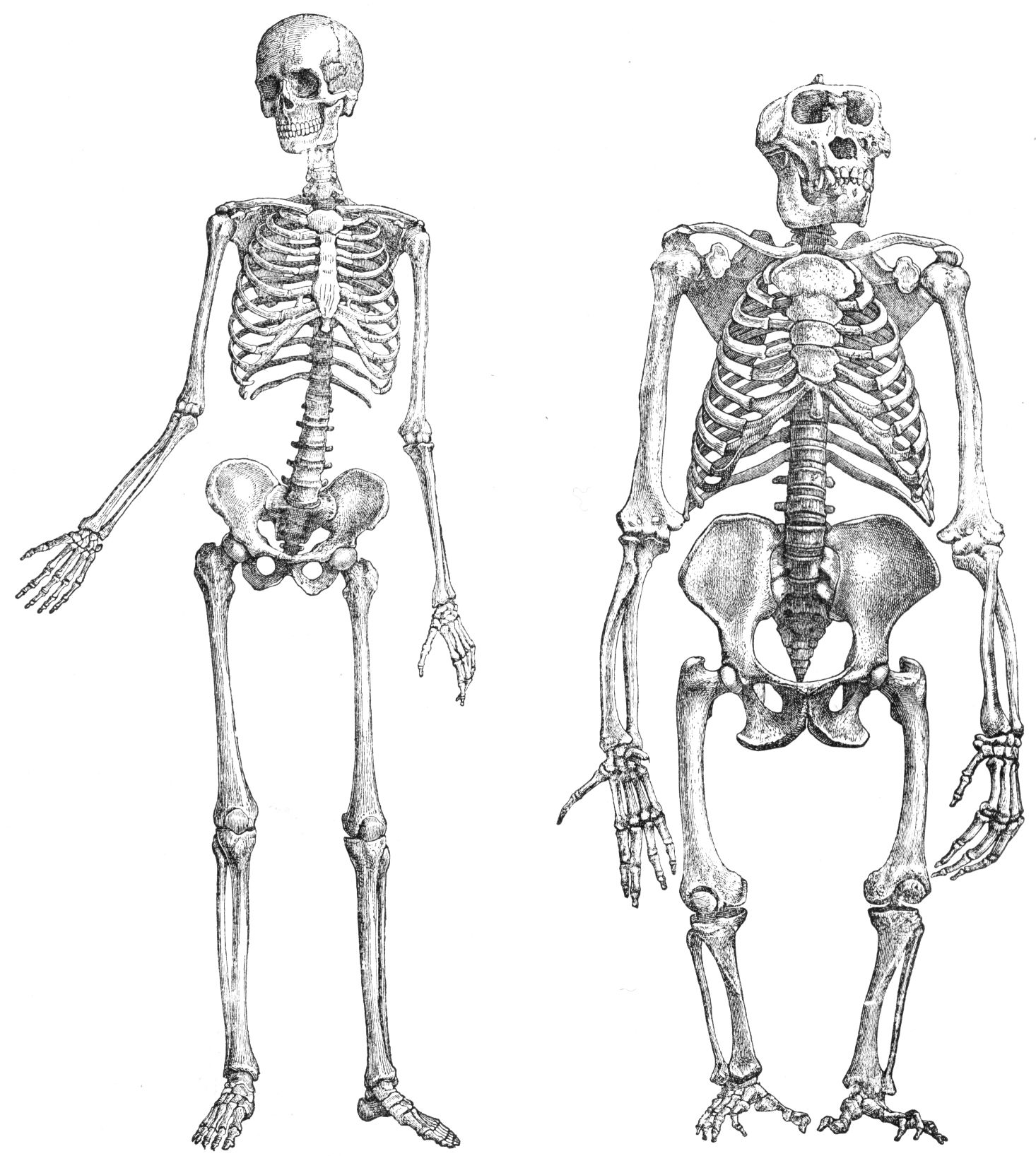
The human body is the entire structure of a Human, human being. It is composed of many different types of Cell (biology), cells that together create Tissue (biology), tissues and subsequently Organ (biology), organs and then Organ system, organ systems. The external human body consists of a human head, head, hair, neck, torso (which includes the thorax and abdomen), Sex organ, genitals, arms, Hand, hands, human leg, legs, and Foot, feet. The internal human body includes organs, Human tooth, teeth, bones, muscle, tendons, ligaments, blood vessels and blood, lymphatic vessels and lymph. The study of the human body includes anatomy, physiology, histology and embryology. The body Anatomical variation, varies anatomically in known ways. Physiology focuses on the systems and organs of the human body and their functions. Many systems and mechanisms interact in order to maintain homeostasis, with safe levels of substances such as sugar, iron, and oxygen in the blood. The body is st ... [...More Info...] [...Related Items...] OR: [Wikipedia] [Google] [Baidu] |
Robert Schleip
Robert Schleip (born 1954) is a German psychologist, human biologist and author, best known for his research in the field of fascia. His work includes scientific papers and books about fascia and its role in musculoskeletal health. He serves as the director of the Fascia Research Group at the University of Ulm and the Technical University of Munich. Schleip is also the founding director of the Fascia Research Society, the research director of the European Rolfing Association and vice president of the Ida P. Rolf Research Foundation. He is involved in the alternative medicine field of rolfing: he is a certified rolfer, and has been involved in rolfing-related boards, committees, associations, and foundations. Education In 1978, Schleip became Germany's first certified rolfer at the Rolf Institute and subsequently in 1983 became a Certified Advanced Rolfer in the field of Structural Integration. Schleip graduated with a degree in psychology from the University of Heide ... [...More Info...] [...Related Items...] OR: [Wikipedia] [Google] [Baidu] |
Scar
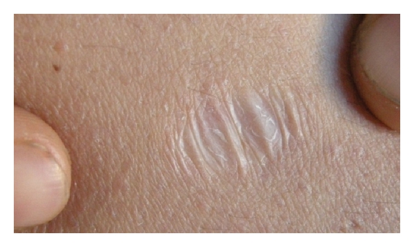
A scar (or scar tissue) is an area of fibrosis, fibrous tissue that replaces normal skin after an injury. Scars result from the biological process of wound repair in the skin, as well as in other Organ (anatomy), organs, and biological tissue, tissues of the body. Thus, scarring is a natural part of the healing process. With the exception of very minor lesions, every wound (e.g., after accident, disease, or surgery) results in some degree of scarring. An exception to this are animals with complete Regeneration (biology), regeneration, which regrow tissue without scar formation. Scar tissue is composed of the same protein (collagen) as the tissue that it replaces, but the fiber composition of the protein is different; instead of a random basketweave formation of the collagen fibers found in normal tissue, in fibrosis the collagen cross-links and forms a pronounced alignment in a single direction. This collagen scar tissue alignment is usually of inferior functional quality to the ... [...More Info...] [...Related Items...] OR: [Wikipedia] [Google] [Baidu] |
Fibrosis
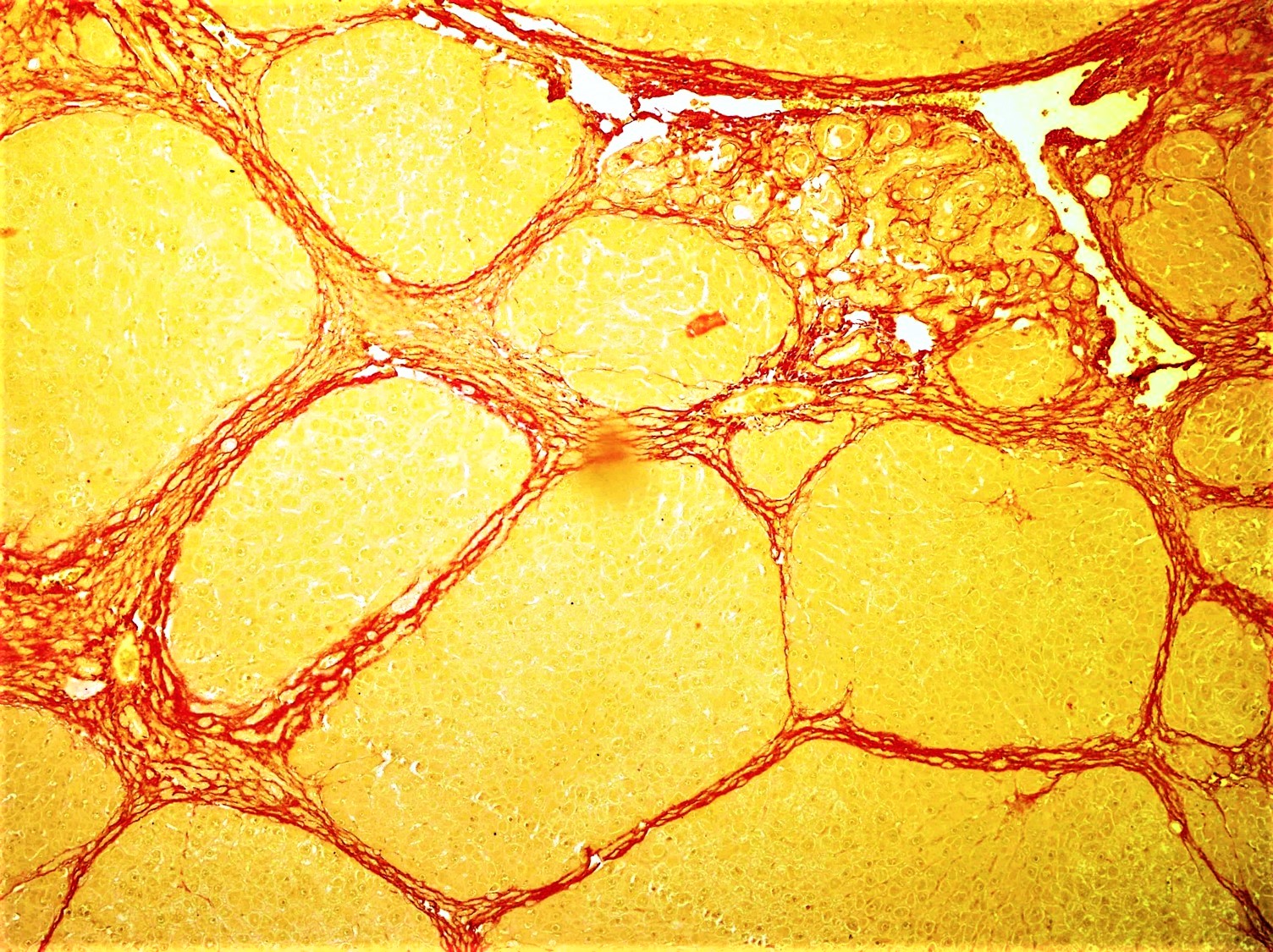
Fibrosis, also known as fibrotic scarring, is the development of fibrous connective tissue in response to an injury. Fibrosis can be a normal connective tissue deposition or excessive tissue deposition caused by a disease. Repeated injuries, chronic inflammation and repair are susceptible to fibrosis, where an accidental excessive accumulation of extracellular matrix components, such as the collagen, is produced by fibroblasts, leading to the formation of a permanent fibrotic scar. In response to injury, this is called scarring, and if fibrosis arises from a single cell line, this is called a fibroma. Physiologically, fibrosis acts to deposit connective tissue, which can interfere with or totally inhibit the normal architecture and function of the underlying organ or tissue. Fibrosis can be used to describe the pathological state of excess deposition of fibrous tissue, as well as the process of connective tissue deposition in healing. Defined by the pathological accumulation of ... [...More Info...] [...Related Items...] OR: [Wikipedia] [Google] [Baidu] |
Fasciitis
Fasciitis is an inflammation of the fascia, which is the connective tissue surrounding muscles, blood vessels and nerves. In particular, it often involves one of the following diseases: * Necrotizing fasciitis * Plantar fasciitis * Ischemic fasciitis, classified by the World Health Organization, 2020, as a specific tumor form in the category of fibroblastic and myofibroblastic tumors. * Eosinophilic fasciitis * Paraneoplastic fasciitis References External links Inflammations {{Musculoskeletal-disease-stub ... [...More Info...] [...Related Items...] OR: [Wikipedia] [Google] [Baidu] |
Fascial Compartments Of Thigh
The fascial compartments of thigh are the three fascial compartments that divide and contain the thigh muscles. The fascia lata is the strong and deep fascia of the thigh that surrounds the thigh muscles and forms the outer limits of the compartments. Internally the muscle compartments are divided by the lateral and medial intermuscular septa. The three groups of muscles contained in the compartments have their own nerve supply: Compartments See also * Compartment syndrome * Fascial compartments of arm References External links *knee/muscles/indexat the Dartmouth Medical School The Geisel School of Medicine is the medical school of Dartmouth College located in Hanover, New Hampshire. The fourth oldest medical school in the United States, it was founded in 1797 by New England physician Nathan Smith. It is one of the sev ...'s Department of Anatomy Lower limb anatomy {{musculoskeletal-stub ... [...More Info...] [...Related Items...] OR: [Wikipedia] [Google] [Baidu] |
Fascial Compartments Of Leg
The fascial compartments of the leg are the four fascial compartments that separate and contain the muscles of the lower leg (from the knee to the ankle). The compartments are divided by septa formed from the fascia. The compartments usually have nerve and blood supplies separate from their neighbours. All of the muscles within a compartment will generally be supplied by the same nerve. Intermuscular septa The lower leg is divided into four compartments by the interosseous membrane of the leg, the anterior intermuscular septum, the transverse intermuscular septum and the posterior intermuscular septum. Each compartment contains connective tissue, nerves and blood vessels. The septa are formed from the fascia which is made up of a strong type of connective tissue. The fascia also separates the skeletal muscles from the subcutaneous tissue. Due to the great pressure placed on the leg, from the column of blood from the heart to the feet, the fascia is very thick in order to sup ... [...More Info...] [...Related Items...] OR: [Wikipedia] [Google] [Baidu] |
Thigh
In anatomy, the thigh is the area between the hip (pelvis) and the knee. Anatomically, it is part of the lower limb. The single bone in the thigh is called the femur. This bone is very thick and strong (due to the high proportion of bone tissue), and forms a ball and socket joint at the hip, and a modified hinge joint at the knee. Structure Bones The femur is the only bone in the thigh and serves as an attachment site for all thigh muscles. The head of the femur articulates with the acetabulum in the pelvic bone forming the hip joint, while the distal part of the femur articulates with the tibia and patella forming the knee. By most measures, the femur is the strongest and longest bone in the body. The femur is categorised as a long bone and comprises a diaphysis, the shaft (or body) and two epiphyses, the lower extremity and the upper extremity of femur, that articulate with adjacent bones in the hip and knee. Muscular compartments In cross-section, the thigh is d ... [...More Info...] [...Related Items...] OR: [Wikipedia] [Google] [Baidu] |
Human Leg
The leg is the entire lower limb (anatomy), limb of the human body, including the foot, thigh or sometimes even the hip or Gluteal muscles, buttock region. The major bones of the leg are the femur (thigh bone), tibia (shin bone), and adjacent fibula. There are 30 bones in each leg. The thigh is located in between the hip and knee. The calf (leg), calf (rear) and Tibia#Structure, shin (front), or shank, are located between the knee and ankle. Legs are used for standing, many forms of human movement, recreation such as dancing, and constitute a significant portion of a person's mass. Evolution has led to the human leg's development into a mechanism specifically adapted for efficient bipedalism, bipedal gait. While the capacity to walk upright is not unique to humans, other primates can only achieve this for short periods and at a great expenditure of energy. In humans, female legs generally have greater hip anteversion and tibiofemoral angles, while male legs have longer femur a ... [...More Info...] [...Related Items...] OR: [Wikipedia] [Google] [Baidu] |
Fascial Compartments Of Arm
A fascia (; : fasciae or fascias; adjective fascial; ) is a generic term for macroscopic membranous bodily structures. Fasciae are classified as superficial, visceral or deep, and further designated according to their anatomical location. The knowledge of fascial structures is essential in surgery, as they create borders for infectious processes (for example Psoas abscess) and haematoma. An increase in pressure may result in a compartment syndrome, where a prompt fasciotomy may be necessary. For this reason, profound descriptions of fascial structures are available in anatomical literature from the 19th century. Function Fasciae were traditionally thought of as passive structures that transmit mechanical tension generated by muscular activities or external forces throughout the body. An important function of muscle fasciae is to reduce friction of muscular force. In doing so, fasciae provide a supportive and movable wrapping for nerves and blood vessels as they pass th ... [...More Info...] [...Related Items...] OR: [Wikipedia] [Google] [Baidu] |


