|
Type XVIII Collagen
Type XVIII collagen is a type of collagen which can be cleaved to form endostatin. The endostatin is from the c terminus end of the collagen XVIII, and is known to have an inhibitory effect on the growth of blood vessels. This is seen with tumors, where endostatin inhibits the growth of the blood vessels of the tumor as well as the overall growth of the tumor. The collagen XVIII is located within the basement membrane, and plays a major role in the integrity of the structure of the basement membrane for both endothelial and epithelial cells. The collagen XVIII has three different isoforms. While each of the isoforms has the same C-terminus end, they have a varying structure on the N-terminus end, which results in the formation of short, medium or long form Collagen XVIII. Type XVIII Collagen and Knobloch Syndrome Knobloch Syndrome is a type of inherited autosomal recessive disorder in which a mutation occurs in the endostatin COL18A1 protein encoding gene, which codes for p ... [...More Info...] [...Related Items...] OR: [Wikipedia] [Google] [Baidu] |
Collagen
Collagen () is the main structural protein in the extracellular matrix found in the body's various connective tissues. As the main component of connective tissue, it is the most abundant protein in mammals, making up from 25% to 35% of the whole-body protein content. Collagen consists of amino acids bound together to form a triple helix of elongated fibril known as a collagen helix. It is mostly found in connective tissue such as cartilage, bones, tendons, ligaments, and skin. Depending upon the degree of mineralization, collagen tissues may be rigid (bone) or compliant (tendon) or have a gradient from rigid to compliant (cartilage). Collagen is also abundant in corneas, blood vessels, the gut, intervertebral discs, and the dentin in teeth. In muscle tissue, it serves as a major component of the endomysium. Collagen constitutes one to two percent of muscle tissue and accounts for 6% of the weight of the skeletal muscle tissue. The fibroblast is the most common cell tha ... [...More Info...] [...Related Items...] OR: [Wikipedia] [Google] [Baidu] |
Endostatin
Endostatin is a naturally occurring, 20-kDa C-terminal fragment derived from type XVIII collagen. It is reported to serve as an anti-angiogenic agent, similar to angiostatin and thrombospondin. Endostatin is a broad-spectrum angiogenesis inhibitor and may interfere with the pro-angiogenic action of growth factors such as basic fibroblast growth factor (bFGF/FGF-2) and vascular endothelial growth factor (VEGF). Background Endostatin is an endogenous inhibitor of angiogenesis. It was first found secreted in the media of non-metastasizing mouse cells from a hemangioendothelioma cell line in 1997 and was subsequently found in humans. It is produced by proteolytic cleavage of collagen XVIII, a member of the multiplexin family that is characterized by interruptions in the triple helix creating multiple domains, by proteases such as cathepsins. Collagen is a component of epithelial and endothelial basement membranes. Endostatin, as a fragment of collagen 18, demonstrates a role of ... [...More Info...] [...Related Items...] OR: [Wikipedia] [Google] [Baidu] |
N-terminus
The N-terminus (also known as the amino-terminus, NH2-terminus, N-terminal end or amine-terminus) is the start of a protein or polypeptide, referring to the free amine group (-NH2) located at the end of a polypeptide. Within a peptide, the amine group is bonded to the carboxylic group of another amino acid, making it a chain. That leaves a free carboxylic group at one end of the peptide, called the C-terminus, and a free amine group on the other end called the N-terminus. By convention, peptide sequences are written N-terminus to C-terminus, left to right (in LTR writing systems). This correlates the translation direction to the text direction, because when a protein is translated from messenger RNA, it is created from the N-terminus to the C-terminus, as amino acids are added to the carboxyl end of the protein. Chemistry Each amino acid has an amine group and a carboxylic group. Amino acids link to one another by peptide bonds which form through a dehydration reaction ... [...More Info...] [...Related Items...] OR: [Wikipedia] [Google] [Baidu] |
Knobloch Syndrome
Knobloch syndrome is a rare genetic disorder presenting severe eyesight problems and often a defect in the skull. It was named after the ophthalmologist William Hunter Knobloch (1926 - 2005), who first described the syndrome in 1971. A usual occurrence is a degeneration of the vitreous humour and the retina, two components of the eye. This breakdown often results in the separation of the retina (the light-sensitive tissue at the back of the eye) from the eye, called retinal detachment, which can be recurrent. Extreme myopia (near-sightedness) is a common feature. The limited evidence available from electroretinography suggests that a cone-rod pattern of dysfunction is also a feature. Knobloch syndrome is caused by mutations in an autosomal recessive inherited gene. These mutations have been found in the COL18A1 gene that instructs for the formation of a protein that builds collagen XVIII. This type of collagen is found in the basement membranes of various body tissues. Its de ... [...More Info...] [...Related Items...] OR: [Wikipedia] [Google] [Baidu] |
The FASEB Journal
''The FASEB Journal'' is a scientific journal related to experimental biosciences, promoting scientific progress and education. It is published by the Federation of American Societies for Experimental Biology The Federation of American Societies for Experimental Biology (FASEB) is a non-profit organization that is the principal umbrella organization of U.S. societies in the field of biological and medical research. This organization organizes academ ..., that was founded in 1912, originally by three societies (29 as of January 2019 ). References Publications established in 1912 Biology journals Monthly journals Academic journals published by learned and professional societies 1912 establishments in the United States {{biology-journal-stub ... [...More Info...] [...Related Items...] OR: [Wikipedia] [Google] [Baidu] |
Laminin
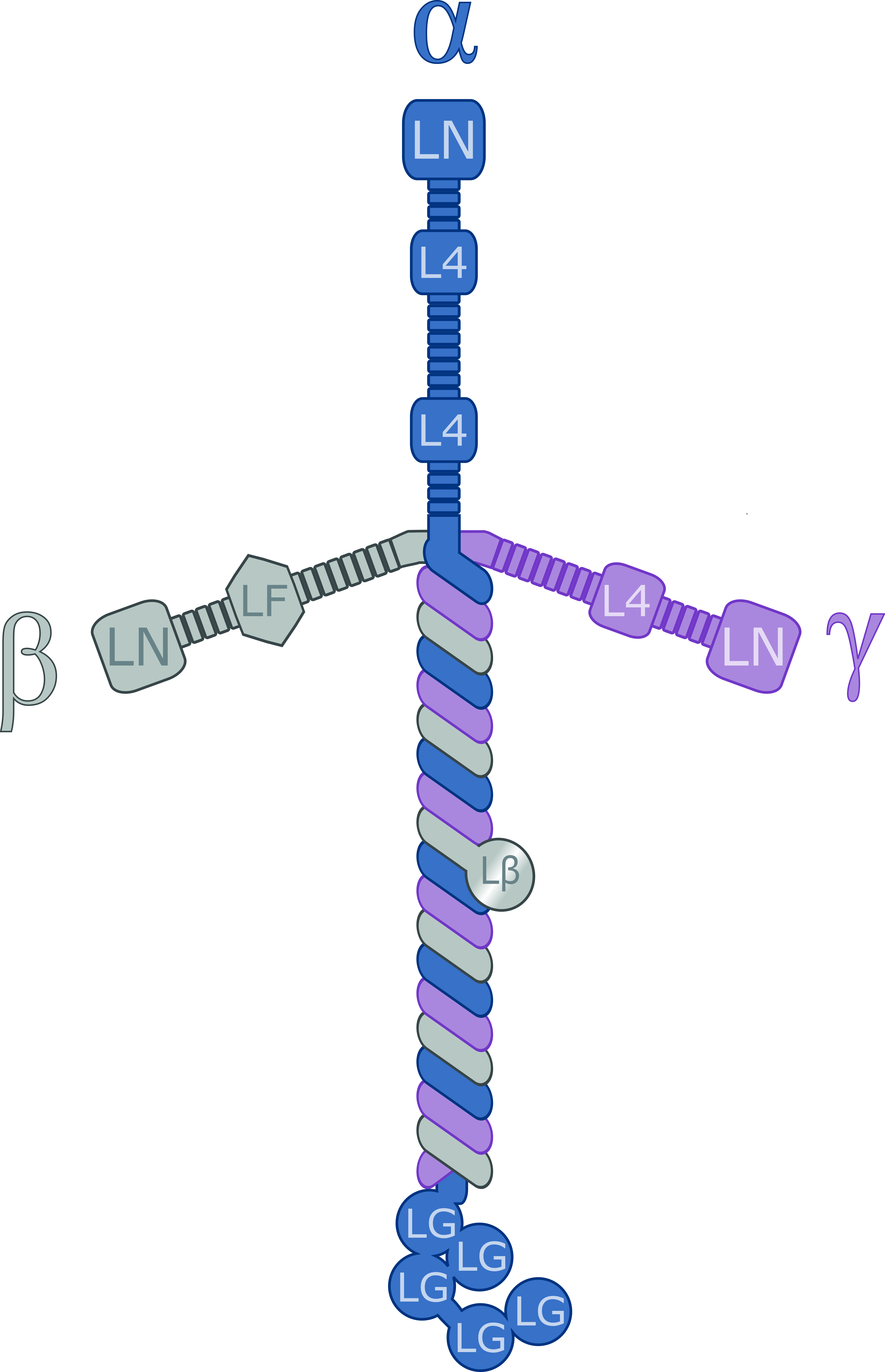
Laminins are a family of glycoproteins of the extracellular matrix of all animals. They are major components of the basal lamina (one of the layers of the basement membrane), the protein network foundation for most cells and organs. The laminins are an important and biologically active part of the basal lamina, influencing cell differentiation, migration, and adhesion. Laminins are heterotrimeric proteins with a high molecular mass (~400 to ~900 kDa). They contain three different chains (α, β and γ) encoded by five, four, and three paralogous genes in humans, respectively. The laminin molecules are named according to their chain composition. Thus, laminin-511 contains α5, β1, and γ1 chains. Fourteen other chain combinations have been identified ''in vivo''. The trimeric proteins intersect to form a cross-like structure that can bind to other cell membrane and extracellular matrix molecules. The three shorter arms are particularly good at binding to other laminin molecule ... [...More Info...] [...Related Items...] OR: [Wikipedia] [Google] [Baidu] |
Occipital Encephalocele
Encephalocele is a neural tube defect characterized by sac-like protrusions of the brain and the membranes that cover it through openings in the skull. These defects are caused by failure of the neural tube to close completely during fetal development. Encephaloceles cause a groove down the middle of the skull, or between the forehead and nose, or on the back side of the skull. The severity of encephalocele varies, depending on its location. Signs and symptoms Encephaloceles are often accompanied by craniofacial abnormalities or other brain malformations. Symptoms may include neurologic problems, hydrocephalus (cerebrospinal fluid accumulated in the brain), spastic quadriplegia (paralysis of the limbs), microcephaly (an abnormally small head), ataxia (uncoordinated muscle movement), developmental delay, vision problems, mental and growth retardation, and seizures. File:Encephalocele of a newborn.JPG, A neonate with a large encephalocele. File:Encephalocele2.jpg, Encephalocele on ... [...More Info...] [...Related Items...] OR: [Wikipedia] [Google] [Baidu] |
Dense Deposit Disease
Membranoproliferative glomerulonephritis (MPGN) is a type of glomerulonephritis caused by deposits in the kidney glomerular mesangium and basement membrane ( GBM) thickening, activating complement and damaging the glomeruli. MPGN accounts for approximately 4% of primary renal causes of nephrotic syndrome in children and 7% in adults. It should not be confused with membranous glomerulonephritis, a condition in which the basement membrane is thickened, but the mesangium is not. Type There are three types of MPGN, but this classification is becoming obsolete as the causes of this pattern are becoming understood. Type I Type I, the most common by far, is caused by immune complexes depositing in the kidney. It is characterised by subendothelial and mesangial immune deposits. It is believed to be associated with the classical complement pathway. Type II Also called recently as ‘C3 nephropathy’ The preferred name is "dense deposit disease." Most cases of dense deposit disease d ... [...More Info...] [...Related Items...] OR: [Wikipedia] [Google] [Baidu] |

