|
Torus Tubarius
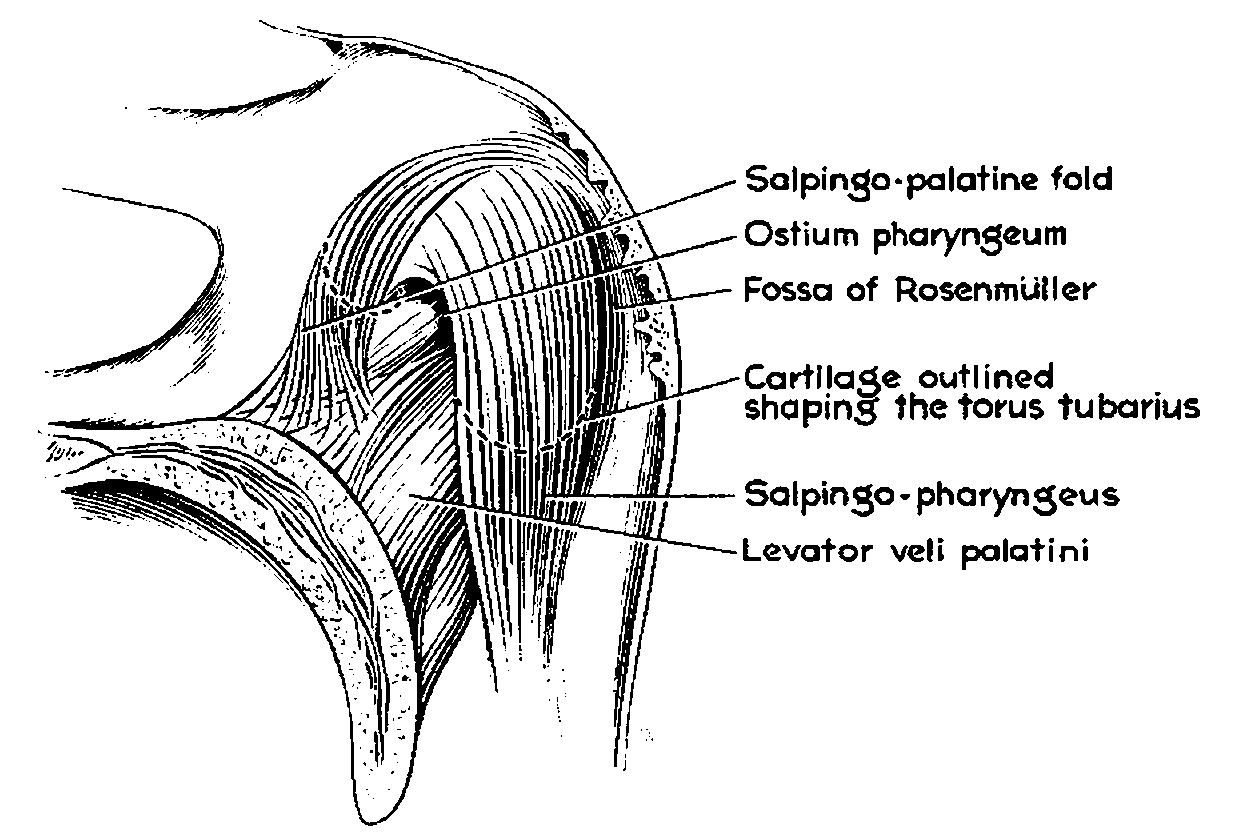
The torus tubarius (or torus of the auditory tube) is an elevation of the mucous membrane of the Pharynx#Nasopharynx, nasal part of the pharynx formed by the underlying base of the Eustachian tube, cartilaginous portion of the Eustachian tube, Eustachian tube (auditory tube). The torus tubarius is situated behind the pharyngeal orifice of the auditory tube. The torus tubarius is very close to the tubal tonsil, which is sometimes also referred to as the ''tonsil of (the) torus tubarius''. Two folds run anteriorly and posteriorly to the torus tubarius: the salpingopalatine fold (anteriorly), and the salpingopharyngeal fold (posteriorly). See also * Tubarial salivary gland References External links * * {{Authority control Ear ... [...More Info...] [...Related Items...] OR: [Wikipedia] [Google] [Baidu] |
Auditory Tube
The Eustachian tube (), also called the auditory tube or pharyngotympanic tube, is a tube that links the nasopharynx to the middle ear, of which it is also a part. In adult humans, the Eustachian tube is approximately long and in diameter. It is named after the sixteenth-century Italian anatomist Bartolomeo Eustachi. In humans and other tetrapods, both the middle ear and the ear canal are normally filled with air. Unlike the air of the ear canal, however, the air of the middle ear is not in direct contact with the atmosphere outside the body; thus, a pressure difference can develop between the atmospheric pressure of the ear canal and the middle ear. Normally, the Eustachian tube is collapsed, but it gapes open with swallowing and with positive pressure, allowing the middle ear's pressure to adjust to the atmospheric pressure. When taking off in an aircraft, the ambient air pressure goes from higher (on the ground) to lower (in the sky). The air in the middle ear Boyle's law, ... [...More Info...] [...Related Items...] OR: [Wikipedia] [Google] [Baidu] |
Mucous Membrane
A mucous membrane or mucosa is a membrane that lines various cavities in the body of an organism and covers the surface of internal organs. It consists of one or more layers of epithelial cells overlying a layer of loose connective tissue. It is mostly of endodermal origin and is continuous with the skin at body openings such as the eyes, eyelids, ears, inside the nose, inside the mouth, lips, the genital areas, the urethral opening and the anus. Some mucous membranes secrete mucus, a thick protective fluid. The function of the membrane is to stop pathogens and dirt from entering the body and to prevent bodily tissues from becoming dehydrated. Structure The mucosa is composed of one or more layers of epithelial cells that secrete mucus, and an underlying lamina propria of loose connective tissue. The type of cells and type of mucus secreted vary from organ to organ and each can differ along a given tract. Mucous membranes line the digestive, respiratory and rep ... [...More Info...] [...Related Items...] OR: [Wikipedia] [Google] [Baidu] |
Pharynx
The pharynx (: pharynges) is the part of the throat behind the human mouth, mouth and nasal cavity, and above the esophagus and trachea (the tubes going down to the stomach and the lungs respectively). It is found in vertebrates and invertebrates, though its structure varies across species. The pharynx carries food to the esophagus and air to the larynx. The flap of cartilage called the epiglottis stops food from entering the larynx. In humans, the pharynx is part of the Digestion, digestive system and the conducting zone of the respiratory system. (The conducting zone—which also includes the nostrils of the Human nose, nose, the larynx, trachea, bronchus, bronchi, and bronchioles—filters, warms, and moistens air and conducts it into the lungs). The human pharynx is conventionally divided into three sections: the nasopharynx, oropharynx, and laryngopharynx (hypopharynx). In humans, two sets of pharyngeal muscles form the pharynx and determine the shape of its lumen (anatomy), ... [...More Info...] [...Related Items...] OR: [Wikipedia] [Google] [Baidu] |
Eustachian Tube
The Eustachian tube (), also called the auditory tube or pharyngotympanic tube, is a tube that links the nasopharynx to the middle ear, of which it is also a part. In adult humans, the Eustachian tube is approximately long and in diameter. It is named after the sixteenth-century Italian anatomist Bartolomeo Eustachi. In humans and other tetrapods, both the middle ear and the ear canal are normally filled with air. Unlike the air of the ear canal, however, the air of the middle ear is not in direct contact with the atmosphere outside the body; thus, a pressure difference can develop between the atmospheric pressure of the ear canal and the middle ear. Normally, the Eustachian tube is collapsed, but it gapes open with swallowing and with positive pressure, allowing the middle ear's pressure to adjust to the atmospheric pressure. When taking off in an aircraft, the ambient air pressure goes from higher (on the ground) to lower (in the sky). The air in the middle ear expands as ... [...More Info...] [...Related Items...] OR: [Wikipedia] [Google] [Baidu] |
Tubal Tonsil
The tubal tonsil, also known as Gerlach tonsil, is one of the four main tonsil groups forming Waldeyer's tonsillar ring. Structure Each tubal tonsil is located posterior to the opening of the Eustachian tube on the lateral wall of the nasopharynx. It is one of the four main tonsil groups forming Waldeyer's tonsillar ring. This ring also includes the palatine tonsils, the lingual tonsils, and the adenoid. Clinical significance The tubal tonsil may be affected by tonsillitis. However, this usually affects only the palatine tonsils. History The tubal tonsil may also be known as the Gerlach tonsil. It is very close to the torus tubarius The torus tubarius (or torus of the auditory tube) is an elevation of the mucous membrane of the Pharynx#Nasopharynx, nasal part of the pharynx formed by the underlying base of the Eustachian tube, cartilaginous portion of the Eustachian tube, Eu ..., which is why this tonsil is sometimes also called the ''tonsil of (the) torus tubarius ... [...More Info...] [...Related Items...] OR: [Wikipedia] [Google] [Baidu] |
Salpingopalatine Fold
The pharynx (: pharynges) is the part of the throat behind the mouth and nasal cavity, and above the esophagus and trachea (the tubes going down to the stomach and the lungs respectively). It is found in vertebrates and invertebrates, though its structure varies across species. The pharynx carries food to the esophagus and air to the larynx. The flap of cartilage called the epiglottis stops food from entering the larynx. In humans, the pharynx is part of the digestive system and the conducting zone of the respiratory system. (The conducting zone—which also includes the nostrils of the nose, the larynx, trachea, bronchi, and bronchioles—filters, warms, and moistens air and conducts it into the lungs). The human pharynx is conventionally divided into three sections: the nasopharynx, oropharynx, and laryngopharynx (hypopharynx). In humans, two sets of pharyngeal muscles form the pharynx and determine the shape of its lumen. They are arranged as an inner layer of longitudinal ... [...More Info...] [...Related Items...] OR: [Wikipedia] [Google] [Baidu] |
Tubarial Salivary Gland
The tubarial salivary glands, also known as the tubarial glands, are a pair of salivary glands found in humans between the nasal cavity and throat. Description The tubarial glands (TGs) are located in the nasopharynx. They are situated proximal to the eustachian tube, superior to the soft palate and posterior to the inferior nasal conchae. The tubarial glands overlay the torus tubarius region and are found on the dorsolateral or posterior lateral wall of the nasopharynx, extending from the skull base down on the inner side of the superior constrictor muscle. The tubarial glands are difficult to visualize on standard radiological images like MRI, appearing as shadowy regions of soft tissue. However, they can be visualized clearly using prostate-specific membrane antigen (PSMA) positron emission tomography—computed tomography ( PET/CT). History The glands were discovered by a group of Dutch scientists at the Netherlands Cancer Institute in September 2020 using PET/CT scans. ... [...More Info...] [...Related Items...] OR: [Wikipedia] [Google] [Baidu] |


