|
TSG-6
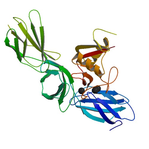
Tumor necrosis factor-inducible gene 6 protein also known as TNF-stimulated gene 6 protein or TSG-6 is a protein that in humans is encoded by the ''TNFAIP6'' (tumor necrosis factor, alpha-induced protein 6) gene. Structure and function TSG-6 is a 30 kDa secreted protein that contains a hyaluronan-binding LINK domain a and thus is a member of the hyaluronan-binding protein family, also called hyaladherins. The hyaluronan-binding domain is known to be involved in extracellular matrix stability and cell migration. This protein has been shown to form a stable, covalent complex with inter-alpha-inhibitor ( IαI), and thus enhance the serine protease inhibitory activity of IαI, which is important in the protease network associated with inflammation. The expression of this gene can be induced by a number of signalling molecules, principally tumor necrosis factor α (TNF-α) and interleukin-1 ( IL-1). The expression can also be induced by mechanical stimuli in vascular smooth muscle ce ... [...More Info...] [...Related Items...] OR: [Wikipedia] [Google] [Baidu] |
PTX3
Pentraxin-related protein PTX3 also known as TNF-inducible gene 14 protein (TSG-14) is a protein that in humans is encoded by the ''PTX3'' gene. Pentraxin 3 (ptx3) is a member of the pentraxin superfamily. This super family characterized by cyclic multimeric structure. PTX3 is rapidly produced and released by several cell types, in particular by mononuclear phagocytes, dendritic cells (DCs), fibroblasts and endothelial cells in response to primary inflammatory signals .g., toll-like receptor (TLR) engagement, TNFα, Interleukin 1">IL-1β]. PTX3 binds with high affinity to the complement component C1q, the extracellular matrix component TNFα induced protein 6 (TNFAIP6; also called TNF-stimulated gene 6, TSG-6) and selected microorganisms, including ''Aspergillus fumigatus'' and ''Pseudomonas aeruginosa''. PTX3 activates the classical pathway of complement activation and facilitates pathogen recognition by macrophages and DCs. Structure Human and murine PTX3, localized in th ... [...More Info...] [...Related Items...] OR: [Wikipedia] [Google] [Baidu] |
Hyaladherin
Hyaladherins, also known as hyaluronan-binding proteins, are proteins capable of binding to hyaluronic acid. Most hyaladherins belong to the Link module superfamily, including its main receptor CD44, hyalectans and TSG-6. In addition there is a diverse group of hyaladherins lacking a Link module; these include the receptor RHAMM, C1QBP (HABP1) and HABP2. The primary roles of hyaladherins are cell adhesion, structural support of the extracellular matrix (ECM) and cell signalling. Due to the role of aberrant hyaluronic acid synthesis and degradation in various cancers, hyaladherins, as well as hyaluronic acid, are considered a promising target for cancer therapy. See also * Hyaluronan synthase *Hyaluronidase Hyaluronidases are a family of enzymes that catalyse the degradation of hyaluronic acid. Karl Meyer classified these enzymes in 1971, into three distinct groups, a scheme based on the enzyme reaction products. The three main types of hyaluroni ... References {{refl ... [...More Info...] [...Related Items...] OR: [Wikipedia] [Google] [Baidu] |
Aggrecan
Aggrecan (ACAN), also known as cartilage-specific proteoglycan core protein (CSPCP) or chondroitin sulfate proteoglycan 1, is a protein that in humans is encoded by the ''ACAN'' gene. This gene is a member of the lectican ( chondroitin sulfate proteoglycan) family. The encoded protein is an integral part of the extracellular matrix in cartilagenous tissue and it withstands compression in cartilage. Aggrecan is a proteoglycan, or a protein modified with large carbohydrates; the human form of the protein is 2316 amino acids long and can be expressed in multiple isoforms due to alternative splicing. Aggrecan was named for its ability to form large aggregates in the cartilage tissue (a large aggregating proteoglycan). Structure Aggrecan is a high molecular weight ( molar mass between 1 million and 3 million) proteoglycan. It exhibits a bottlebrush structure, in which chondroitin sulfate and keratan sulfate glycosaminoglycan (GAG) chains are attached to an extended protein ... [...More Info...] [...Related Items...] OR: [Wikipedia] [Google] [Baidu] |
Tumor Necrosis Factor-alpha
Tumor necrosis factor (TNF), formerly known as TNF-α, is a chemical messenger produced by the immune system that induces inflammation. TNF is produced primarily by activated macrophages, and induces inflammation by binding to its receptors on other cells. It is a member of the tumor necrosis factor superfamily, a family of transmembrane proteins that are cytokines, chemical messengers of the immune system. Excessive production of TNF plays a critical role in several inflammatory diseases, and TNF-blocking drugs are often employed to treat these diseases. TNF is produced primarily by macrophages but is also produced in several other cell types, such as T cells, B cells, dendritic cells, and mast cells. It is produced rapidly in response to pathogens, cytokines, and environmental stressors. TNF is initially produced as a type II transmembrane protein (tmTNF), which is then cleaved by TNF alpha converting enzyme (TACE) into a soluble form (sTNF) and secreted from the cel ... [...More Info...] [...Related Items...] OR: [Wikipedia] [Google] [Baidu] |
Thrombospondin
Thrombospondins (TSPs) are a family of secreted glycoproteins with antiangiogenic functions. Due to their dynamic role within the extracellular matrix they are considered matricellular proteins. The first member of the family, thrombospondin 1 (THBS1), was discovered in 1971 by Nancy L. Baenziger. Types The thrombospondins are a family of multifunctional proteins. The family consists of thrombospondins 1–5 and can be divided into two subgroups: A, which contains TSP-1 and TSP-2, and B, which contains TSP-3, TSP-4 and TSP-5 (also designated cartilage oligomeric protein or COMP). TSP-1 and TSP-2 are homotrimers, consisting of three identical subunits, whereas TSP-3, TSP-4 and TSP-5 are homopentamers. TSP-1 and TSP-2 are produced by immature astrocytes during brain development, which promotes the development of new synapses. Thrombospondin 1 Thrombospondin 1 (TSP-1) is encoded by THBS1. It was first isolated from platelets that had been stimulated with thrombin, and ... [...More Info...] [...Related Items...] OR: [Wikipedia] [Google] [Baidu] |
Versican
Versican is a large extracellular matrix proteoglycan that is present in a variety of human tissues. It is encoded by the ''VCAN'' gene. Versican is a large chondroitin sulfate proteoglycan with an apparent molecular mass of more than 1000kDa. In 1989, Zimmermann and Ruoslahti cloned and sequenced the core protein of fibroblast chondroitin sulfate proteoglycan. They designated it versican in recognition of its versatile modular structure. Versican belongs to the lectican protein family, with aggrecan (abundant in cartilage), brevican and neurocan (nervous system proteoglycans) as other members. Versican is also known as chondroitin sulfate proteoglycan core protein 2 or chondroitin sulfate proteoglycan 2 (CSPG2), and PG-M. Structure These proteoglycans share a homology (biology), homologous globular N-terminal, C-terminal, and glycosaminoglycan (GAG) binding regions. The N-terminal (G1) globular domain consists of Antibody, Ig-like loop and two link modules, and has Hyalur ... [...More Info...] [...Related Items...] OR: [Wikipedia] [Google] [Baidu] |
Proteoglycan
Proteoglycans are proteins that are heavily glycosylated. The basic proteoglycan unit consists of a "core protein" with one or more covalently attached glycosaminoglycan (GAG) chain(s). The point of attachment is a serine (Ser) residue to which the glycosaminoglycan is joined through a tetrasaccharide bridge (e.g. chondroitin sulfate- GlcA- Gal-Gal- Xyl-PROTEIN). The Ser residue is generally in the sequence -Ser- Gly-X-Gly- (where X can be any amino acid residue but proline), although not every protein with this sequence has an attached glycosaminoglycan. The chains are long, linear carbohydrate polymers that are negatively charged under physiological conditions due to the occurrence of sulfate and uronic acid groups. Proteoglycans occur in connective tissue. Types Proteoglycans are categorized by their relative size (large and small) and the nature of their glycosaminoglycan chains. Types include: Certain members are considered members of the "small leucine-rich pr ... [...More Info...] [...Related Items...] OR: [Wikipedia] [Google] [Baidu] |
Interleukin 1
The Interleukin-1 family (IL-1 family) is a group of 11 cytokines that plays a central role in the regulation of immune and inflammatory responses to infections or sterile insults. Discovery Discovery of these cytokines began with studies on the pathogenesis of fever. The studies were performed by Eli Menkin and Paul Beeson in 1943–1948 on the fever-producing properties of proteins released from rabbit peritoneal exudate cells. These studies were followed by contributions of several investigators, who were primarily interested in the link between fever and infection/inflammation. The basis for the term "interleukin" was to streamline the growing number of biological properties attributed to soluble factors from macrophages and lymphocytes. IL-1 was the name given to the macrophage product, whereas IL-2 was used to define the lymphocyte product. At the time of the assignment of these names, there was no amino acid sequence analysis known and the terms were used to define b ... [...More Info...] [...Related Items...] OR: [Wikipedia] [Google] [Baidu] |
Protein
Proteins are large biomolecules and macromolecules that comprise one or more long chains of amino acid residue (biochemistry), residues. Proteins perform a vast array of functions within organisms, including Enzyme catalysis, catalysing metabolic reactions, DNA replication, Cell signaling, responding to stimuli, providing Cytoskeleton, structure to cells and Fibrous protein, organisms, and Intracellular transport, transporting molecules from one location to another. Proteins differ from one another primarily in their sequence of amino acids, which is dictated by the Nucleic acid sequence, nucleotide sequence of their genes, and which usually results in protein folding into a specific Protein structure, 3D structure that determines its activity. A linear chain of amino acid residues is called a polypeptide. A protein contains at least one long polypeptide. Short polypeptides, containing less than 20–30 residues, are rarely considered to be proteins and are commonly called pep ... [...More Info...] [...Related Items...] OR: [Wikipedia] [Google] [Baidu] |
Serine Protease
Serine proteases (or serine endopeptidases) are enzymes that cleave peptide bonds in proteins. Serine serves as the nucleophilic amino acid at the (enzyme's) active site. They are found ubiquitously in both eukaryotes and prokaryotes. Serine proteases fall into two broad categories based on their structure: chymotrypsin-like (trypsin-like) or subtilisin-like. Classification The MEROPS protease classification system counts 16 protein superfamily, superfamilies (as of 2013) each containing many protein family, families. Each superfamily uses the catalytic triad or dyad in a different protein fold and so represent convergent evolution of the catalytic mechanism. The majority belong to the S1 family of the PA clan (superfamily) of proteases. For protein superfamily, superfamilies, P: superfamily, containing a mixture of nucleophile class families, S: purely serine proteases. superfamily. Within each superfamily, protein family, families are designated by their catalytic nucl ... [...More Info...] [...Related Items...] OR: [Wikipedia] [Google] [Baidu] |
Gene
In biology, the word gene has two meanings. The Mendelian gene is a basic unit of heredity. The molecular gene is a sequence of nucleotides in DNA that is transcribed to produce a functional RNA. There are two types of molecular genes: protein-coding genes and non-coding genes. During gene expression (the synthesis of Gene product, RNA or protein from a gene), DNA is first transcription (biology), copied into RNA. RNA can be non-coding RNA, directly functional or be the intermediate protein biosynthesis, template for the synthesis of a protein. The transmission of genes to an organism's offspring, is the basis of the inheritance of phenotypic traits from one generation to the next. These genes make up different DNA sequences, together called a genotype, that is specific to every given individual, within the gene pool of the population (biology), population of a given species. The genotype, along with environmental and developmental factors, ultimately determines the phenotype ... [...More Info...] [...Related Items...] OR: [Wikipedia] [Google] [Baidu] |
Covalent
A covalent bond is a chemical bond that involves the sharing of electrons to form electron pairs between atoms. These electron pairs are known as shared pairs or bonding pairs. The stable balance of attractive and repulsive forces between atoms, when they share electrons, is known as covalent bonding. For many molecules, the sharing of electrons allows each atom to attain the equivalent of a full valence shell, corresponding to a stable electronic configuration. In organic chemistry, covalent bonding is much more common than ionic bonding. Covalent bonding also includes many kinds of interactions, including σ-bonding, π-bonding, metal-to-metal bonding, agostic interactions, bent bonds, three-center two-electron bonds and three-center four-electron bonds. The term "covalence" was introduced by Irving Langmuir in 1919, with Nevil Sidgwick using "co-valent link" in the 1920s. Merriam-Webster dates the specific phrase ''covalent bond'' to 1939, recognizing its first known ... [...More Info...] [...Related Items...] OR: [Wikipedia] [Google] [Baidu] |


