|
Subcallosal Area
The subcallosal area (parolfactory area of Broca) is a small triangular field on the medial surface of the hemisphere in front of the subcallosal gyrus, from which it is separated by the posterior parolfactory sulcus; it is continuous below with the olfactory trigone, and above and in front with the cingulate gyrus; it is limited anteriorly by the anterior parolfactory sulcus. The subcallosal area is also known as "Zuckerkandl's gyrus", for Emil Zuckerkandl. The parahippocampal gyrus, subcallosal area, and cingulate gyrus have been described together as the periarcheocortex. The "subcallosal area" and "parolfactory area" are considered equivalent in BrainInfo, but in Terminologia Anatomica they are considered distinct structures. Additional images File:Subcallosal area.gif, 3D view of the subcallosal area in an average human brain References Cerebrum {{neuroanatomy-stub ... [...More Info...] [...Related Items...] OR: [Wikipedia] [Google] [Baidu] |
Rhinencephalon
In animal anatomy, the rhinencephalon (from the Greek, ῥίς, ''rhis'' = "nose", and ἐγκέφαλος, ''enkephalos'' = "brain"), also called the smell-brain or olfactory brain, is a part of the brain involved with smell (i.e. olfaction). It forms the paleocortex in the human brain. Components The term ''rhinencephalon'' has been used to describe different structures at different points in time. One definition includes the olfactory bulb, olfactory tract, anterior olfactory nucleus, anterior perforated substance, medial olfactory stria, lateral olfactory stria, parts of the amygdala and prepyriform area. Some references classify other areas of the brain related to perception of smell as rhinencephalon, but areas of the human brain that receive fibers strictly from the olfactory bulb are limited to those of the paleopallium. As such, the rhinencephalon includes the olfactory bulb, the olfactory tract, the olfactory tubercle and striae, the anterior olfactory nucleus and pa ... [...More Info...] [...Related Items...] OR: [Wikipedia] [Google] [Baidu] |
Subcallosal Gyrus
The subcallosal gyrus (paraterminal gyrus, peduncle of the corpus callosum) is a narrow lamina on the medial surface of the hemisphere in front of the lamina terminalis, behind the parolfactory area, and below the rostrum of the corpus callosum. It is continuous around the genu of the corpus callosum with the indusium griseum. It is also considered a part of limbic system The limbic system, also known as the paleomammalian cortex, is a set of brain structures located on both sides of the thalamus, immediately beneath the medial temporal lobe of the cerebrum primarily in the forebrain.Schacter, Daniel L. 2012. ''P ... of the brain. References External links * Limbic system Gyri {{neuroanatomy-stub ... [...More Info...] [...Related Items...] OR: [Wikipedia] [Google] [Baidu] |
Posterior Parolfactory Sulcus
Posterior may refer to: * Posterior (anatomy), the end of an organism opposite to anterior ** Buttocks, as a euphemism * Posterior horn (other) * Posterior probability The posterior probability is a type of conditional probability that results from updating the prior probability with information summarized by the likelihood via an application of Bayes' rule. From an epistemological perspective, the posteri ..., the conditional probability that is assigned when the relevant evidence is taken into account * Posterior tense, a relative future tense {{disambiguation ... [...More Info...] [...Related Items...] OR: [Wikipedia] [Google] [Baidu] |
Olfactory Trigone
The olfactory trigone is a small triangular area in front of the anterior perforated substance. Its apex, directed forward, occupies the posterior part of the olfactory sulcus, and is brought into view by throwing back the olfactory tract The olfactory tract (olfactory peduncle or olfactory stalk) is a bilateral bundle of afferent nerve fibers from the mitral and tufted cells of the olfactory bulb that connects to several target regions in the brain, including the piriform cort .... It is part of the olfactory pathway. References Olfactory system {{Neuroanatomy-stub ... [...More Info...] [...Related Items...] OR: [Wikipedia] [Google] [Baidu] |
Cingulate Gyrus
The cingulate cortex is a part of the brain situated in the medial aspect of the cerebral cortex. The cingulate cortex includes the entire cingulate gyrus, which lies immediately above the corpus callosum, and the continuation of this in the cingulate sulcus. The cingulate cortex is usually considered part of the limbic lobe. It receives inputs from the thalamus and the neocortex, and projects to the entorhinal cortex via the cingulum (anatomy), cingulum. It is an integral part of the limbic system, which is involved with emotion formation and processing, learning, and memory. The combination of these three functions makes the cingulate gyrus highly influential in linking motivational outcomes to behavior (e.g. a certain action induced a positive emotional response, which results in learning). This role makes the cingulate cortex highly important in disorders such as Major depressive disorder, depression and schizophrenia. It also plays a role in executive function and respirator ... [...More Info...] [...Related Items...] OR: [Wikipedia] [Google] [Baidu] |
Anterior Parolfactory Sulcus
Standard anatomical terms of location are used to describe unambiguously the anatomy of humans and other animals. The terms, typically derived from Latin or Greek roots, describe something in its standard anatomical position. This position provides a definition of what is at the front ("anterior"), behind ("posterior") and so on. As part of defining and describing terms, the body is described through the use of anatomical planes and axes. The meaning of terms that are used can change depending on whether a vertebrate is a biped or a quadruped, due to the difference in the neuraxis, or if an invertebrate is a non-bilaterian. A non-bilaterian has no anterior or posterior surface for example but can still have a descriptor used such as proximal or distal in relation to a body part that is nearest to, or furthest from its middle. International organisations have determined vocabularies that are often used as standards for subdisciplines of anatomy. For example, ''Terminologia Anato ... [...More Info...] [...Related Items...] OR: [Wikipedia] [Google] [Baidu] |
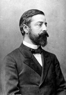
Emil Zuckerkandl
Emil Zuckerkandl (1 September 1849 Győr, Hungary – 28 May 1910, Vienna, Austria-Hungary) was an Austrian-Hungarian anatomist who held the first chair for anatomy at the University of Vienna as of 1888. Biography Zuckerkandl was born in Győr on 1 September 1849, to a Jewish family. He had two brothers: the industrialist Victor Zuckerkandl, and the urologist Otto Zuckerkandl (1861–1921). Until his 16th year, Emil wanted to become a violin virtuoso. Having not attended school, he is reported to have subsequently self-studied the entire upper level gymnasium material in a year. He was educated at the University of Vienna ( MD, 1874) and was an admiring student of Josef Hyrtl, and an anatomical assistant to Karl von Rokitansky (1804–1878) and Karl Langer (1819–1887). In 1875, he became privatdozent of anatomy at the University of Utrecht, and he was appointed assistant professor at the University of Vienna in 1879, being made professor at Graz in 1882. Beginning i ... [...More Info...] [...Related Items...] OR: [Wikipedia] [Google] [Baidu] |
Parahippocampal Gyrus
The parahippocampal gyrus (or hippocampal gyrus') is a grey matter cortical region, a gyrus of the brain that surrounds the hippocampus and is part of the limbic system. The region plays an important role in memory encoding and retrieval. It has been involved in some cases of hippocampal sclerosis. Asymmetry has been observed in schizophrenia. Structure The anterior part of the gyrus includes the perirhinal and entorhinal cortices. The term parahippocampal cortex is used to refer to an area that encompasses both the posterior parahippocampal gyrus and the medial portion of the fusiform gyrus. Function Scene recognition The parahippocampal place area (PPA) is a sub-region of the parahippocampal cortex that lies medially in the inferior temporo-occipital cortex. PPA plays an important role in the encoding and recognition of environmental scenes (rather than faces). fMRI studies indicate that this region of the brain becomes highly active when human subjects view topograp ... [...More Info...] [...Related Items...] OR: [Wikipedia] [Google] [Baidu] |
BrainInfo
''NeuroNames'' is an integrated nomenclature for structures in the brain and spinal cord of the four species most studied by neuroscientists: human, macaque, rat and mouse. It offers a standard, controlled vocabulary of common names for structures, which is suitable for unambiguous neuroanatomical indexing of information in digital databases. Terms in the standard vocabulary have been selected for ease of pronunciation, mnemonic value, and frequency of use in recent neuroscientific publications. Structures and their relations to each other are defined in terms of the standard vocabulary. Currently NeuroNames contains standard names, synonyms and definitions of some 2,500 neuroanatomical entities. The nomenclature is maintained by the University of Washington and is the core component of a tool called "BrainInfo". BrainInfo helps one identify structures in the brain. One can either search by a structure name or locate the structure in a brain atlas and get information such as its ... [...More Info...] [...Related Items...] OR: [Wikipedia] [Google] [Baidu] |
Terminologia Anatomica
''Terminologia Anatomica'' (commonly abbreviated TA) is the international standard for human anatomy, human anatomical terminology. It is developed by the Federative International Programme on Anatomical Terminology (FIPAT) a program of the International Federation of Associations of Anatomists (IFAA). History The sixth edition of the previous standard, ''Nomina Anatomica'', was released in 1989. The first edition of ''Terminologia Anatomica'', superseding Nomina Anatomica, was developed by the Federative Committee on Anatomical Terminology (FCAT) and the International Federation of Associations of Anatomists (IFAA) and released in 1998. In April 2011, this edition was published online by the Federative International Programme on Anatomical Terminologies (FIPAT), the successor of FCAT. The first edition contained 7635 Latin items. The second edition was released online by FIPAT in 2019 and approved and adopted by the IFAA General Assembly in 2020. The latest errata is dated Au ... [...More Info...] [...Related Items...] OR: [Wikipedia] [Google] [Baidu] |


