|
Strabismus Hook
This is a list of instruments used in ophthalmology.Ophthalmology Oral & Practical 3rd edition, by Dr. Danesh Instrument list A complete list of ophthalmic instruments can be found below: Image gallery File:ASICO Akahoshi Combo II Prechopper.jpg, Akahoshi Combo II Prechopper File:Briller2.JPG, Glasses File:ColorContactLens.JPG, Contact lenses File:Medical Instrument Eye Plain dissecting forceps.jpg, Plain dissecting forceps File:Medical instrument Artery forceps or Haemostat.jpg, Artery forceps or Haemostat File:Medical Instrument Mosquito forceps.jpg, Mosquito forceps File:Medical Instrument Linen holding forceps.jpg, Linen holding forceps File:Medical Instrument Bowman's lacrimal probe.jpg, Bowman's lacrimal probe File:Medical Instrument Saint Martin's forceps.jpg, Saint Martin's forceps File:Medical Instrument Eye Lens expressor.jpg, Eye Lens expressor File:Medical instrument Eye ENT Nettleship's Punctum dilator.jpg, Nettleship's punctum dilator File:Medical Instrument Eye ... [...More Info...] [...Related Items...] OR: [Wikipedia] [Google] [Baidu] |
Ophthalmology
Ophthalmology ( ) is a surgical subspecialty within medicine that deals with the diagnosis and treatment of eye disorders. An ophthalmologist is a physician who undergoes subspecialty training in medical and surgical eye care. Following a medical degree, a doctor specialising in ophthalmology must pursue additional postgraduate residency training specific to that field. This may include a one-year integrated internship that involves more general medical training in other fields such as internal medicine or general surgery. Following residency, additional specialty training (or fellowship) may be sought in a particular aspect of eye pathology. Ophthalmologists prescribe medications to treat eye diseases, implement laser therapy, and perform surgery when needed. Ophthalmologists provide both primary and specialty eye care - medical and surgical. Most ophthalmologists participate in academic research on eye diseases at some point in their training and many include research as p ... [...More Info...] [...Related Items...] OR: [Wikipedia] [Google] [Baidu] |
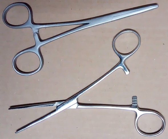
Artery Forceps
A hemostat (also called a hemostatic clamp, arterial forceps, or pean after Jules-Émile Péan) is a surgical tool used in many surgical procedures to control bleeding. For this reason, it is common in the initial phases of surgery for the initial incision to be lined with hemostats which close blood vessels awaiting ligation. Hemostats belong to a group of instruments that pivot (similar to scissors, and including needle holders, tissue holders and various clamps) where the structure of the tip determines its function. The hemostat has handles that can be held in place by their locking mechanism. The locking mechanism is typically a series of interlocking teeth, a few on each handle, that allow the user to adjust the clamping force of the pliers. When locked together, the force between the tips is approximately 40 N (9 lbf). History The earliest known drawing of a pivoting surgical instrument dates back to 1500 BC on a tomb at Thebes, Egypt. Later Roman bronze and steel pivo ... [...More Info...] [...Related Items...] OR: [Wikipedia] [Google] [Baidu] |
Chalazion
A chalazion (; plural chalazia or chalazions) or meibomian cyst is a cyst in the eyelid usually due to a blocked meibomian gland, typically in the middle of the eyelid, red, and not painful. They tend to come on gradually over a few weeks. A chalazion may occur following a stye or from hardened oils blocking the gland. The blocked gland is usually the meibomian gland, but can also be the gland of Zeis. A stye and cellulitis may appear similar. A stye, however, is usually more sudden in onset, painful, and occurs at the edge of the eyelid. Cellulitis is also typically painful. Treatment is initiated with warm compresses. In addition, antibiotic/corticosteroid eyedrops or ointment may be used. If this is not effective, injecting corticosteroids into the lesion may be tried. If large, incision and drainage may be recommended. While relatively common, the frequency of the condition is unknown. It is most common in people 30–50 years of age, and equally common in males and fe ... [...More Info...] [...Related Items...] OR: [Wikipedia] [Google] [Baidu] |
Intraocular Lens
Intraocular lens (IOL) is a lens (optics), lens implanted in the human eye, eye as part of a treatment for cataracts or myopia. If the natural lens is left in the eye, the IOL is known as Phakic intraocular lens, phakic, otherwise it is a pseudophakic, or false lens. Such a lens is typically implanted during cataract surgery, after the eye's cloudy lens (anatomy), natural lens (cataract) has been removed. The pseudophakic IOL provides the same light-focusing function as the natural crystalline lens. The phakic type of IOL is placed over the existing natural lens and is used in refractive surgery to change the eye's optical power as a treatment for myopia (nearsightedness). This is an alternative to LASIK. IOLs usually consist of a small plastic lens with plastic side struts, called haptics, to hold the lens in place in the capsular bag inside the eye. IOLs were conventionally made of an inflexible material (Polymethyl methacrylate, PMMA), although this has largely been superseded ... [...More Info...] [...Related Items...] OR: [Wikipedia] [Google] [Baidu] |
Surgical Incision
In surgery, a surgical incision is a cut made through the skin and soft tissue to facilitate an operation or procedure. Often, multiple incisions are possible for an operation. In general, a surgical incision is made as small and unobtrusive as possible to facilitate safe and timely operating conditions. Anatomy Surgical incisions are planned based on the expected extent of exposure needed for the specific operation planned. Within each region of the body, several incisions are common. Head and neck * Wilde's incision – This post-aural incision is used for a variant mastoiditis drainage, and was named after Sir William Wilde, an ENT surgeon in Dublin who first described it at the end of the nineteenth century. His son, Oscar Wilde's, death was stated by his doctors to be due to meningitis stemming from an ear infection. He had recently had an operation, believed by some to be a mastoidectomy. Chest * Median sternotomy – This is the primary incision used for cardiac p ... [...More Info...] [...Related Items...] OR: [Wikipedia] [Google] [Baidu] |
Glaucoma
Glaucoma is a group of eye diseases that result in damage to the optic nerve (or retina) and cause vision loss. The most common type is open-angle (wide angle, chronic simple) glaucoma, in which the drainage angle for fluid within the eye remains open, with less common types including closed-angle (narrow angle, acute congestive) glaucoma and normal-tension glaucoma. Open-angle glaucoma develops slowly over time and there is no pain. Peripheral vision may begin to decrease, followed by central vision, resulting in blindness if not treated. Closed-angle glaucoma can present gradually or suddenly. The sudden presentation may involve severe eye pain, blurred vision, mid-dilated pupil, redness of the eye, and nausea. Vision loss from glaucoma, once it has occurred, is permanent. Eyes affected by glaucoma are referred to as being glaucomatous. Risk factors for glaucoma include increasing age, high pressure in the eye, a family history of glaucoma, and use of steroid medicatio ... [...More Info...] [...Related Items...] OR: [Wikipedia] [Google] [Baidu] |
Lens (anatomy)
The lens, or crystalline lens, is a transparent biconvex structure in the eye that, along with the cornea, helps to refract light to be focused on the retina. By changing shape, it functions to change the focal length of the eye so that it can focus on objects at various distances, thus allowing a sharp real image of the object of interest to be formed on the retina. This adjustment of the lens is known as '' accommodation'' (see also below). Accommodation is similar to the focusing of a photographic camera via movement of its lenses. The lens is flatter on its anterior side than on its posterior side. In humans, the refractive power of the lens in its natural environment is approximately 18 dioptres, roughly one-third of the eye's total power. Structure The lens is part of the anterior segment of the human eye. In front of the lens is the iris, which regulates the amount of light entering into the eye. The lens is suspended in place by the suspensory ligament of the l ... [...More Info...] [...Related Items...] OR: [Wikipedia] [Google] [Baidu] |
Human Eye
The human eye is a sensory organ, part of the sensory nervous system, that reacts to visible light and allows humans to use visual information for various purposes including seeing things, keeping balance, and maintaining circadian rhythm. The eye can be considered as a living optical device. It is approximately spherical in shape, with its outer layers, such as the outermost, white part of the eye (the sclera) and one of its inner layers (the pigmented choroid) keeping the eye essentially light tight except on the eye's optic axis. In order, along the optic axis, the optical components consist of a first lens (the cornea—the clear part of the eye) that accomplishes most of the focussing of light from the outside world; then an aperture (the pupil) in a diaphragm (the iris—the coloured part of the eye) that controls the amount of light entering the interior of the eye; then another lens (the crystalline lens) that accomplishes the remaining focussing of light in ... [...More Info...] [...Related Items...] OR: [Wikipedia] [Google] [Baidu] |
Superior Rectus
The superior rectus muscle is a muscle in the orbit. It is one of the extraocular muscles. It is innervated by the superior division of the oculomotor nerve (III). In the primary position (looking straight ahead), its primary function is elevation, although it also contributes to intorsion and adduction. It is associated with a number of medical conditions, and may be weak, paralysed, overreactive, or even congenitally absent in some people. Structure The superior rectus muscle originates from the annulus of Zinn. It inserts into the anterosuperior surface of the eye. This insertion has a width of around 11 mm. It is around 8 mm from the corneal limbus. Nerve supply The superior rectus muscle is supplied by the superior division of the oculomotor nerve (III). Relations The superior rectus muscle is related to the other extraocular muscles, particularly to the medial rectus muscle and the lateral rectus muscle. The insertion of the superior rectus muscle is around 7.5 mm ... [...More Info...] [...Related Items...] OR: [Wikipedia] [Google] [Baidu] |
Sclera
The sclera, also known as the white of the eye or, in older literature, as the tunica albuginea oculi, is the opaque, fibrous, protective, outer layer of the human eye containing mainly collagen and some crucial elastic fiber. In humans, and some other vertebrates, the whole sclera is white, contrasting with the coloured iris, but in most mammals, the visible part of the sclera matches the colour of the iris, so the white part does not normally show while other vertebrates have distinct colors for both of them. In the development of the embryo, the sclera is derived from the neural crest. In children, it is thinner and shows some of the underlying pigment, appearing slightly blue. In the elderly, fatty deposits on the sclera can make it appear slightly yellow. People with dark skin can have naturally darkened sclerae, the result of melanin pigmentation. The human eye is relatively rare for having a pale sclera (relative to the iris). This makes it easier for one individual to ... [...More Info...] [...Related Items...] OR: [Wikipedia] [Google] [Baidu] |
Cornea
The cornea is the transparent front part of the eye that covers the iris, pupil, and anterior chamber. Along with the anterior chamber and lens, the cornea refracts light, accounting for approximately two-thirds of the eye's total optical power. In humans, the refractive power of the cornea is approximately 43 dioptres. The cornea can be reshaped by surgical procedures such as LASIK. While the cornea contributes most of the eye's focusing power, its focus is fixed. Accommodation (the refocusing of light to better view near objects) is accomplished by changing the geometry of the lens. Medical terms related to the cornea often start with the prefix "'' kerat-''" from the Greek word κέρας, ''horn''. Structure The cornea has unmyelinated nerve endings sensitive to touch, temperature and chemicals; a touch of the cornea causes an involuntary reflex to close the eyelid. Because transparency is of prime importance, the healthy cornea does not have or need blood vess ... [...More Info...] [...Related Items...] OR: [Wikipedia] [Google] [Baidu] |
Cataract
A cataract is a cloudy area in the lens of the eye that leads to a decrease in vision. Cataracts often develop slowly and can affect one or both eyes. Symptoms may include faded colors, blurry or double vision, halos around light, trouble with bright lights, and trouble seeing at night. This may result in trouble driving, reading, or recognizing faces. Poor vision caused by cataracts may also result in an increased risk of falling and depression. Cataracts cause 51% of all cases of blindness and 33% of visual impairment worldwide. Cataracts are most commonly due to aging but may also occur due to trauma or radiation exposure, be present from birth, or occur following eye surgery for other problems. Risk factors include diabetes, longstanding use of corticosteroid medication, smoking tobacco, prolonged exposure to sunlight, and alcohol. The underlying mechanism involves accumulation of clumps of protein or yellow-brown pigment in the lens that reduces transmission of ... [...More Info...] [...Related Items...] OR: [Wikipedia] [Google] [Baidu] |


.jpg)


_PHIL_4284_lores.jpg)