|
Sclera
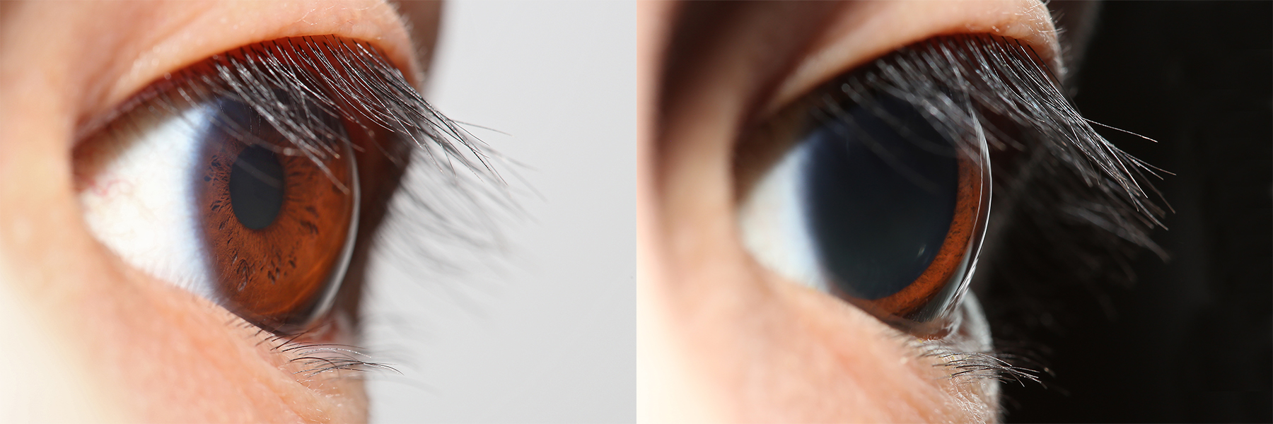
The sclera, also known as the white of the eye or, in older literature, as the tunica albuginea oculi, is the opaque, fibrous, protective outer layer of the eye containing mainly collagen and some crucial elastic fiber. In the development of the embryo, the sclera is derived from the neural crest. In children, it is thinner and shows some of the underlying pigment, appearing slightly blue. In the elderly, fatty deposits on the sclera can make it appear slightly yellow. People with dark skin can have naturally darkened sclerae, the result of melanin pigmentation. In humans, and some other vertebrates, the whole sclera is white or pale, contrasting with the coloured iris (anatomy), iris. The cooperative eye hypothesis suggests that the pale sclera evolved as a method of nonverbal communication that makes it easier for one individual to identify where another individual is looking. Other mammals with white or pale sclera include chimpanzees, many orangutans, some gorillas, and bon ... [...More Info...] [...Related Items...] OR: [Wikipedia] [Google] [Baidu] |
Globe (human Eye)
The human eye is a sensory organ in the visual system that reacts to visible light allowing eyesight. Other functions include maintaining the circadian rhythm, and keeping balance. The eye can be considered as a living optical device. It is approximately spherical in shape, with its outer layers, such as the outermost, white part of the eye (the sclera) and one of its inner layers (the pigmented choroid) keeping the eye essentially light tight except on the eye's optic axis. In order, along the optic axis, the optical components consist of a first lens (the cornea—the clear part of the eye) that accounts for most of the optical power of the eye and accomplishes most of the focusing of light from the outside world; then an aperture (the pupil) in a diaphragm (the iris—the coloured part of the eye) that controls the amount of light entering the interior of the eye; then another lens (the crystalline lens) that accomplishes the remaining focusing of light into imag ... [...More Info...] [...Related Items...] OR: [Wikipedia] [Google] [Baidu] |
Anterior Ciliary Arteries
The anterior ciliary arteries are seven arteries in each eye-socket that arise from muscular branches of the ophthalmic artery and supply the conjunctiva, sclera, rectus muscles, and the ciliary body. The arteries end by anastomosing with branches of the long posterior ciliary arteries to form the circulus arteriosus major. Anatomy There are seven anterior ciliary arteries on each side of the body; two anterior ciliary arteries are associated with the superior, the medial, and the inferior rectus muscles, whereas the lateral rectus muscle is associated with only a single anterior ciliary artery. Origin The anterior ciliary arteries arise from muscular branches of the ophthalmic artery supplying the rectus muscles of the eye. Course and relations The anterior ciliary arteries exit the muscles near the muscles' insertions, passing anterior-ward alongside the rectus muscles' tendons before turning inward to perforate the sclera near the corneal limbus to reach the ci ... [...More Info...] [...Related Items...] OR: [Wikipedia] [Google] [Baidu] |
Cornea
The cornea is the transparency (optics), transparent front part of the eyeball which covers the Iris (anatomy), iris, pupil, and Anterior chamber of eyeball, anterior chamber. Along with the anterior chamber and Lens (anatomy), lens, the cornea Refraction, refracts light, accounting for approximately two-thirds of the eye's total optical power. In humans, the refractive power of the cornea is approximately 43 dioptres. The cornea can be reshaped by surgical procedures such as LASIK. While the cornea contributes most of the eye's focusing power, its Focus (optics), focus is fixed. Accommodation (eye), Accommodation (the refocusing of light to better view near objects) is accomplished by changing the geometry of the lens. Medical terms related to the cornea often start with the prefix "''wikt:kerat-, kerat-''" from the Ancient Greek, Greek word κέρας, ''horn''. Structure The cornea has myelinated, unmyelinated nerve endings sensitive to touch, temperature and chemicals; a to ... [...More Info...] [...Related Items...] OR: [Wikipedia] [Google] [Baidu] |
Lamina Cribrosa Sclerae
The nerve fibers forming the optic nerve exit the eye posteriorly through a hole in the sclera that is occupied by a mesh-like structure called the lamina cribrosa. It is formed by a multilayered network of collagen fibers that extend from the scleral canal wall. The nerve fibers that comprise the optic nerve run through pores formed by these collagen beams. In humans, a central retinal artery is located slightly off-center in the nasal direction. The lamina cribrosa is thought to help support the retinal ganglion cell axons as they traverse the scleral canal. Being structurally weaker than the much thicker and denser sclera, the lamina cribrosa is more sensitive to changes in the intraocular pressure and tends to react to increased pressure through posterior displacement. This is thought to be one of the causes of nerve damage in glaucoma Glaucoma is a group of eye diseases that can lead to damage of the optic nerve. The optic nerve transmits visual information from the eye ... [...More Info...] [...Related Items...] OR: [Wikipedia] [Google] [Baidu] |
Corneal Limbus
The corneal limbus (''Latin'': corneal border) is a highly vascularized and pigmented zone between the cornea, conjunctiva, and the sclera (the white of the eye) that protects and heals the cornea. The cornea is composed of three primary cell types: epithelial cells, corneal fibroblasts, and endothelial cells. The corneal surface is one of the body's most specialized structures that undergoes continuous cellular renewal and regeneration. It contains limbal epithelial stem cells (LESCs) in the palisades of Vogt. Limbal stem cell deficiency (LSCD) can lead to disorders where limbal stem cells are damaged or absent. Additional disorders involving the corneal limbus are caused by deficiencies in interactions between ocular structures, developmental anomalies, and cancer. This article explores the structure, functions, disorders, and clinical significance of the corneal limbus. Etymology The word "limbus" comes from the Latin meaning "border." Structure The corneal limbus is th ... [...More Info...] [...Related Items...] OR: [Wikipedia] [Google] [Baidu] |
Cooperative Eye Hypothesis
The cooperative eye hypothesis is a proposed explanation for the appearance of the human eye. It suggests that the eye's distinctive visible characteristics evolved to make it easier for humans to follow another's gaze while communicating or while working together on tasks.Michael Tomasello, Brian Hare, Hagen Lehmann and Josep Call (2007). Reliance on head versus eyes in the gaze following of great apes and human infants: the cooperative eye hypothesis. ''Journal of Human Evolution'' 52: 314-320 Differences in primate eyes Unlike other primates, all human beings have eyes with a distinct colour contrast between the white sclera, the coloured iris, and the black pupil. This is due to a lack of pigment in the sclera. Other primates mostly have pigmented sclerae that are brown or dark in colour. There is also a higher contrast between human skin, sclera, and irises. Human eyes are also larger in proportion to body size, and are longer horizontally. Among primates, humans are the onl ... [...More Info...] [...Related Items...] OR: [Wikipedia] [Google] [Baidu] |
Iris (anatomy)
The iris (: irides or irises) is a thin, annular structure in the eye in most mammals and birds that is responsible for controlling the diameter and size of the pupil, and thus the amount of light reaching the retina. In optical terms, the pupil is the eye's aperture, while the iris is the diaphragm (optics), diaphragm. Eye color is defined by the iris. Etymology The word "iris" is derived from the Greek word for "rainbow", also Iris (mythology), its goddess plus messenger of the gods in the ''Iliad'', because of the many eye color, colours of this eye part. Structure The iris consists of two layers: the front pigmented Wikt:fibrovascular, fibrovascular layer known as a stroma of iris, stroma and, behind the stroma, pigmented epithelial cells. The stroma is connected to a sphincter muscle (sphincter pupillae), which contracts the pupil in a circular motion, and a set of dilator muscles (dilator pupillae), which pull the iris radially to enlarge the pupil, pulling it in folds. ... [...More Info...] [...Related Items...] OR: [Wikipedia] [Google] [Baidu] |
Long Posterior Ciliary Arteries
The long posterior ciliary arteries are arteries of the orbit. There are long posterior ciliary arteries two on each side of the body. They are branches of the ophthalmic artery. They pass forward within the eye to reach the ciliary body where they ramify and anastomose with the anterior ciliary arteries, thus forming the major arterial circle of the iris.The long posterior ciliary arteries contribute arterial supply to the choroid, ciliary body, and iris. Anatomy There are two long ciliary arteries. They are branches of the ophthalmic artery. Course and relations The long posterior ciliary arteries first run near the optic nerve before piercing the posterior sclera near the optic nerve. They pass anterior-ward - one along each side of the eyeball - between the sclera and choroid to reach the ciliary muscle where they divide into two branches which go on to form the major arterial circle of the iris. Anastomoses Non-terminal branches of the long posterior ciliary arteries ... [...More Info...] [...Related Items...] OR: [Wikipedia] [Google] [Baidu] |
Short Posterior Ciliary Arteries
The short posterior ciliary arteries are a number of branches of the ophthalmic artery. They pass forward with the optic nerve to reach the eyeball, piercing the sclera around the entry of the optic nerve into the eyeball. Anatomy The number of short posterior ciliary arteries varies between individuals; one or more short posterior ciliary arteries initially branch off the ophthalmic artery, subsequently dividing to form up to 20 short posterior ciliary arteries. Origin The short posterior ciliary arteries branch off the ophthalmic artery as it crosses the optic nerve medially. Course and relations About 7 short posterior ciliary arteries accompany the optic nerve, passing anterior-ward to reach the posterior part of the eyeball, where they divide into 15-20 branches and pierce the sclera around the entrance of the optic nerve. Distribution The short posterior ciliary arteries contribute arterial supply to the choroid, ciliary processes, optic disc, the outer retina, and ... [...More Info...] [...Related Items...] OR: [Wikipedia] [Google] [Baidu] |
Choroid
The choroid, also known as the choroidea or choroid coat, is a part of the uvea, the vascular layer of the eye. It contains connective tissues, and lies between the retina and the sclera. The human choroid is thickest at the far extreme rear of the eye (at 0.2 mm), while in the outlying areas it narrows to 0.1 mm. The choroid provides oxygen and nourishment to the outer layers of the retina. Along with the ciliary body and iris, the choroid forms the uveal tract. The structure of the choroid is generally divided into four layers (classified in order of furthest away from the retina to closest): *Haller's layer – outermost layer of the choroid consisting of larger diameter blood vessels; * Sattler's layer – layer of medium diameter blood vessels; * Choriocapillaris – layer of capillaries; and * Bruch's membrane (synonyms: Lamina basalis, Complexus basalis, Lamina vitra) – innermost layer of the choroid. Blood supply There are two circulations of the eye: ... [...More Info...] [...Related Items...] OR: [Wikipedia] [Google] [Baidu] |
Neural Crest
The neural crest is a ridge-like structure that is formed transiently between the epidermal ectoderm and neural plate during vertebrate development. Neural crest cells originate from this structure through the epithelial-mesenchymal transition, and in turn give rise to a diverse cell lineage—including melanocytes, craniofacial cartilage and bone, smooth muscle, dentin, peripheral and enteric neurons, adrenal medulla and glia. After gastrulation, the neural crest is specified at the border of the neural plate and the non-neural ectoderm. During neurulation, the borders of the neural plate, also known as the neural folds, converge at the dorsal midline to form the neural tube. Subsequently, neural crest cells from the roof plate of the neural tube undergo an epithelial to mesenchymal transition, delaminating from the neuroepithelium and migrating through the periphery, where they differentiate into varied cell types. The emergence of the neural crest was important in v ... [...More Info...] [...Related Items...] OR: [Wikipedia] [Google] [Baidu] |
Visual System
The visual system is the physiological basis of visual perception (the ability to perception, detect and process light). The system detects, phototransduction, transduces and interprets information concerning light within the visible range to construct an imaging, image and build a mental model of the surrounding environment. The visual system is associated with the eye and functionally divided into the optics, optical system (including cornea and crystalline lens, lens) and the nervous system, neural system (including the retina and visual cortex). The visual system performs a number of complex tasks based on the ''image forming'' functionality of the eye, including the formation of monocular images, the neural mechanisms underlying stereopsis and assessment of distances to (depth perception) and between objects, motion perception, pattern recognition, accurate motor coordination under visual guidance, and colour vision. Together, these facilitate higher order tasks, such as ... [...More Info...] [...Related Items...] OR: [Wikipedia] [Google] [Baidu] |


