|
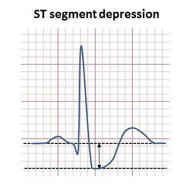
ST Depression
ST depression refers to a finding on an electrocardiogram, wherein the trace in the ST segment is abnormally low below the baseline. Causes It is often a sign of myocardial ischemia, of which coronary insufficiency is a major cause. Other ischemic heart diseases causing ST depression include: * Subendocardial ischemia or even infarction. Subendocardial means non full thickness ischemia. In contrast, ST elevation is transmural (or full thickness) ischemia * Non Q-wave myocardial infarction * Reciprocal changes in acute Q-wave myocardial infarction (e.g., ST depression in leads I & aVL with acute inferior myocardial infarction) * ST segment depression and T-wave changes may be seen in patients with unstable angina Depressed but ''upsloping'' ST segment generally rules out ischemia as a cause. Also, it can be a normal variant or artifacts, such as: * Pseudo-ST-depression, which is a wandering baseline due to poor skin contact of the electrode MicroEKG ManualRetrieved September ... [...More Info...] [...Related Items...] OR: [Wikipedia] [Google] [Baidu] |
Left Bundle Branch Block
Left bundle branch block (LBBB) is a conduction abnormality in the heart that can be seen on an electrocardiogram (ECG). In this condition, activation of the left ventricle of the heart is delayed, which causes the left ventricle to contract later than the right ventricle. Causes Among the causes of LBBB are: * Aortic stenosis * Dilated cardiomyopathy * Acute myocardial infarction * Extensive coronary artery disease * Primary disease of the cardiac electrical conduction system * Long standing hypertension leading to aortic root dilatation and subsequent aortic regurgitation * Lyme disease Mechanisms Slow or absent conduction through the left bundle branch means that it takes longer than normal for the left ventricle to fully depolarise. This can be due to a damaged bundle branch that is completely unable to conduct, but may represent intact conduction that is slower than normal. LBBB may be fixed, present at all times, but may be intermittent for example occurring only duri ... [...More Info...] [...Related Items...] OR: [Wikipedia] [Google] [Baidu] |
QRS Complex
The QRS complex is the combination of three of the graphical deflections seen on a typical electrocardiogram (ECG or EKG). It is usually the central and most visually obvious part of the tracing. It corresponds to the depolarization of the right and left ventricles of the heart and contraction of the large ventricular muscles. In adults, the QRS complex normally lasts ; in children it may be shorter. The Q, R, and S waves occur in rapid succession, do not all appear in all leads, and reflect a single event and thus are usually considered together. A Q wave is any downward deflection immediately following the P wave. An R wave follows as an upward deflection, and the S wave is any downward deflection after the R wave. The T wave follows the S wave, and in some cases, an additional U wave follows the T wave. To measure the QRS interval start at the end of the PR interval (or beginning of the Q wave) to the end of the S wave. Normally this interval is 0.08 to 0.10 seconds. W ... [...More Info...] [...Related Items...] OR: [Wikipedia] [Google] [Baidu] |
Quinidine
Quinidine is a class I antiarrhythmic agent, class IA antiarrhythmic agent used to treat heart rhythm disturbances. It is a diastereomer of Antimalarial medication, antimalarial agent quinine, originally derived from the bark of the cinchona tree. The drug causes increased action potential duration, as well as a prolonged QT interval. As of 2019, its IV formulation is no longer being manufactured for use in the United States. Medical uses Quinidine is occasionally used as a class I antiarrhythmic agent to prevent ventricular arrhythmias, particularly in Brugada Syndrome, although its safety in this indication is uncertain. It reduces the recurrence of atrial fibrillation after patients undergo cardioversion, but it has proarrhythmic agent, proarrhythmic effects and trials suggest that it may lead to an overall increased mortality in these patients. Quinidine is also used to treat short QT syndrome. Eli Lilly has discontinued manufacture of parenteral quinidine gluconate in the ... [...More Info...] [...Related Items...] OR: [Wikipedia] [Google] [Baidu] |
Digoxin
Digoxin (better known as digitalis), sold under the brand name Lanoxin among others, is a medication used to treat various heart disease, heart conditions. Most frequently it is used for atrial fibrillation, atrial flutter, and heart failure. Digoxin is one of the oldest medications used in the field of cardiology. It works by increasing myocardial contractility, increasing stroke volume and blood pressure, reducing heart rate, and somewhat extending the time frame of the Muscle contraction, contraction. Digoxin is taken by mouth or by intravenous, injection into a vein. Digoxin has a half life of approximately 36 hours given at average doses in patients with normal renal function. It is excreted mostly unchanged in the urine. Common side effects include gynecomastia, breast enlargement with other side effects generally due to an excessive dose. These side effects may include loss of appetite, nausea, trouble seeing, confusion, and an Heart arrhythmia, irregular heartbeat. Gre ... [...More Info...] [...Related Items...] OR: [Wikipedia] [Google] [Baidu] |
Dilated Cardiomyopathy
Dilated cardiomyopathy (DCM) is a condition in which the heart becomes enlarged and cannot pump blood effectively. Symptoms vary from none to feeling tired, leg swelling, and shortness of breath. It may also result in chest pain or fainting. Complications can include heart failure, heart valve disease, or an irregular heartbeat. Causes include genetics, alcohol, cocaine, certain toxins, complications of pregnancy, and certain infections. Coronary artery disease and high blood pressure may play a role, but are not the primary cause. In many cases the cause remains unclear. It is a type of cardiomyopathy, a group of diseases that primarily affects the heart muscle. The diagnosis may be supported by an electrocardiogram, chest X-ray, or echocardiogram. In those with heart failure, treatment may include medications in the ACE inhibitor, beta blocker, and diuretic families. A low salt diet may also be helpful. In those with certain types of irregular heartbeat, blood thinner ... [...More Info...] [...Related Items...] OR: [Wikipedia] [Google] [Baidu] |
Pulmonary Embolism
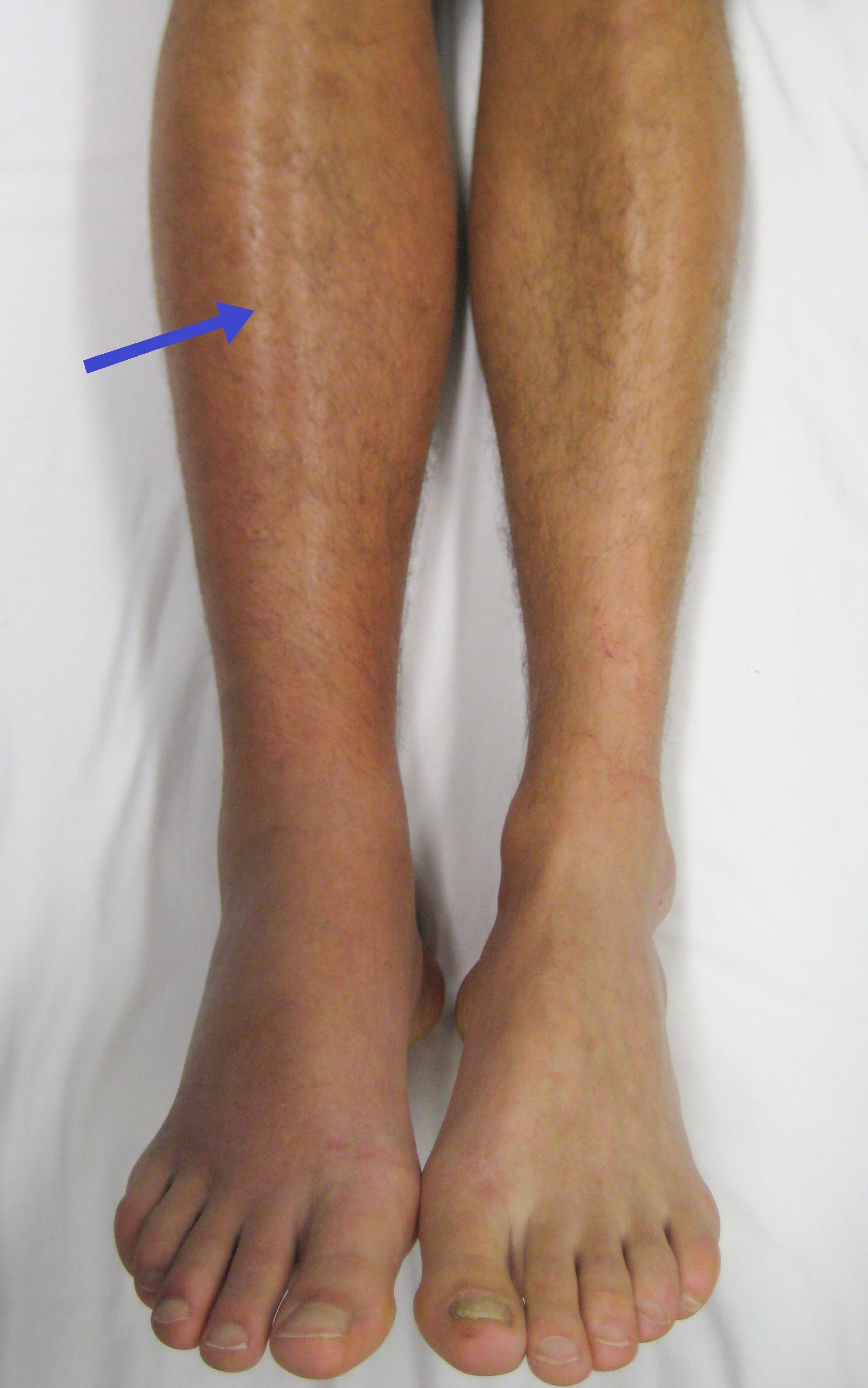
Pulmonary embolism (PE) is a blockage of an pulmonary artery, artery in the lungs by a substance that has moved from elsewhere in the body through the bloodstream (embolism). Symptoms of a PE may include dyspnea, shortness of breath, chest pain particularly upon breathing in, and coughing up blood. Symptoms of a deep vein thrombosis, blood clot in the leg may also be present, such as a erythema, red, warm, swollen, and painful leg. Signs of a PE include low blood oxygen saturation, oxygen levels, tachypnea, rapid breathing, tachycardia, rapid heart rate, and sometimes a mild fever. Severe cases can lead to Syncope (medicine), passing out, shock (circulatory), abnormally low blood pressure, obstructive shock, and cardiac arrest, sudden death. PE usually results from a blood clot in the leg that travels to the lung. The risk of blood clots is increased by advanced age, cancer, prolonged bed rest and immobilization, smoking, stroke, long-haul travel over 4 hours, certain genetics, ... [...More Info...] [...Related Items...] OR: [Wikipedia] [Google] [Baidu] |
Mitral Valve Prolapse
Mitral valve prolapse (MVP) is a valvular heart disease characterized by the displacement of an abnormally thickened mitral valve leaflet into the atria of the heart, left atrium during Systole (medicine), systole. It is the primary form of myxomatous degeneration of the valve. There are various types of MVP, broadly classified as classic and nonclassic. In severe cases of classic MVP, complications include mitral regurgitation, infective endocarditis, congestive heart failure, and, in rare circumstances, cardiac arrest. The diagnosis of MVP primarily relies on echocardiography, which uses ultrasound to visualize the mitral valve. MVP is the most common valvular abnormality, and is estimated to affect 2–3% of the population and 1 in 40 people might have it. The condition was first described by John Brereton Barlow in 1966. It was subsequently termed ''mitral valve prolapse'' by J. Michael Criley. Although mid-systolic click (the sound produced by the prolapsing mitral leaflet) ... [...More Info...] [...Related Items...] OR: [Wikipedia] [Google] [Baidu] |
Mnemonic
A mnemonic device ( ), memory trick or memory device is any learning technique that aids information retention or retrieval in the human memory, often by associating the information with something that is easier to remember. It makes use of elaborative encoding, retrieval cues and imagery as specific tools to encode information in a way that allows for efficient storage and retrieval. It aids original information in becoming associated with something more accessible or meaningful—which in turn provides better retention of the information. Commonly encountered mnemonics are often used for lists and in auditory system, auditory form such as Acrostic, short poems, acronyms, initialisms or memorable phrases. They can also be used for other types of information and in visual or kinesthetic forms. Their use is based on the observation that the human mind more easily remembers spatial, personal, surprising, physical, sexual, humorous and otherwise "relatable" information rather tha ... [...More Info...] [...Related Items...] OR: [Wikipedia] [Google] [Baidu] |
Stroke
Stroke is a medical condition in which poor cerebral circulation, blood flow to a part of the brain causes cell death. There are two main types of stroke: brain ischemia, ischemic, due to lack of blood flow, and intracranial hemorrhage, hemorrhagic, due to bleeding. Both cause parts of the brain to stop functioning properly. Signs and symptoms of stroke may include an hemiplegia, inability to move or feel on one side of the body, receptive aphasia, problems understanding or expressive aphasia, speaking, dizziness, or homonymous hemianopsia, loss of vision to one side. Signs and symptoms often appear soon after the stroke has occurred. If symptoms last less than 24 hours, the stroke is a transient ischemic attack (TIA), also called a mini-stroke. subarachnoid hemorrhage, Hemorrhagic stroke may also be associated with a thunderclap headache, severe headache. The symptoms of stroke can be permanent. Long-term complications may include pneumonia and Urinary incontinence, loss of b ... [...More Info...] [...Related Items...] OR: [Wikipedia] [Google] [Baidu] |
Central Nervous System Disease
Central nervous system diseases or central nervous system disorders are a group of neurological disorders that affect the structure or function of the brain or spinal cord, which collectively form the central nervous system (CNS). These disorders may be caused by such things as infection, injury, blood clots, age related degeneration, cancer, autoimmune disfunction, and birth defects. The symptoms vary widely, as do the treatments. Central nervous system tumors are the most common forms of pediatric cancer. Brain tumors are the most frequent and have the highest mortality. Some disorders, such as substance addiction, autism, and ADHD may be regarded as CNS disorders, though the classifications are not without dispute. Signs and symptoms Every disease has different signs and symptoms. Some of them are persistent headache; pain in the face, back, arms, or legs; an inability to concentrate; loss of feeling; memory loss; loss of muscle strength; tremors; seizures; increased reflex ... [...More Info...] [...Related Items...] OR: [Wikipedia] [Google] [Baidu] |
Mitral Valve Prolapse
Mitral valve prolapse (MVP) is a valvular heart disease characterized by the displacement of an abnormally thickened mitral valve leaflet into the atria of the heart, left atrium during Systole (medicine), systole. It is the primary form of myxomatous degeneration of the valve. There are various types of MVP, broadly classified as classic and nonclassic. In severe cases of classic MVP, complications include mitral regurgitation, infective endocarditis, congestive heart failure, and, in rare circumstances, cardiac arrest. The diagnosis of MVP primarily relies on echocardiography, which uses ultrasound to visualize the mitral valve. MVP is the most common valvular abnormality, and is estimated to affect 2–3% of the population and 1 in 40 people might have it. The condition was first described by John Brereton Barlow in 1966. It was subsequently termed ''mitral valve prolapse'' by J. Michael Criley. Although mid-systolic click (the sound produced by the prolapsing mitral leaflet) ... [...More Info...] [...Related Items...] OR: [Wikipedia] [Google] [Baidu] |


