|
Left Bundle Branch Block
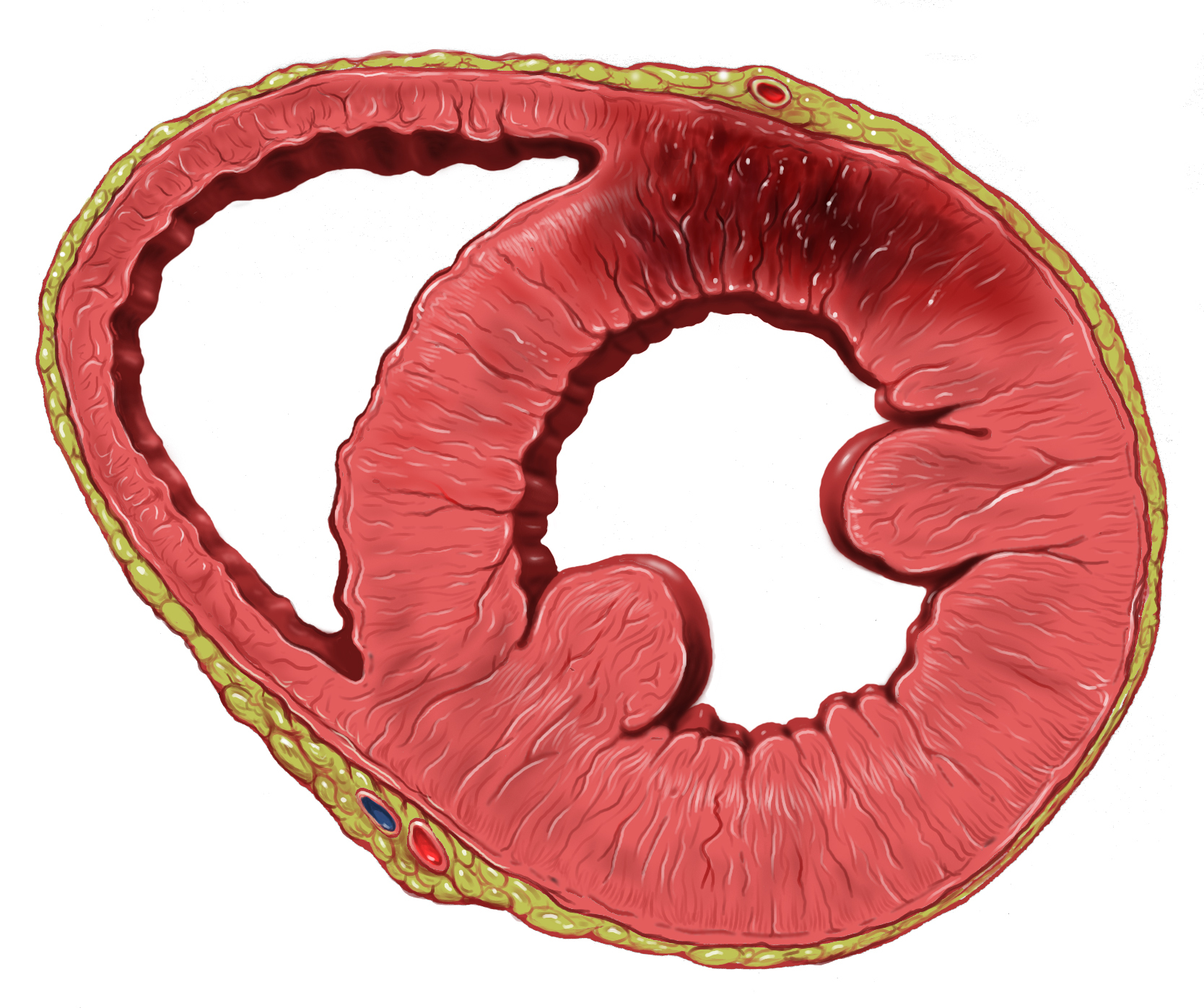
Left bundle branch block (LBBB) is a conduction abnormality in the heart that can be seen on an electrocardiogram (ECG). In this condition, activation of the left ventricle of the heart is delayed, which causes the left ventricle to contract later than the right ventricle. Causes Among the causes of LBBB are: * Aortic stenosis * Dilated cardiomyopathy * Acute myocardial infarction * Extensive coronary artery disease * Primary disease of the cardiac electrical conduction system * Long standing hypertension leading to aortic root dilatation and subsequent aortic regurgitation * Lyme disease Mechanisms Slow or absent conduction through the left bundle branch means that it takes longer than normal for the left ventricle to fully depolarise. This can be due to a damaged bundle branch that is completely unable to conduct, but may represent intact conduction that is slower than normal. LBBB may be fixed, present at all times, but may be intermittent for example occurring only duri ... [...More Info...] [...Related Items...] OR: [Wikipedia] [Google] [Baidu] |
Cardiology
Cardiology () is the study of the heart. Cardiology is a branch of medicine that deals with disorders of the heart and the cardiovascular system. The field includes medical diagnosis and treatment of congenital heart defects, coronary artery disease, heart failure, valvular heart disease, and electrophysiology. Physicians who specialize in this field of medicine are called cardiologists, a sub-specialty of internal medicine. Pediatric cardiologists are pediatricians who specialize in cardiology. Physicians who specialize in cardiac surgery are called cardiothoracic surgeons or cardiac surgeons, a specialty of general surgery. Specializations All cardiologists in the branch of medicine study the disorders of the heart, but the study of adult and child heart disorders each require different training pathways. Therefore, an adult cardiologist (often simply called "cardiologist") is inadequately trained to take care of children, and pediatric cardiologists are not trained to treat ... [...More Info...] [...Related Items...] OR: [Wikipedia] [Google] [Baidu] |
LBBB2009
Left bundle branch block (LBBB) is a conduction abnormality in the heart that can be seen on an electrocardiogram (ECG). In this condition, activation of the left ventricle of the heart is delayed, which causes the left ventricle to contract later than the right ventricle. Causes Among the causes of LBBB are: * Aortic stenosis * Dilated cardiomyopathy * Acute myocardial infarction * Extensive coronary artery disease * Primary disease of the cardiac electrical conduction system * Long standing hypertension leading to aortic root dilatation and subsequent aortic regurgitation * Lyme disease Mechanisms Slow or absent conduction through the left bundle branch means that it takes longer than normal for the left ventricle to fully depolarise. This can be due to a damaged bundle branch that is completely unable to conduct, but may represent intact conduction that is slower than normal. LBBB may be fixed, present at all times, but may be intermittent for example occurring only durin ... [...More Info...] [...Related Items...] OR: [Wikipedia] [Google] [Baidu] |
Sensitivity And Specificity
In medicine and statistics, sensitivity and specificity mathematically describe the accuracy of a test that reports the presence or absence of a medical condition. If individuals who have the condition are considered "positive" and those who do not are considered "negative", then sensitivity is a measure of how well a test can identify true positives and specificity is a measure of how well a test can identify true negatives: * Sensitivity (true positive rate) is the probability of a positive test result, conditioned on the individual truly being positive. * Specificity (true negative rate) is the probability of a negative test result, conditioned on the individual truly being negative. If the true status of the condition cannot be known, sensitivity and specificity can be defined relative to a " gold standard test" which is assumed correct. For all testing, both diagnoses and screening, there is usually a trade-off between sensitivity and specificity, such that higher sensiti ... [...More Info...] [...Related Items...] OR: [Wikipedia] [Google] [Baidu] |
Sgarbossa's Criteria
Sgarbossa's criteria are a set of electrocardiographic findings generally used to identify myocardial infarction (also called ''acute myocardial infarction or a "heart attack"'') in the presence of a left bundle branch block (LBBB) or a ventricular paced rhythm. Myocardial infarction (MI) is often difficult to detect when LBBB is present on ECG. A large clinical trial of thrombolytic therapy for MI (GUSTO-1) evaluated the electrocardiographic diagnosis of evolving MI in the presence of LBBB. The rule was defined by Dr. Elena Sgarbossa, Argentine- born American cardiologist. Among 26,003 North American patients who had a myocardial infarction confirmed by enzyme studies, 131 (0.5%) had LBBB. A scoring system, now commonly called Sgarbossa criteria, was developed from the coefficients assigned by a logistic model for each independent criterion, on a scale of 0 to 5. A minimal score of 3 was required for a specificity of 90%. Sgarbossa's criteria Three criteria are included in ... [...More Info...] [...Related Items...] OR: [Wikipedia] [Google] [Baidu] |
Southern Illinois University School Of Medicine
Southern Illinois University School of Medicine is a medical school located in Springfield, the capital of the U.S. state of Illinois. It is part of the Southern Illinois University system, which includes a campus in Edwardsville as well as the flagship in Carbondale. The medical school was founded in 1970 and achieved full accreditation in 1972. It was founded to relieve a chronic shortage of physicians in downstate Illinois. Notability SIU was once the only medical school in Illinois with its main campus outside of Chicago or its suburbs until the Carle Illinois College of Medicine was formed in 2018 (although the University of Illinois at Chicago does maintain satellites in Peoria and Rockford). SIU was early to incorporate problem-based learning (PBL) into their curricula (see below) and "standardized patients" for medical student testing purposes. SIU students begin care of patients in a clinical setting within the first two weeks of classes. By the end of their ... [...More Info...] [...Related Items...] OR: [Wikipedia] [Google] [Baidu] |
Myocardial Infarction
A myocardial infarction (MI), commonly known as a heart attack, occurs when Ischemia, blood flow decreases or stops in one of the coronary arteries of the heart, causing infarction (tissue death) to the heart muscle. The most common symptom is retrosternal Angina, chest pain or discomfort that classically radiates to the left shoulder, arm, or jaw. The pain may occasionally feel like heartburn. This is the dangerous type of acute coronary syndrome. Other symptoms may include shortness of breath, nausea, presyncope, feeling faint, a diaphoresis, cold sweat, Fatigue, feeling tired, and decreased level of consciousness. About 30% of people have atypical symptoms. Women more often present without chest pain and instead have neck pain, arm pain or feel tired. Among those over 75 years old, about 5% have had an MI with little or no history of symptoms. An MI may cause heart failure, an Cardiac arrhythmia, irregular heartbeat, cardiogenic shock or cardiac arrest. Most MIs occur d ... [...More Info...] [...Related Items...] OR: [Wikipedia] [Google] [Baidu] |
Left Ventricular Hypertrophy
Left ventricular hypertrophy (LVH) is thickening of the heart muscle of the left ventricle of the heart, that is, left-sided ventricular hypertrophy and resulting increased left ventricular mass. Causes While ventricular hypertrophy occurs naturally as a reaction to aerobic exercise and strength training, it is most frequently referred to as a pathological reaction to cardiovascular disease, or high blood pressure. It is one aspect of ventricular remodeling. While LVH itself is not a disease, it is usually a marker for disease involving the heart. Disease processes that can cause LVH include any disease that increases the afterload that the heart has to contract against, and some primary diseases of the muscle of the heart. Causes of increased afterload that can cause LVH include aortic stenosis, aortic insufficiency and hypertension. Primary disease of the muscle of the heart that cause LVH are known as hypertrophic cardiomyopathies, which can lead into heart failure. ... [...More Info...] [...Related Items...] OR: [Wikipedia] [Google] [Baidu] |
Electrocardiography
Electrocardiography is the process of producing an electrocardiogram (ECG or EKG), a recording of the heart's electrical activity through repeated cardiac cycles. It is an electrogram of the heart which is a graph of voltage versus time of the electrical activity of the heart using electrodes placed on the skin. These electrodes detect the small electrical changes that are a consequence of cardiac muscle depolarization followed by repolarization during each cardiac cycle (heartbeat). Changes in the normal ECG pattern occur in numerous cardiac abnormalities, including: * Cardiac rhythmicity, Cardiac rhythm disturbances, such as atrial fibrillation and ventricular tachycardia; * Inadequate coronary artery blood flow, such as myocardial ischemia and myocardial infarction; * and electrolyte disturbances, such as hypokalemia. Traditionally, "ECG" usually means a 12-lead ECG taken while lying down as discussed below. However, other devices can record the electrical activity of ... [...More Info...] [...Related Items...] OR: [Wikipedia] [Google] [Baidu] |
Left Posterior Fascicular Block
A left posterior fascicular block (LPFB), also known as left posterior hemiblock (LPH), is a condition where the left posterior fascicle, which travels to the inferior and posterior portion of the left ventricle, does not conduct the electrical impulses from the atrioventricular node. The wave-front instead moves more quickly through the left anterior fascicle and right bundle branch, leading to a right axis deviation seen on the ECG. Definition The American Heart Association The American Heart Association (AHA) is a nonprofit organization in the United States that funds cardiovascular medical research, educates consumers on healthy living and fosters appropriate Heart, cardiac care in an effort to reduce disability ... has defined a LPFB as: * Frontal plane axis between 90° and 180° in adults * rS pattern in leads I and aVL * qR pattern in leads III and aVF * QRS duration less than 120 ms The broad nature of the posterior bundle as well as its dual blood supply makes i ... [...More Info...] [...Related Items...] OR: [Wikipedia] [Google] [Baidu] |
Left Anterior Fascicular Block
Left anterior fascicular block (LAFB) is an abnormal condition of the left ventricle of the heart, related to, but distinguished from, left bundle branch block (LBBB). It is caused by only the left anterior fascicle – one half of the left bundle branch being defective. It is manifested on the ECG by left axis deviation. It is much more common than left posterior fascicular block. Mechanism Normal activation of the left ventricle (LV) proceeds down the left bundle branch, which consist of three fascicles, the left anterior fascicle, the left posterior fascicle, and the septal fascicle. The posterior fascicle supplies the posterior and inferoposterior walls of the LV, the anterior fascicle supplies the upper and anterior parts of the LV and the septal fascicle supplies the septal wall with innervation. LAFB — which is also known as left anterior hemiblock (LAHB) — occurs when a cardiac impulse spreads first through the left posterior fascicle, causing a delay in activation ... [...More Info...] [...Related Items...] OR: [Wikipedia] [Google] [Baidu] |
ST Segment
In electrocardiography, the ST segment connects the QRS complex and the T wave and has a duration of 0.005 to 0.150 sec (5 to 150 ms). It starts at the J point (junction between the QRS complex and ST segment) and ends at the beginning of the T wave. However, since it is usually difficult to determine exactly where the ST segment ends and the T wave begins, the relationship between the ST segment and T wave should be examined together. The typical ST segment duration is usually around 0.08 sec (80 ms). It should be essentially level with the PR and TP segments. The ST segment represents the isoelectric period when the ventricles are in between depolarization and repolarization. Interpretation * The normal ST segment has a slight upward concavity. * Flat, downsloping, or depressed ST segments may indicate coronary ischemia. * ST elevation may indicate transmural myocardial infarction. An elevation of >1mm and longer than 80 milliseconds following the J-point. This measure has a ... [...More Info...] [...Related Items...] OR: [Wikipedia] [Google] [Baidu] |
T Wave
In electrocardiography, the T wave represents the repolarization of the ventricles. The interval from the beginning of the QRS complex to the apex of the T wave is referred to as the ''absolute refractory period''. The last half of the T wave is referred to as the ''relative refractory period'' or ''vulnerable period''. The T wave contains more information than the QT interval. The T wave can be described by its symmetry, skewness, slope of ascending and descending limbs, amplitude and subintervals like the Tpeak–Tend interval. In most leads, the T wave is positive. This is due to the repolarization of the membrane. During ventricle contraction (QRS complex), the heart depolarizes. Repolarization of the ventricle happens in the opposite direction of depolarization and is negative current, signifying the relaxation of the cardiac muscle of the ventricles. But this negative flow causes a positive T wave; although the cell becomes more negatively charged, the net effect is in ... [...More Info...] [...Related Items...] OR: [Wikipedia] [Google] [Baidu] |


