|
ST Segment
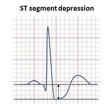
In electrocardiography, the ST segment connects the QRS complex and the T wave and has a duration of 0.005 to 0.150 sec (5 to 150 ms). It starts at the J point (junction between the QRS complex and ST segment) and ends at the beginning of the T wave. However, since it is usually difficult to determine exactly where the ST segment ends and the T wave begins, the relationship between the ST segment and T wave should be examined together. The typical ST segment duration is usually around 0.08 sec (80 ms). It should be essentially level with the PR and TP segments. The ST segment represents the isoelectric period when the ventricles are in between depolarization and repolarization. Interpretation * The normal ST segment has a slight upward concavity. * Flat, downsloping, or depressed ST segments may indicate coronary ischemia. * ST elevation may indicate transmural myocardial infarction. An elevation of >1mm and longer than 80 milliseconds following the J-point. This measure has a ... [...More Info...] [...Related Items...] OR: [Wikipedia] [Google] [Baidu] |
False Positive
A false positive is an error in binary classification in which a test result incorrectly indicates the presence of a condition (such as a disease when the disease is not present), while a false negative is the opposite error, where the test result incorrectly indicates the absence of a condition when it is actually present. These are the two kinds of errors in a binary test, in contrast to the two kinds of correct result (a and a ). They are also known in medicine as a false positive (or false negative) diagnosis, and in statistical classification as a false positive (or false negative) error. In statistical hypothesis testing, the analogous concepts are known as type I and type II errors, where a positive result corresponds to rejecting the null hypothesis, and a negative result corresponds to not rejecting the null hypothesis. The terms are often used interchangeably, but there are differences in detail and interpretation due to the differences between medical testing and sta ... [...More Info...] [...Related Items...] OR: [Wikipedia] [Google] [Baidu] |
Base Deficit
In physiology, base excess and base deficit refer to an excess or deficit, respectively, in the amount of base present in the blood. The value is usually reported as a concentration in units of mEq/L (mmol/L), with positive numbers indicating an excess of base and negative a deficit. A typical reference range for base excess is −2 to +2 mEq/L. Comparison of the base excess with the reference range assists in determining whether an acid/base disturbance is caused by a respiratory, metabolic, or mixed metabolic/respiratory problem. While carbon dioxide defines the respiratory component of acid–base balance, base excess defines the metabolic component. Accordingly, measurement of base excess is defined, under a standardized pressure of carbon dioxide, by titrating back to a standardized blood pH of 7.40. The predominant base contributing to base excess is bicarbonate. Thus, a deviation of serum bicarbonate from the reference range is ordinarily mirrored by a deviation in base ... [...More Info...] [...Related Items...] OR: [Wikipedia] [Google] [Baidu] |
Fetal Electrocardiography
Electrocardiography is the process of producing an electrocardiogram (ECG or EKG), a recording of the heart's electrical activity through repeated cardiac cycles. It is an electrogram of the heart which is a graph of voltage versus time of the electrical activity of the heart using electrodes placed on the skin. These electrodes detect the small electrical changes that are a consequence of cardiac muscle depolarization followed by repolarization during each cardiac cycle (heartbeat). Changes in the normal ECG pattern occur in numerous cardiac abnormalities, including: * Cardiac rhythm disturbances, such as atrial fibrillation and ventricular tachycardia; * Inadequate coronary artery blood flow, such as myocardial ischemia and myocardial infarction; * and electrolyte disturbances, such as hypokalemia. Traditionally, "ECG" usually means a 12-lead ECG taken while lying down as discussed below. However, other devices can record the electrical activity of the heart such as a ... [...More Info...] [...Related Items...] OR: [Wikipedia] [Google] [Baidu] |
Digitalis Toxicity
Digoxin toxicity, also known as digoxin poisoning, is a type of poisoning that occurs in people who take too much of the medication digoxin or eat plants such as foxglove that contain a similar substance. Symptoms are typically vague. They may include vomiting, loss of appetite, confusion, blurred vision, changes in color perception, and decreased energy. Potential complications include an irregular heartbeat, which can be either too fast or too slow. Toxicity may occur over a short period of time following an overdose or gradually during long-term treatment. Risk factors include low potassium, low magnesium, and high calcium. Digoxin is a medication used for heart failure or atrial fibrillation. An electrocardiogram is a routine part of diagnosis. Blood levels are only useful more than six hours following the last dose. Activated charcoal may be used if it can be given within two hours of the person taking the medication. Atropine may be used if the heart rate is slow while ... [...More Info...] [...Related Items...] OR: [Wikipedia] [Google] [Baidu] |
Hypokalemia
Hypokalemia is a low level of potassium (K+) in the blood serum. Mild low potassium does not typically cause symptoms. Symptoms may include feeling tired, leg cramps, weakness, and constipation. Low potassium also increases the risk of an abnormal heart rhythm, which is often too slow and can cause cardiac arrest. Causes of hypokalemia include vomiting, diarrhea, medications like furosemide and steroids, dialysis, diabetes insipidus, hyperaldosteronism, hypomagnesemia, and not enough intake in the diet. Normal potassium levels in humans are between 3.5 and 5.0 mmol/L (3.5 and 5.0 mEq/L) with levels below 3.5 mmol/L defined as hypokalemia. It is classified as severe when levels are less than 2.5 mmol/L. Low levels may also be suspected based on an electrocardiogram (ECG). The opposite state is called hyperkalemia that means high level of potassium in the blood serum. The speed at which potassium should be replaced depends on whether or not there are symptom ... [...More Info...] [...Related Items...] OR: [Wikipedia] [Google] [Baidu] |
ST Depression
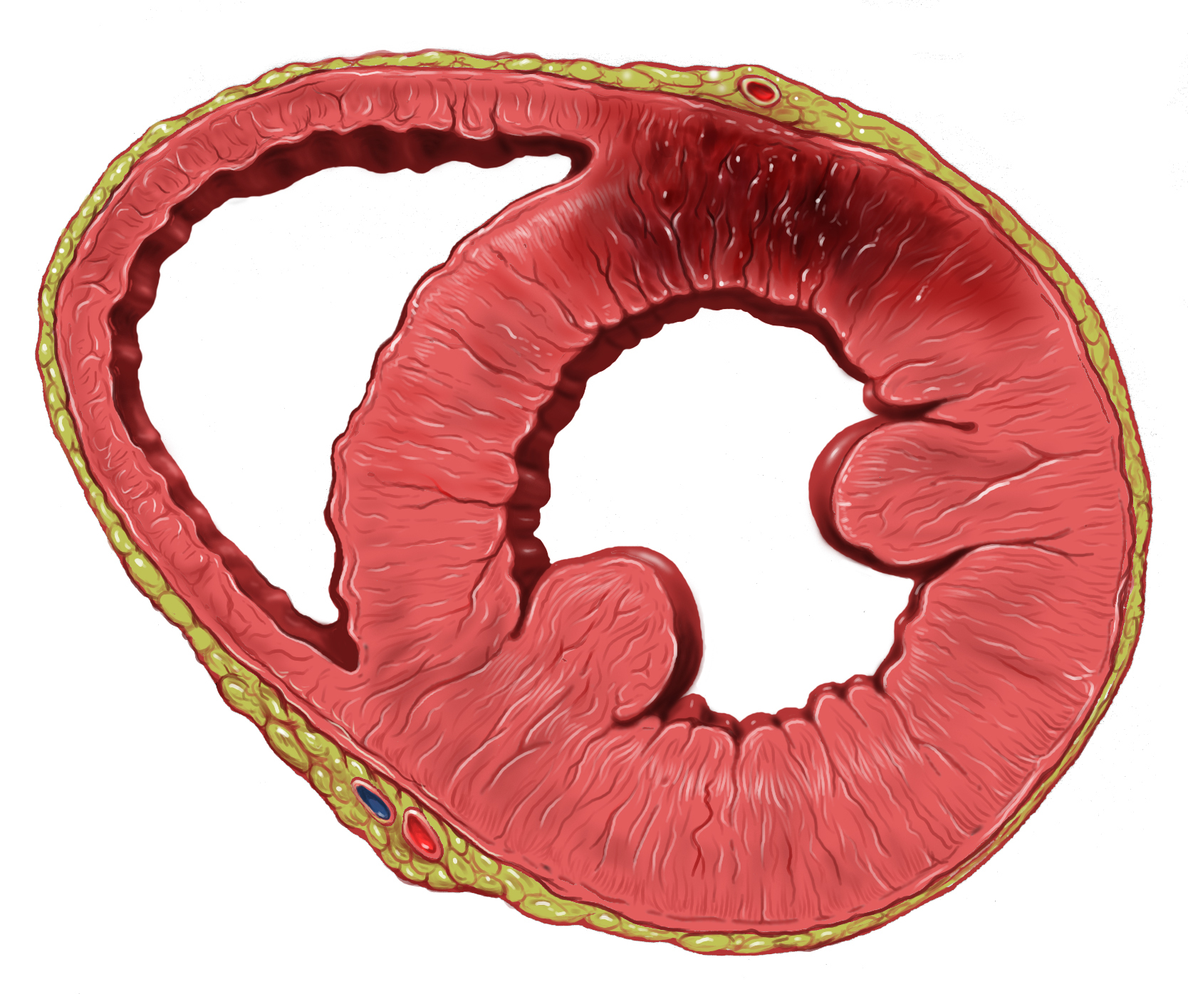
ST depression refers to a finding on an electrocardiogram, wherein the trace in the ST segment is abnormally low below the baseline. Causes It is often a sign of myocardial ischemia, of which coronary insufficiency is a major cause. Other ischemic heart diseases causing ST depression include: * Subendocardial ischemia or even infarction. Subendocardial means non full thickness ischemia. In contrast, ST elevation is transmural (or full thickness) ischemia * Non Q-wave myocardial infarction * Reciprocal changes in acute Q-wave myocardial infarction (e.g., ST depression in leads I & aVL with acute inferior myocardial infarction) * ST segment depression and T-wave changes may be seen in patients with unstable angina Depressed but ''upsloping'' ST segment generally rules out ischemia as a cause. Also, it can be a normal variant or artifacts, such as: * Pseudo-ST-depression, which is a wandering baseline due to poor skin contact of the electrode MicroEKG ManualRetrieved September ... [...More Info...] [...Related Items...] OR: [Wikipedia] [Google] [Baidu] |
False Negative
A false positive is an error in binary classification in which a test result incorrectly indicates the presence of a condition (such as a disease when the disease is not present), while a false negative is the opposite error, where the test result incorrectly indicates the absence of a condition when it is actually present. These are the two kinds of errors in a binary test, in contrast to the two kinds of correct result (a and a ). They are also known in medicine as a false positive (or false negative) diagnosis, and in statistical classification as a false positive (or false negative) error. In statistical hypothesis testing, the analogous concepts are known as type I and type II errors, where a positive result corresponds to rejecting the null hypothesis, and a negative result corresponds to not rejecting the null hypothesis. The terms are often used interchangeably, but there are differences in detail and interpretation due to the differences between medical testing and sta ... [...More Info...] [...Related Items...] OR: [Wikipedia] [Google] [Baidu] |
J-point
The QRS complex is the combination of three of the graphical deflections seen on a typical electrocardiogram (ECG or EKG). It is usually the central and most visually obvious part of the tracing. It corresponds to the depolarization of the right and left ventricles of the heart and contraction of the large ventricular muscles. In adults, the QRS complex normally lasts ; in children it may be shorter. The Q, R, and S waves occur in rapid succession, do not all appear in all leads, and reflect a single event and thus are usually considered together. A Q wave is any downward deflection immediately following the P wave. An R wave follows as an upward deflection, and the S wave is any downward deflection after the R wave. The T wave follows the S wave, and in some cases, an additional U wave follows the T wave. To measure the QRS interval start at the end of the PR interval (or beginning of the Q wave) to the end of the S wave. Normally this interval is 0.08 to 0.10 seconds. Whe ... [...More Info...] [...Related Items...] OR: [Wikipedia] [Google] [Baidu] |
Electrocardiography
Electrocardiography is the process of producing an electrocardiogram (ECG or EKG), a recording of the heart's electrical activity through repeated cardiac cycles. It is an electrogram of the heart which is a graph of voltage versus time of the electrical activity of the heart using electrodes placed on the skin. These electrodes detect the small electrical changes that are a consequence of cardiac muscle depolarization followed by repolarization during each cardiac cycle (heartbeat). Changes in the normal ECG pattern occur in numerous cardiac abnormalities, including: * Cardiac rhythmicity, Cardiac rhythm disturbances, such as atrial fibrillation and ventricular tachycardia; * Inadequate coronary artery blood flow, such as myocardial ischemia and myocardial infarction; * and electrolyte disturbances, such as hypokalemia. Traditionally, "ECG" usually means a 12-lead ECG taken while lying down as discussed below. However, other devices can record the electrical activity of ... [...More Info...] [...Related Items...] OR: [Wikipedia] [Google] [Baidu] |
Myocardial Infarction
A myocardial infarction (MI), commonly known as a heart attack, occurs when Ischemia, blood flow decreases or stops in one of the coronary arteries of the heart, causing infarction (tissue death) to the heart muscle. The most common symptom is retrosternal Angina, chest pain or discomfort that classically radiates to the left shoulder, arm, or jaw. The pain may occasionally feel like heartburn. This is the dangerous type of acute coronary syndrome. Other symptoms may include shortness of breath, nausea, presyncope, feeling faint, a diaphoresis, cold sweat, Fatigue, feeling tired, and decreased level of consciousness. About 30% of people have atypical symptoms. Women more often present without chest pain and instead have neck pain, arm pain or feel tired. Among those over 75 years old, about 5% have had an MI with little or no history of symptoms. An MI may cause heart failure, an Cardiac arrhythmia, irregular heartbeat, cardiogenic shock or cardiac arrest. Most MIs occur d ... [...More Info...] [...Related Items...] OR: [Wikipedia] [Google] [Baidu] |


